Primary Intracranial Ewing Sarcoma With EWSR1-FLI1 Gene Translocation Mimicking a Meningioma and a Multidisciplinary Therapeutic Approach: A Case Report and Systematic Review of Literatures
- Affiliations
-
- 1Department of Premedicine, Seoul National University College of Medicine, Seoul, Korea
- 2Departments of Neurosurgery, Seoul National University Bundang Hospital, Seoul National University College of Medicine, Seongnam, Korea
- 3Departments of Internal Medicine, Seoul National University Bundang Hospital, Seoul National University College of Medicine, Seongnam, Korea
- 4Departments of Radiology, Seoul National University Bundang Hospital, Seoul National University College of Medicine, Seongnam, Korea
- 5Departments of Pathology, Seoul National University Bundang Hospital, Seoul National University College of Medicine, Seongnam, Korea
- KMID: 2547418
- DOI: http://doi.org/10.14791/btrt.2023.0030
Abstract
- Ewing sarcoma and peripheral primitive neuroectodermal tumor (ES/pPNET) is an undifferentiated malignant tumor that is most prevalent in children and young adults and often radiologically mimics a meningioma. A 38-year-old female patient visited our hospital with complaints of right-sided tinnitus, right hemiparesis, and imbalance. She underwent preoperative imaging and was subsequently diagnosed as having a meningioma on the petrous ridge. After partial resection, EWSR1-FLI1 gene fusion was confirmed, and she was diagnosed with ES/pPNET. The tumor was successfully treated using a multidisciplinary approach of adjuvant chemo- and radiotherapy. This case is noteworthy because it is an extremely rare case of an intracranial ES/pPNET, and it is worth sharing our clinical experience that the tumor was successfully treated through a multidisciplinary therapeutic approach even though complete resection was not achieved.
Figure
-

Fig. 1 Preoperative images of the patient: T1-weighted image (A), T2-weighted image (B), contrast-enhanced T1-weighted image (C), ADC map (D), and CBV map (E) of MRI; and 18F-FDG PET/CT (F). ADC, apparent diffusion coefficient; CBV, cerebral blood volume; FDG, fluorodeoxyglucose; PET/CT, positron emission tomography/computed tomography.

Fig. 2 Pathologic results of the patient. Tissue stained by hematoxylin and eosin (A), immunohistochemically stained by GFAP (B), VMA (C), CD99 (D), and vimentin (E), and tissue with fluorescence in situ hybridization using dual-color break-apart rearrangement probes which flank the EWSR1 breakpoint (F). Pseudorosette formation and rearranged chromosome 22 are annotated by white triangles (A) and arrows (B), respectively. GFAP, glial fibrillary acidic protein; VMA, vimentin antibodies; CD, cluster of differentiation.
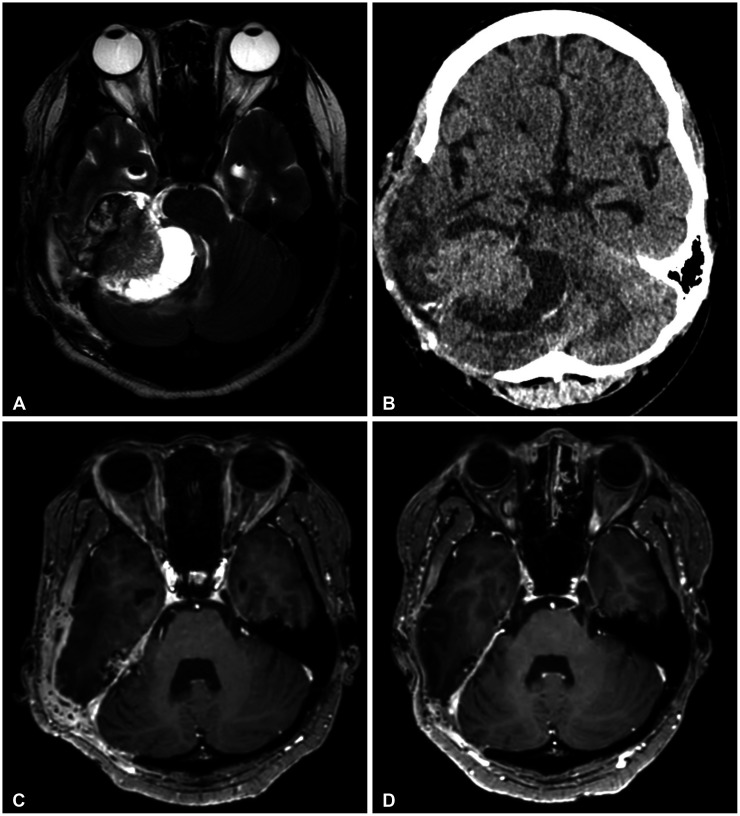
Fig. 3 Postoperative images of the patient: T2-weighted image at POD 15 (A), CT at POD 31 (B), contrast-enhanced T1-weighted image 3 months after surgery (C), and contrast-enhanced T1-weighted image 10 months after surgery (D). POD, postoperative day.
Reference
-
1. Chen J, Cheng R, Fan F, Zheng Y, Li Y, Chen Y, et al. Cranial Ewing sarcoma/peripheral primitive neuroectodermal tumors: a retrospective study focused on prognostic factors and long-term outcomes. Front Oncol. 2019; 9:1023. PMID: 31649882.
Article2. Deshpande G, Epari S, Gupta C, Shetty O, Gurav M, Chinnaswamy G, et al. Primary intracranial Ewing sarcoma/peripheral primitive neuroectodermal tumor, an entity of unacquaintance: a series of 8 cases. Childs Nerv Syst. 2021; 37:839–849. PMID: 32761378.
Article3. Geng Z, Gao W, Cheng W, Wu A. Primary intracranial Ewing sarcoma invading the superior sagittal sinus with EWSR1-FLI1 gene fusion and EWSR1 gene mutation: a case report and literature review. World Neurosurg. 2023; 175:1–10. PMID: 36990350.
Article4. Jiang Y, Zhao L, Wang Y, Liu X, Wu X, Li Y. Primary intracranial Ewing sarcoma/peripheral primitive neuroectodermal tumor mimicking meningioma: a case report and literature review. Front Oncol. 2020; 10:528073. PMID: 33123461.
Article5. Cherif El Asri A, Benzagmout M, Chakour K, Chaoui MF, Laaguili J, Chahdi H, et al. Primary intracranial pPNET/Ewing sarcoma: diagnosis, management, and prognostic factors dilemma–a systematic review of the literature. World Neurosurg. 2018; 115:346–356. PMID: 29729469.
Article6. Yim J, Lee WS, Kim SK, Kang HJ, Bae J, Park SH. Intracranial Ewing sarcoma with whole genome study. Childs Nerv Syst. 2019; 35:547–552. PMID: 30406421.
Article7. Haveman LM, Ranft A, Berg HVD, Klco-Brosius S, Ladenstein R, Paulussen M, et al. Primary and metastatic intracranial Ewing sarcoma at diagnosis: retrospective international study and systematic review. Cancers (Basel). 2020; 12:1675. PMID: 32599807.
Article8. Mazur MA, Gururangan S, Bridge JA, Cummings TJ, Mukundan S, Fuchs H, et al. Intracranial Ewing sarcoma. Pediatr Blood Cancer. 2005; 45:850–856. PMID: 15929128.
Article9. Tanboon J, Sitthinamsuwan B, Paruang T, Marrano P, Thorner PS. Primary intracranial Ewing sarcoma with an unusually aggressive course: a case report and review of the literature. Neuropathology. 2012; 32:293–300. PMID: 22007764.
Article10. Ke Ch, Duan Q, Yang H, Zhu F, Yan M, Xu SP, et al. Meningeal Ewing sarcoma/peripheral PNET: clinicopathological, immunohistochemical and FISH study of four cases. Neuropathology. 2017; 37:35–44. PMID: 27500883.
Article11. Kumar V, Singh A, Sharma V, Kumar M. Primary intracranial dural-based Ewing sarcoma/peripheral primitive neuroectodermal tumor mimicking a meningioma: a rare tumor with review of literature. Asian J Neurosurg. 2017; 12:351–357. PMID: 28761507.
Article12. VandenHeuvel KA, Al-Rohil RN, Stevenson ME, Qian J, Gross NL, McNall-Knapp R, et al. Primary intracranial Ewing’s sarcoma with unusual features. Int J Clin Exp Pathol. 2015; 8:260–274. PMID: 25755713.13. Salunke P, Sharma M, Gupta K. Ewing sarcoma of the occipital bone in an elderly patient. World Neurosurg. 2014; 81:e10–e12.
Article14. Cole M, Parajuli S, Laske D, Goldstein L, Morrison T, Mukherjee A, et al. Peripheral primitive neuroectodermal tumor of the dura in a 51-year-old woman following intensive treatment for breast cancer. Am J Case Rep. 2014; 15:294–299. PMID: 25045413.
Article15. Antonelli M, Caltabiano R, Chiappetta C, Oliva MA, Giangaspero F, Lanzafame S. Primary peripheral PNET/Ewing’s sarcoma arising in the meninges, confirmed by the presence of the rare translocation t(21;22) (q22;q12). Neuropathology. 2011; 31:549–555. PMID: 21284749.
Article16. Mellai M, Caldera V, Comino A, Fortunato M, Bernucci C, Schiffer D. PNET/ESFT of the cranial vault: a case report. Clin Neuropathol. 2010; 29:372–377. PMID: 21073841.
Article17. Attabib NA, West M, Rhodes RH. Peripheral primitive neuroectodermal tumor of the cavernous sinus: case report. Neurosurgery. 2006; 58:E992. PMID: 16639307.
Article18. Mobley BC, Roulston D, Shah GV, Bijwaard KE, McKeever PE. Peripheral primitive neuroectodermal tumor/Ewing’s sarcoma of the craniospinal vault: case reports and review. Hum Pathol. 2006; 37:845–853. PMID: 16784984.
Article19. D’Antonio A, Caleo A, Garcia JF, Marsilia GM, De Dominicis G, Boscaino A. Primary peripheral PNET/Ewing’s sarcoma of the dura with FISH analysis. Histopathology. 2004; 45:651–654. PMID: 15569062.
Article20. Simmons MA, Luff DA, Banerjee SS, Ramsden RT. Peripheral primitive neuroectodermal tumour (pPNET) of the cerebellopontine angle presenting in adult life. J Laryngol Otol. 2001; 115:848–852. PMID: 11668007.
Article21. Kalamarides M, Dewolf E, Couvelard A, Shahidi A, Bouccara D, Cyna-Gorse F, et al. Extraaxial primitive neuroectodermal tumor mimicking a vestibular schwannoma: diagnostic and therapeutic difficulties. Report of two cases. J Neurosurg. 2001; 94:612–616. PMID: 11302660.
Article22. Papotti M, Abbona G, Pagani A, Monga G, Bussolati G. Primitive neuroectodermal tumor of the meninges: an histological, immunohistochemical, ultrastructural, and cytogenetic study. Endocr Pathol. 1998; 9:275–280. PMID: 12114719.
Article
