J Yeungnam Med Sci.
2023 Oct;40(4):423-425. 10.12701/jyms.2022.00549.
Porokeratosis ptychotropica: a case report
- Affiliations
-
- 1Department of Dermatology, Keimyung University School of Medicine, Daegu, Korea
- KMID: 2547367
- DOI: http://doi.org/10.12701/jyms.2022.00549
Abstract
- Porokeratosis ptychotropica is an uncommon form of porokeratosis, which was initially described in 1995. It is clinically characterized by symmetrical reddish to brown-colored hyperkeratotic, verrucous, or psoriasiform plaques on the perianal and gluteal regions. The lesions tend to integrate and expand centrally, with small peripheral satellite lesions. Early skin biopsy and appropriate diagnosis are essential because malignant change occurs in 7.5% of porokeratotic lesions. Conventional treatment options include topical steroid, retinoid, imiquimod, 5-fluorouracil, isotretinoin, excimer laser, photodynamic therapy, intralesional steroid or bleomycin injection, cryotherapy, carbon dioxide (CO2) laser, and dermatome and excision, but none seem to achieve complete clearance. A 68-year-old woman presented with diffuse hyperkeratotic scaly lichenoid plaques on the buttocks that had persisted for several years. A skin biopsy of the buttocks revealed multiple cornoid lamellae and intense hyperkeratosis. There were some dyskeratotic cells beneath the cornoid lamellae and the granular layer was absent. Porokeratosis ptychotropica was diagnosed based on the characteristic clinical appearance and typical histopathological manifestations. She was treated with a CO2 laser in one session and topical application of urea and imiquimod cream for 1 month. The lesions slightly improved at the 1-month follow-up. We herein present a rare case of porokeratosis ptychotropica.
Keyword
Figure
-
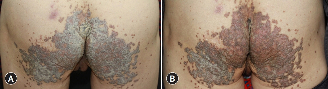
Fig. 1. (A) Asymptomatic diffuse hyperkeratotic scaly lichenoid plaques on the buttock that had persisted for several years. (B) The lesions are slightly improved at the 1-month follow-up.
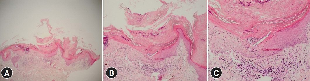
Fig. 2. (A) Skin biopsy of the buttocks reveals multiple cornoid lamellae and intense hyperkeratosis (hematoxylin and eosin [H&E] stain, x40). (B, C) Beneath the cornoid lamellae, there are some dyskeratotic cells and the granular layer is absent (H&E stain; [B] x100, [C] x200).
Reference
-
References
1. Lucker GP, Happle R, Steijlen PM. An unusual case of porokeratosis involving the natal cleft: porokeratosis ptychotropica? Br J Dermatol. 1995; 132:150–1.
Article2. Yeo J, Winhoven S, Tallon B. Porokeratosis ptychotropica: a rare and evolving variant of porokeratosis. J Cutan Pathol. 2013; 40:1042–7.
Article3. McGuigan K, Shurman D, Campanelli C, Lee JB. Porokeratosis ptychotropica: a clinically distinct variant of porokeratosis. J Am Acad Dermatol. 2009; 60:501–3.
Article4. Wang Q, Wan H, Liu W, Zhang L. Porokeratosis ptychotropica: a giant lesion in a Chinese man. Australas J Dermatol. 2017; 58:e149–50.
Article5. Liu W, Liu JW, Ma DL. Porokeratosis ptychotropica. JAMA Dermatol. 2019; 155:845.
Article6. Mazori DR, Shvartsbeyn M, Meehan SA, Tarsis SL. Transformation of porokeratosis ptychotropica into invasive squamous cell carcinoma. Int J Dermatol. 2017; 56:679–80.
Article7. Kawakami Y, Mitsui S. A case of porokeratosis ptychotropica: successful treatment with topical 5% imiquimod cream. Clin Exp Dermatol. 2017; 42:839–41.
Article8. Fustà-Novell X, Podlipnik S, Combalia A, Morgado-Carrasco D, Ferrando J, Mascaró JM Jr, et al. Porokeratosis ptychotropica responding to photodynamic therapy: an alternative treatment for a refractory disease. Photodermatol Photoimmunol Photomed. 2017; 33:271–4.9. Ho T, Schwentker AR, Barron DR, Lucky AW. Clinical course of porokeratosis ptychotropica over 7 years in an otherwise healthy child. Pediatr Dermatol. 2020; 37:248–50.10. Pitney L, Weedon D, Pitney M. Porokeratosis ptychotropica: a rare variant with discrete lesions. Australas J Dermatol. 2015; 56:e28–9.
Article11. Choi HM, Kim SM, Kang JW, Ro BI, Cho HK. Porokeratosis ptychotropica: a lesser-known variant of porokeratosis. Korean J Dermatol. 2019; 57:562–3.12. Cho YM, Lee JS, Koo DW, Jung KE. Porokeratosis ptychotropica—a rare variant of porokeratosis: a case report. Korean J Dermatol. 2018; 56:653–5.13. Lee S, Choe SJ, Ahn SK. Porokeratosis ptychotropica coexisting with tinea corporis. Ann Dermatol. 2017; 29:506–8.
Article
- Full Text Links
-
- Actions
-
Cited
- CITED
-
- Close
- Share
-
- Similar articles
-
- Porokeratosis Ptychotropica: A Lesser-known Variant of Porokeratosis
- Porokeratosis Ptychotropica—a Rare Variant of Porokeratosis: A Case Report
- Porokeratosis Ptychotropica Coexisting with Tinea Corporis
- A Case of Porokeratosis Palmaris et Plantaris Disseminata
- Coexistence of Porokeratosis of Mibelli and Disseminated Superficial Actinic Porokeratosis
