Anat Cell Biol.
2023 Sep;56(3):367-373. 10.5115/acb.23.024.
Forensic age-at-death estimation using the sternal junction in Thai adults: an autopsy study
- Affiliations
-
- 1Department of Forensic Medicine, Faculty of Medicine Siriraj Hospital, Mahidol University, Bangkok, Thailand
- 2Department of Forensic Medicine, Faculty of Medicine, Naresuan University, Phitsanulok, Thailand
- KMID: 2546465
- DOI: http://doi.org/10.5115/acb.23.024
Abstract
- One of the main parameters in the analysis of skeletal remains in forensic anthropological cases is the estimation of age. This study aimed to investigate the correlation between age and the fusion status of the sternal junction. This crosssectional study was carried out on 184 sterna from 94 females and 90 males obtained from known-age cadavers in the Thai population. By direct observation, the fusion stage of the manubrio-sternal and sterno-xiphoidal junctions was studied and divided into unfused and fused joints. The results showed that a large proportion of the sterna remain unfused throughout adulthood, with fusion observed in both young and old cadavers. Insignificant differences in the rate of fusion, the sexes and ages were observed. None of the sterna under 30 years of age in females and 32 years of age in males showed fusion of the manubrio-sternal and sterno-xiphoidal junctions. Based on the variability of the sternal fusions observed in this study, we highlighted a very limited role of the sternum alone in the estimation of age in the Thai population.
Figure
-
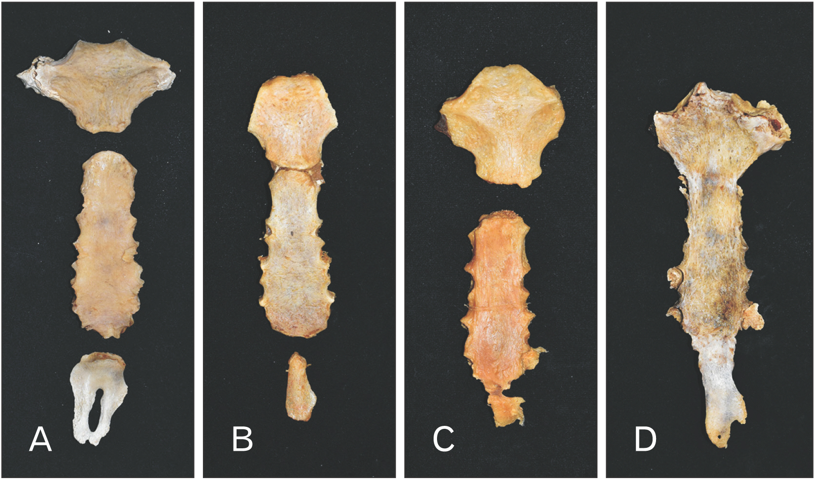
Fig. 1 Photograph showing the sterna with (A) no fusion at both junctions, (B) fusion at manubrio-sternal junction but no fusion at sterno-xiphoidal junction, (C) no fusion at manubrio-sternal junction but fusion at sterno-xiphoidal junction, and (D) fusion at both junctions.
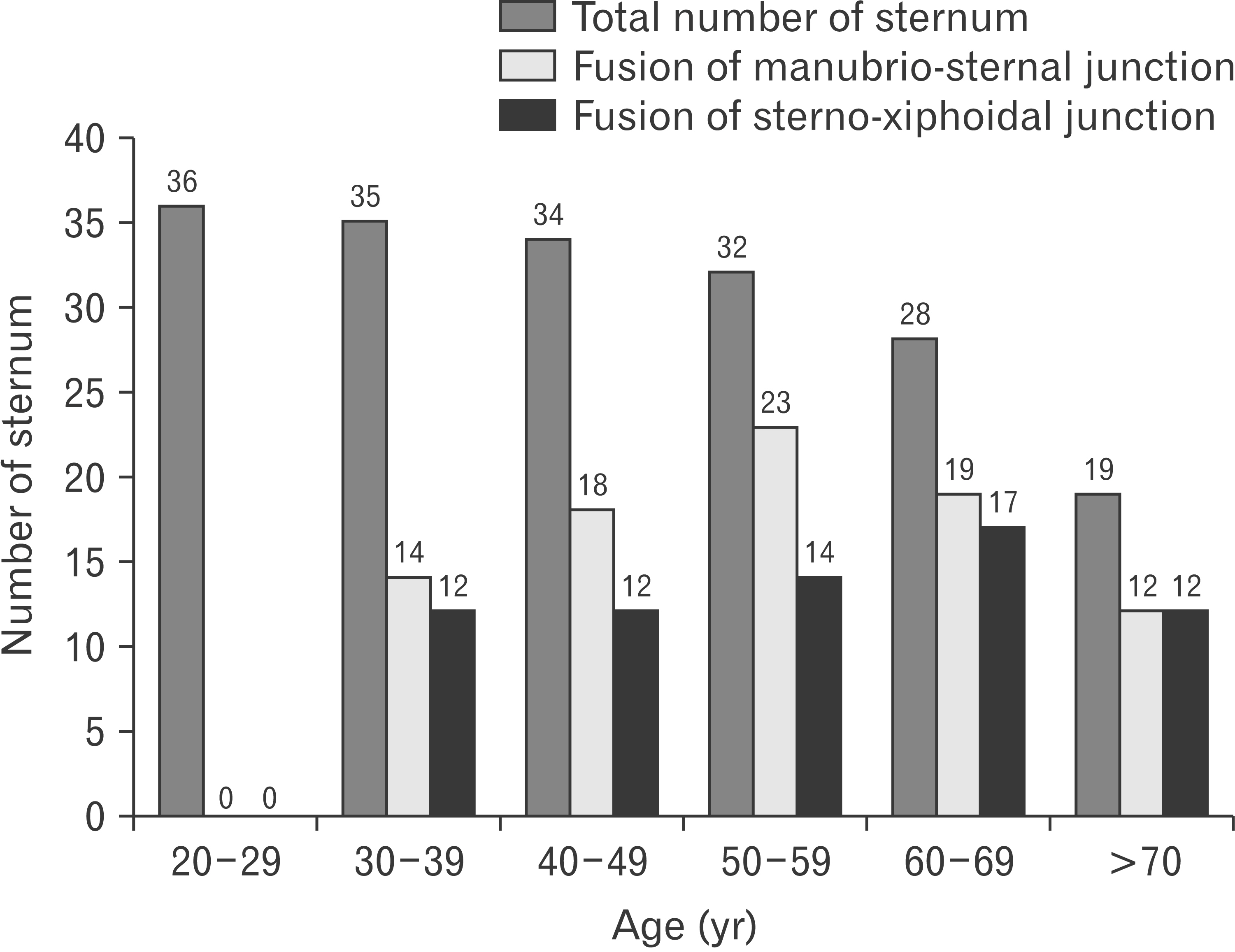
Fig. 2 Age distribution of the study population and the proportion of sterna with the fusion of manubrio-sternal and sterno-xiphoidal junctions.
Reference
-
References
1. Buikstra JE, Ubelaker DH. 1994. Standards for data collection from human skeletal remains: proceedings of a seminar at the field museum of natural history (Arkansas Archeological Survey Research Report). 12154th ed. Arkansas Archeological Survey.2. Franklin D. 2010; Forensic age estimation in human skeletal remains: current concepts and future directions. Leg Med (Tokyo). 12:1–7. DOI: 10.1016/j.legalmed.2009.09.001. PMID: 19853490.
Article3. Brooks S, Suchey JM. 1990; Skeletal age determination based on the os pubis: a comparison of the Acsádi-Nemeskéri and Suchey-Brooks methods. Hum Evol. 5:227–38. DOI: 10.1007/BF02437238.
Article4. Mulhern DM, Jones EB. 2005; Test of revised method of age estimation from the auricular surface of the ilium. Am J Phys Anthropol. 126:61–5. DOI: 10.1002/ajpa.10410. PMID: 15386244.
Article5. Bassed RB, Briggs C, Drummer OH. 2010; Analysis of time of closure of the spheno-occipital synchondrosis using computed tomography. Forensic Sci Int. 200:161–4. DOI: 10.1016/j.forsciint.2010.04.009. PMID: 20451338.
Article6. Işcan MY, Loth SR, Wright RK. 1984; Age estimation from the rib by phase analysis: white males. J Forensic Sci. 29:1094–104. DOI: 10.1520/JFS11776J. PMID: 6502109.7. Webb PA, Suchey JM. 1985; Epiphyseal union of the anterior iliac crest and medial clavicle in a modern multiracial sample of American males and females. Am J Phys Anthropol. 68:457–66. DOI: 10.1002/ajpa.1330680402. PMID: 4083337.
Article8. Chantharawetchakun T, Vachirawongsakorn V. 2021; Age estimation in the Thai male population using epiphyseal union of the medial clavicle. Chiang Mai Med J. 60:149–55. DOI: 10.12982/CMUMEDJ.2021.13.
Article9. Jeamamornrat V, Monum T, Keereewan W, Mahakkanukrauh P. 2022; Stature estimation using the sternum in a Thai population. Anat Cell Biol. 55:170–8. DOI: 10.5115/acb.22.045. PMID: 35773219. PMCID: PMC9256492.
Article10. Ashley GT. 1954; The morphological and pathological significance of synostosis at the manubrio-sternal joint. Thorax. 9:159–66. DOI: 10.1136/thx.9.2.159. PMID: 13179129. PMCID: PMC1019362.11. Sun YX, Zhao GC, Yan W. 1995; Age estimation on the female sternum by quantification theory I and stepwise regression analysis. Forensic Sci Int. 74:57–62. DOI: 10.1016/0379-0738(95)01737-4. PMID: 7665133.
Article12. Chandrakanth HV, Kanchan T, Krishan K, Arun M, Pramod Kumar GN. 2012; Estimation of age from human sternum: an autopsy study on a sample from South India. Int J Legal Med. 126:863–8. DOI: 10.1007/s00414-012-0752-0. PMID: 22875076.
Article13. Singh J, Pathak RK. 2013; Sex and age related non-metric variation of the human sternum in a Northwest Indian postmortem sample: a pilot study. Forensic Sci Int. 228:181.e1–12. DOI: 10.1016/j.forsciint.2013.02.002. PMID: 23453187.
Article14. Chopra M, Singh H, Kohli K, Aggarwal OP. 2014; Age estimation from sternum for age group 25 years onwards. J Indian Acad Forensic Med. 36:340–2.15. Manoharan C, Dhanalakshmi V, Thangam D, Joe AE. 2016; Estimation of age from human sternum- an autopsy study. Indian J Forensic Community Med. 3:128–32. DOI: 10.5958/2394-6776.2016.00029.1.16. Sahu MR, Tripathy PR, Mohanty MK, Padhi KS, Naveen A. 2022; Age estimation from sternum of eastern Indian population: autopsy based study. Indian J Forensic Community Med. 9:59–64. DOI: 10.18231/j.ijfcm.2022.014.
Article17. Bacci N, Nchabeleng EK, Billings BK. 2018; Forensic age-at-death estimation from the sternum in a black South African population. Forensic Sci Int. 282:233.e1–7. DOI: 10.1016/j.forsciint.2017.11.002. PMID: 29195663.
Article18. Oktay C, Aytaç G. 2022; Evaluation of manubriosternal joint fusion and second costal cartilage calcification: are they useful for estimating advanced age groups? J Forensic Sci. 67:450–9. DOI: 10.1111/1556-4029.14951. PMID: 34893986.
Article19. Bolatlı G, Ünver Doğan N, Koplay M, Fazlıoğulları Z, Karabulut AK. 2020; Evaluation of sternal morphology according to age and sex with multidetector computerized tomography. Anatomy. 14:29–38. DOI: 10.2399/ana.20.016.
Article20. Ali MIM, Mosallam W, Mostafa EMA, Aly SM, Ali NM. 2021; Sternum as an indicator for sex and age estimation using multidetector computed tomography in an Egyptian population. Forensic Imaging. 26:200457. DOI: 10.1016/j.fri.2021.200457.
Article21. Macaluso PJ, Lucena J. 2014; Morphological variations of the anterior thoracic skeleton and their forensic significance: radiographic findings in a Spanish autopsy sample. Forensic Sci Int. 241:220.e1–7. DOI: 10.1016/j.forsciint.2014.05.009. PMID: 24933632.
Article22. Monum T, Makino Y, Prasitwattanaseree S, Yajima D, Chiba F, Torimitsu S, Hoshioka Y, Yoshida M, Urabe S, Oya Y, Iwase H. 2020; Age estimation from ossification of sternum and true ribs using 3D post-mortem CT images in a Japanese population. Leg Med (Tokyo). 43:101663. DOI: 10.1016/j.legalmed.2019.101663. PMID: 31954957.
Article23. Monum T, Mekjaidee K, Pattamapaspong N, Prasitwattanaseree S. 2017; Age estimation by chest plate radiographs in a Thai male population. Sci Justice. 57:169–73. DOI: 10.1016/j.scijus.2017.02.003. PMID: 28454625.
Article24. O'Neal ML, Dwornik JJ, Ganey TM, Ogden JA. 1998; Postnatal development of the human sternum. J Pediatr Orthop. 18:398–405. DOI: 10.1097/01241398-199805000-00024. PMID: 9600571.25. Bayaroğulları H, Yengil E, Davran R, Ağlagül E, Karazincir S, Balcı A. 2014; Evaluation of the postnatal development of the sternum and sternal variations using multidetector CT. Diagn Interv Radiol. 20:82–9. DOI: 10.5152/dir.2013.13121. PMID: 24100061. PMCID: PMC4463249. PMID: 78dd171619aa4c48957fa983e336a0e9.26. Cunningham C, Scheuer L, Black S, Liversidge H, Christie A. 2016. Developmental juvenile osteology. 2nd ed. Elsevier Science;p. 235–7. DOI: 10.1016/B978-0-12-382106-5.00003-7.27. Parreira VF, Bueno CJ, França DC, Vieira DS, Pereira DR, Britto RR. 2010; Breathing pattern and thoracoabdominal motion in healthy individuals: influence of age and sex. Rev Bras Fisioter. 14:411–6. DOI: 10.1590/S1413-35552010000500010. PMID: 21180867.28. Kaneko H, Horie J. 2012; Breathing movements of the chest and abdominal wall in healthy subjects. Respir Care. 57:1442–51. DOI: 10.4187/respcare.01655. PMID: 22348414.29. Jordan S, Lim L, Seubsman SA, Bain C, Sleigh A. 2012; Secular changes and predictors of adult height for 86 105 male and female members of the Thai Cohort Study born between 1940 and 1990. J Epidemiol Community Health. 66:75–80. DOI: 10.1136/jech.2010.113043. PMID: 20805198. PMCID: PMC3230828.30. Saunders SR, Fitzgerald C, Rogers T, Dudar C, McKillop H. 1992; A test of several methods of skeletal age estimation using a documented archaeological sample. Can Soc Forensic Sci J. 25:97–118. DOI: 10.1080/00085030.1992.10757005.
Article31. Baccino E, Ubelaker DH, Hayek LA, Zerilli A. 1999; Evaluation of seven methods of estimating age at death from mature human skeletal remains. J Forensic Sci. 44:931–6. DOI: 10.1520/JFS12019J. PMID: 10486944.
Article
- Full Text Links
-
- Actions
-
Cited
- CITED
-
- Close
- Share
-
- Similar articles
-
- Stature estimation using the sternum in a Thai population
- Profile of Forensic Autopsy Practices in Small Towns and Cities in Rural Areas
- Examination of Abdominal Organ in Forensic Autopsy
- Discrepancies in the Cause and Manner of Death Reported in Postmortem Inspection and Autopsy
- Sudden Death associated with Thyrotoxicosis: Report of Three Autopsy Cases
