Morphology of middle cerebral artery using computed tomography angiographic study in a tertiary care hospital
- Affiliations
-
- 1Department of Anatomy, All India Institute of Medical Sciences, Raipur, India
- 2Department of Anatomy, Jawaharlal Institute of Postgraduate Medical Education and Research, Puducherry, India
- 3Department of Radiodiagnosis, Jawaharlal Institute of Postgraduate Medical Education and Research, Puducherry, India
- KMID: 2546464
- DOI: http://doi.org/10.5115/acb.22.242
Abstract
- Increased tortuosity of vessel is associated with high incidence of plaque formation leading to atherosclerosis. Surgical procedures are done after analyzing morphology of middle cerebral artery (MCA). However, literature describing MCA morphology using computed tomography angiography (CTA) is limited, so this study was planned to determine its incidence in Indian population. Datasets of CTA from 289 patients (180 males and 109 females), average age: 49.29±16.16 years (range: 11 to 85 years), from a tertiary care hospital were systematically reviewed for morphology of MCA. Cases involving aneurysms and infarcts were excluded. Four shapes of MCA were recognized: straight, U, inverted U, and S-shaped. MCA was straight in 44% (254/578), U-shaped in 37% (215/578), S shaped in 15% (89/578) and inverted U-shaped in 3% (20/578) cases. In males, MCA was straight in 46% (166/360), U-shaped in 37% (134/360), S-shaped in 16% (58/360) and inverted U-shaped in 4% (14/360) cases. In females, MCA was straight in 42% cases (92/218), U-shaped in 37% (81/218), S-shaped in 17% (36/218) and inverted U-shaped in 4% (9/218). On comparing shape with various age groups using chi square test, U shaped (P≤0.001) and S-shaped (P=0.003) MCA were found to be statistically significant. The incidence of straight shape was higher in advanced age group (>60 years). Knowledge of MCA shape will be useful for clinicians and surgeons in successful endovascular recanalization. Also, this data would help surgeons during neurointerventional procedures.
Keyword
Figure
-
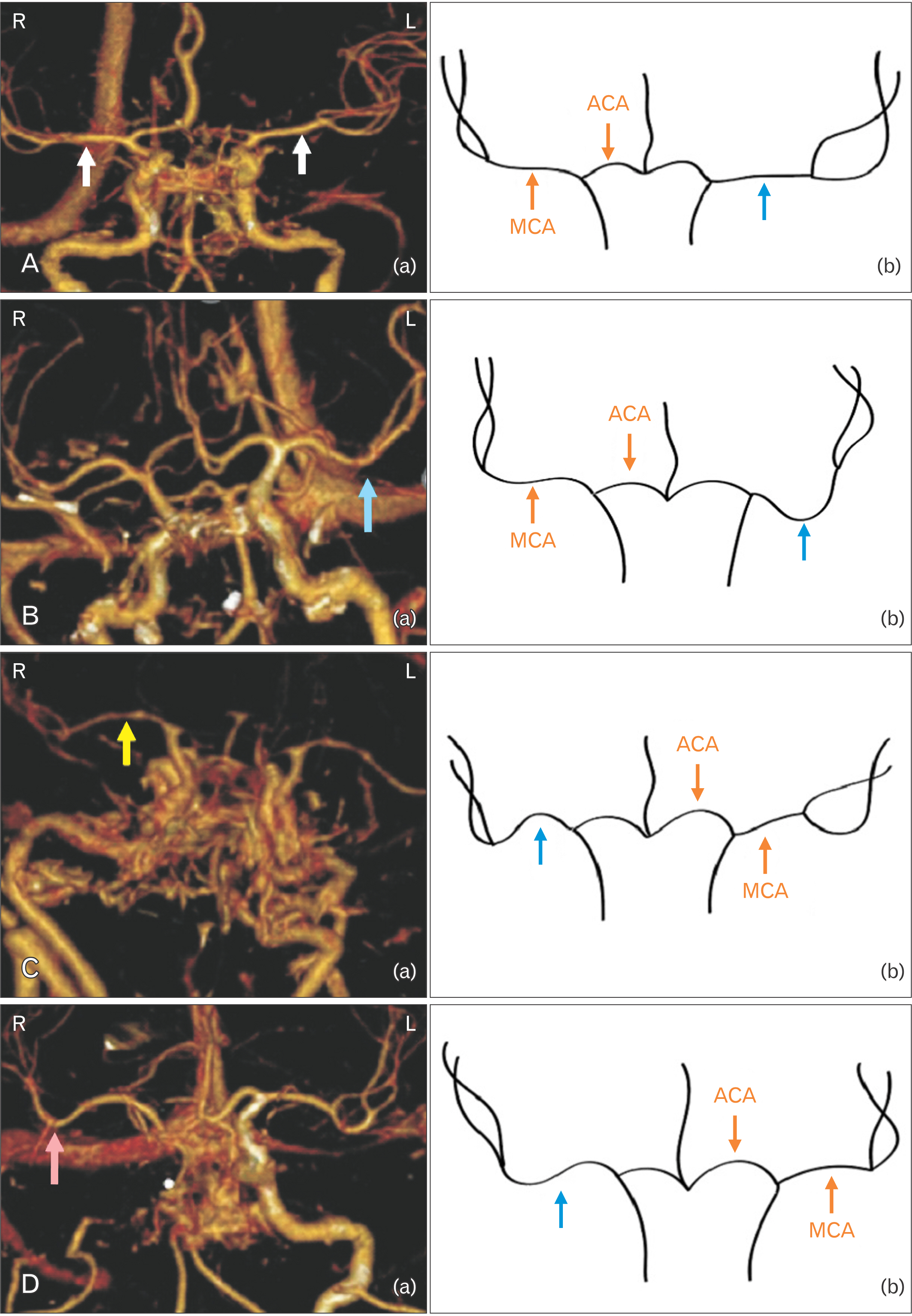
Fig. 1 VRT images (3D) and schematic diagrams showing shapes of middle cerebral artery. (A) Straight shape. (a) VRT image (3D) showing bilateral straight-shaped middle cerebral artery on both sides. White arrow: single trunk middle cerebral artery. (b) Schematic diagram showing bilateral straight-shaped middle cerebral artery on both sides. Blue arrow: single trunk middle cerebral artery. (B) U-shape. (a) VRT image (3D) showing unilateral U-shaped middle cerebral artery on left side. (b) Schematic diagram showing U-shaped middle cerebral artery on left side. Blue arrow: U-shaped middle cerebral artery. (C) Inverted U-shape. (a) VRT image (3D) showing inverted U-shaped middle cerebral artery on right side. Yellow arrow: Inverted U-shaped middle cerebral artery. (b) Schematic diagram showing inverted U-shaped middle cerebral artery on right side. Blue arrow: Inverted U- shaped middle cerebral artery. (D) S-shape. (a) VRT image (3D) showing S-shaped middle cerebral artery on right side. Pink arrow: S-shaped middle cerebral artery. (b) Schematic diagram showing S-shaped middle cerebral artery on right side. Blue arrow: S-shaped middle cerebral artery. VRT, volume rendered technique; R, right; L, left; ACA, anterior cerebral artery, MCA, middle cerebral artery.
Reference
-
References
1. Shalom DE, Trevisan MA, Mallela A, Nuñez M, Goldschmidt E. 2021; Brain folding shapes the branching pattern of the middle cerebral artery. PLoS One. 16:e0245167. DOI: 10.1371/journal.pone.0245167. PMID: 33411825. PMCID: PMC7790398. PMID: aea8835576414283859b7916dc0e1fb2.
Article2. Han J, Qiao H, Li X, Li X, He Q, Wang Y, Cheng Z. 2014; The three-dimensional shape analysis of the M1 segment of the middle cerebral artery using MRA at 3T. Neuroradiology. 56:995–1005. DOI: 10.1007/s00234-014-1414-3. PMID: 25119254.
Article3. Mazighi M, Tanasescu R, Ducrocq X, Vicaut E, Bracard S, Houdart E, Woimant F. 2006; Prospective study of symptomatic atherothrombotic intracranial stenoses: the GESICA study. Neurology. 66:1187–91. DOI: 10.1212/01.wnl.0000208404.94585.b2. PMID: 16636236.
Article4. Sánchez-Sánchez C, Egido JA, González-Gutiérrez JL, Mera-Campillo J, Carneado-Ruiz J, Díaz-Otero F. 2004; Stroke and intracranial stenosis: clinical profile in a series of 134 patients in Spain. Rev Neurol. 39:305–11. Spanish. DOI: 10.33588/rn.3904.2004147. PMID: 15340886.5. Malek AM, Alper SL, Izumo S. 1999; Hemodynamic shear stress and its role in atherosclerosis. JAMA. 282:2035–42. DOI: 10.1001/jama.282.21.2035. PMID: 10591386.
Article6. Fuster V, Badimon JJ, Chesebro JH. 1998; Atherothrombosis: mechanisms and clinical therapeutic approaches. Vasc Med. 3:231–9. DOI: 10.1177/1358836X9800300310. PMID: 9892516.7. Zhang C, Xie S, Li S, Pu F, Deng X, Fan Y, Li D. 2012; Flow patterns and wall shear stress distribution in human internal carotid arteries: the geometric effect on the risk for stenoses. J Biomech. 45:83–9. DOI: 10.1016/j.jbiomech.2011.10.001. PMID: 22079384.
Article8. Datir P, Lee AY, Lamm SD, Han HC. 2011; Effects of geometric variations on the buckling of arteries. Int J Appl Mech. 3:385–406. DOI: 10.1142/S1758825111001044. PMID: 22287983. PMCID: PMC3266375.
Article9. Smedby O, Johansson J, Mölgaard J, Olsson AG, Walldius G, Erikson U. 1995; Predilection of atherosclerosis for the inner curvature in the femoral artery. A digitized angiography study. Arterioscler Thromb Vasc Biol. 15:912–7. DOI: 10.1161/01.ATV.15.7.912. PMID: 7600123.10. Kim BJ, Yoon Y, Lee DH, Kang DW, Kwon SU, Kim JS. 2015; The shape of middle cerebral artery and plaque location: high-resolution MRI finding. Int J Stroke. 10:856–60. DOI: 10.1111/ijs.12497. PMID: 25846063.
Article11. Reçi V, Bexheti S. 2019; Morphologic variations of end trunks of M1 segment of middle cerebral artery. J Alzheimers Neurodegener Dis. 5:030. DOI: 10.24966/AND-9608/100030.
Article12. Yu YN, Li ML, Xu YY, Meng Y, Trieu H, Villablanca JP, Gao S, Feng F, Liebeskind DS, Xu WH. 2018; Middle cerebral artery geometric features are associated with plaque distribution and stroke. Neurology. 91:e1760–9. DOI: 10.1212/WNL.0000000000006468. PMID: 30291186.
Article13. Xu WH, Li ML, Gao S, Ni J, Zhou LX, Yao M, Peng B, Feng F, Jin ZY, Cui LY. 2011; Plaque distribution of stenotic middle cerebral artery and its clinical relevance. Stroke. 42:2957–9. DOI: 10.1161/STROKEAHA.111.618132. PMID: 21799160.
Article14. Kiresi D, Gumus S, Cengiz SL, Cicekcibasi A. 2009; The morphometric analysis of the V2 and V3 segments of the vertebral artery: normal values on MDCT. Comput Med Imaging Graph. 33:399–407. DOI: 10.1016/j.compmedimag.2009.03.006. PMID: 19394795.
Article15. Jeyakumar R, Veerapandian R. 2018; Study of anatomical variations in middle cerebral artery. Int J Sci Study. 5:5–10.16. Gibo H, Carver CC, Rhoton AL Jr, Lenkey C, Mitchell RJ. 1981; Microsurgical anatomy of the middle cerebral artery. J Neurosurg. 54:151–69. DOI: 10.3171/jns.1981.54.2.0151. PMID: 7452329.
Article17. Herman LH, Ostrowski AZ, Gurdjian ES. 1963; Perforating branches of the middle cerebral artery. An anatomical study. Arch Neurol. 8:32–4. DOI: 10.1001/archneur.1963.00460010048005. PMID: 13961097.18. Vuillier F, Medeiros E, Moulin T, Cattin F, Bonneville JF, Tatu L. 2008; Main anatomical features of the M1 segment of the middle cerebral artery: a 3D time-of-flight magnetic resonance angiography at 3 T study. Surg Radiol Anat. 30:509–14. DOI: 10.1007/s00276-008-0360-3. PMID: 18465079.
Article19. Lazorthes G, Gouazé A, Salamon G. 1976. Vascularisation et circulation de l'encéphale. Les veines de l'encéphale. Masson.20. Rosner SS, Rhoton AL Jr, Ono M, Barry M. 1984; Microsurgical anatomy of the anterior perforating arteries. J Neurosurg. 61:468–85. DOI: 10.3171/jns.1984.61.3.0468. PMID: 6747683.
Article21. Nomura T. 1970. Atlas of cerebral angiography. Springer;DOI: 10.1007/978-3-030-16095-1_1.22. Benetos A, Waeber B, Izzo J, Mitchell G, Resnick L, Asmar R, Safar M. 2002; Influence of age, risk factors, and cardiovascular and renal disease on arterial stiffness: clinical applications. Am J Hypertens. 15:1101–8. DOI: 10.1016/S0895-7061(02)03029-7. PMID: 12460708.
Article23. Stone PH, Coskun AU, Kinlay S, Clark ME, Sonka M, Wahle A, Ilegbusi OJ, Yeghiazarians Y, Popma JJ, Orav J, Kuntz RE, Feldman CL. 2003; Effect of endothelial shear stress on the progression of coronary artery disease, vascular remodeling, and in-stent restenosis in humans: in vivo 6-month follow-up study. Circulation. 108:438–44. DOI: 10.1161/01.CIR.0000080882.35274.AD. PMID: 12860915.
Article24. Asakura T, Karino T. 1990; Flow patterns and spatial distribution of atherosclerotic lesions in human coronary arteries. Circ Res. 66:1045–66. DOI: 10.1161/01.RES.66.4.1045. PMID: 2317887.
Article25. Chistiakov DA, Orekhov AN, Bobryshev YV. 2017; Effects of shear stress on endothelial cells: go with the flow. Acta Physiol (Oxf). 219:382–408. DOI: 10.1111/apha.12725. PMID: 27246807.
Article26. Dolan JM, Kolega J, Meng H. 2013; High wall shear stress and spatial gradients in vascular pathology: a review. Ann Biomed Eng. 41:1411–27. DOI: 10.1007/s10439-012-0695-0. PMID: 23229281. PMCID: PMC3638073.
Article27. Schwaiger BJ, Gersing AS, Zimmer C, Prothmann S. 2015; The curved MCA: influence of vessel anatomy on recanalization results of mechanical thrombectomy after acute ischemic stroke. AJNR Am J Neuroradiol. 36:971–6. DOI: 10.3174/ajnr.A4222. PMID: 25634721. PMCID: PMC7990584.
Article28. Mori T, Fukuoka M, Kazita K, Mori K. 1998; Follow-up study after intracranial percutaneous transluminal cerebral balloon angioplasty. AJNR Am J Neuroradiol. 19:1525–33.
Article29. Zhu L, Liebeskind DS, Jahan R, Starkman S, Salamon N, Duckwiler G, Vinuela F, Tateshima S, Gonzalez N, Villablanca P, Ali LK, Kim D, Ovbiagele B, Froehler M, Tenser M, Saver JL. 2012; Thrombus branching and vessel curvature are important determinants of middle cerebral artery trunk recanalization with Merci thrombectomy devices. Stroke. 43:787–92. DOI: 10.1161/STROKEAHA.110.612986. PMID: 22282888. PMCID: PMC3288443.
Article30. Patti G, Pasceri V, Melfi R, Goffredo C, Chello M, D'Ambrosio A, Montesanti R, Di Sciascio G. 2005; Impaired flow-mediated dilation and risk of restenosis in patients undergoing coronary stent implantation. Circulation. 111:70–5. DOI: 10.1161/01.CIR.0000151308.06673.D2. PMID: 15630038.
Article31. Papafaklis MI, Chatzizisis YS, Naka KK, Giannoglou GD, Michalis LK. 2012; Drug-eluting stent restenosis: effect of drug type, release kinetics, hemodynamics and coating strategy. Pharmacol Ther. 134:43–53. DOI: 10.1016/j.pharmthera.2011.12.006. PMID: 22212618.
Article32. Santamarina A, Weydahl E, Siegel JM Jr, Moore JE Jr. 1998; Computational analysis of flow in a curved tube model of the coronary arteries: effects of time-varying curvature. Ann Biomed Eng. 26:944–54. DOI: 10.1114/1.113. PMID: 9846933.
Article33. Hademenos GJ, Massoud TF. 1997; Biophysical mechanisms of stroke. Stroke. 28:2067–77. DOI: 10.1161/01.STR.28.10.2067. PMID: 9341720.
Article34. Kliś KM, Krzyżewski RM, Kwinta BM, Łasocha B, Brzegowy P, Stachura K, Popiela TJ, Borek R, Gąsowski J. 2020; Increased tortuosity of basilar artery might be associated with higher risk of aneurysm development. Eur Radiol. 30:5625–32. DOI: 10.1007/s00330-020-06917-3. PMID: 32405752. PMCID: PMC7476915.
Article35. National Institute of Neurological Disorders and Stroke. 1950. Cerebral arteriosclerosis [Internet]. National Institutes of Health;Available from: https://www.ninds.nih.gov/health-information/disorders/cerebral-arteriosclerosis. cited 2023 Jan 23.
- Full Text Links
-
- Actions
-
Cited
- CITED
-
- Close
- Share
-
- Similar articles
-
- Angiographic Analysis of Middle Cerebral Artery Bifurcation Aneurysm
- Traumatic Occlusion of the Middle Cerebral Artery
- A Case bilateral Persistent Primitive Trigeminal Artery Combined with Cerebral Rete Mirabile
- Sphenoid Ridge Meningioma Presenting as Acute Cerebral Infarction
- Accessory Middle Cerebral Artery
