Anatomical knowledge of zygomatico-buccal plexus in a cadaveric study
- Affiliations
-
- 1Department of Anatomy, Faculty of Medicine, Chulalongkorn University and King Chulalongkorn Memorial Hospital, Bangkok, Thailand
- 2Preclinical Department, Faculty of Medicine, Siam University, Bangkok, Thailand
- 3Department of Biomedical Engineering, College of Health Sciences, Christian University of Thailand, Nakhonpathom, Thailand
- KMID: 2546327
- DOI: http://doi.org/10.5115/acb.23.040
Abstract
- The details of the facial nerve pattern were clearly explained in the parotid gland (PG), lateral area of the face, and periorbital areas to prevent the unexpected outcome of medical intervention. However, it remains unclear whether information about the zygomatico-buccal plexus (ZBP) in the masseteric and buccal regions. Therefore, this study aimed to help clinicians avoid this ZBP injury by predicting their common location. This study was conducted in forty-two hemifaces of twenty-nine embalmed cadavers by conventional dissection. The characteristics of the buccal branch (BB) and the ZBP were investigated in the mid-face region. The results presented that the BB gave 2–5 branches to emerge from the PG. According to the masseteric and buccal regions, the BB were arranged into ZBP in three patterns including an incomplete loop (11.9%), a single-loop (31.0%), and a multi-loop (57.1%). The mean distance and diameter of the medial line of the ZBP at the corner of the mouth level were 31.6 (6.7) and 1.5 (0.6) mm respectively, while at the alar base level were 22.5 (4.3) and 1.1 (0.6) mm respectively. Moreover, the angular nerve arose from the superior portion of the ZBP at the alar base level. The BB formed a multiloop mostly and showed a constant medial line of ZBP in an area approximately 30 mm lateral to the corner of the mouth, and 20 mm lateral to the alar base. Therefore, it is recommended that physicians should be very careful when performing facial rejuvenation in the mid-face region.
Figure
-

Fig. 1 The location of the emerging point of the BB is sporadically distributed throughout the anterior border of the PG. Then, it branched and arranged into the ZBP in masseteric and buccal regions, and presented the medial line of the ZBP branched into small twigs to innervate muscles around the mouth and nose. BB, buccal branch; ZBP, zygomatico-buccal plexus; TB, temporal branch; ZB, zygomatic branch; BB1, first buccal branch; BB2, second buccal branch; BB3, third buccal branch; MMB, marginal mandibular branch; CB, cervical branch; AN, angular nerve; PG, parotid gland; PD, parotid duct; MM, masseter muscle; LLS, levator labii superioris muscle; OO, orbicularis oculi muscle; OOr, orbicularis oris muscle; DAO, depressor anguli oris muscle; red arrowhead, the medial line of the ZBP; yellow arrowhead, small twigs of the BB; blue arrow, ZB gave branch to connect the BB; FHL, Frankfort’s horizontal line; midFHL, midpoint of the FHL; ATL, anterior tragal line; red circle, emerging point of the BB.

Fig. 2 The area for investigation the pattern of ZBP (red area). ZBP, zygomatico-buccal plexus; SAC, supra-alar crest; CM, the corner of the mouth; midFHL, midpoint of the Frankfort’s horizontal line. TB, temporal branch; ZB, zygomatic branch; BB1, first buccal branch; BB2, second buccal branch; BB3, third buccal branch; PG, parotid gland; PD, parotid duct; LLS, levator labii superioris muscle; OO, orbicularis oculi muscle; OOr, orbicularis oris muscle; red arrowhead, the medial line of the ZBP.

Fig. 3 Three patterns of the ZBP; incomplete (A, a), single (B, b), and multi-loop patterns (C, c). ZBP, zygomatico-buccal plexus; SAC, supra-alar crest; CM, corner of the mouth; PD, parotid duct; FA, facial artery; FV, facial vein; red arrowhead, ZBP; BB1, first buccal branch; BB2, second buccal branch; BB3, third buccal branch; LLS, levator labii superioris muscle; OOr, orbicularis oris muscle; ZMj, zygomaticus major muscle.
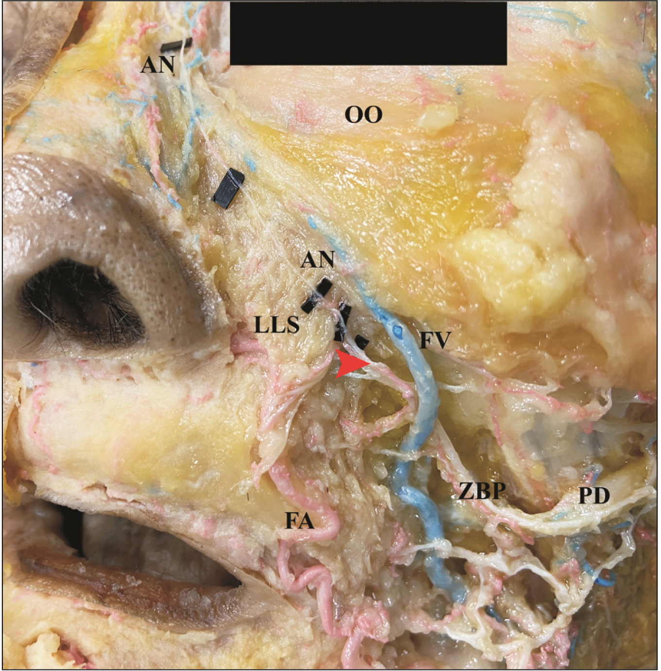
Fig. 4 The origin and location of the AN that arose from superior part of the ZBP. ZBP, zygomatico-buccal plexus; AN, angular nerve; PD, parotid duct; LLS, levator labii superioris muscle; OO, orbicularis oculi muscle; red arrowhead, the origin of the AN; FV, facial vein; FA, facial artery.
Reference
-
References
1. Bendella H, Spacca B, Rink S, Stoffels HJ, Nakamura M, Scaal M, Heinen H, Guntinas-Lichius O, Goldbrunner R, Grosheva M, Angelov DN. 2017; Anastomotic patterns of the facial parotid plexus (PP): a human cadaver study. Ann Anat. 213:52–61. DOI: 10.1016/j.aanat.2017.06.001. PMID: 28662373.
Article2. Hovland N, Phuong A, Lu GN. 2021; Anatomy of the facial nerve. Oper Tech Otolaryngol Head Neck Surg. 32:190–6. DOI: 10.1016/j.otot.2021.10.009.
Article3. Liu AT, Yu DZ, Chen G, Dang RS, Zhang YF, Zhang WJ, Liu BL, Jiang H. 2010; Profiling of innervations of mimetic muscles in fresh human cadavers using a modified Sihler's technique. Muscle Nerve. 42:88–94. DOI: 10.1002/mus.21622. PMID: 20544911.
Article4. Al-Shaikh KMS, Mutwakil M, Ahmed M, Zaghloul S. 2015; Anatomical study of the facial nerve. World J Zool. 10:267–73.5. Hwang K. 2010; Surgical anatomy of the lower eyelid relating to lower blepharoplasty. Anat Cell Biol. 43:15–24. DOI: 10.5115/acb.2010.43.1.15. PMID: 21190001. PMCID: PMC2998777.
Article6. Kehrer A, Engelmann S, Ruewe M, Geis S, Taeger C, Kehrer M, Prantl L, Tamm E, Bleys RRLAW, Mandlik V. 2019; Anatomical study of the zygomatic and buccal branches of the facial nerve: application to facial reanimation procedures. Clin Anat. 32:480–8. DOI: 10.1002/ca.23332. PMID: 30663808.
Article7. Jirawatnotai S, Kaewpichai K, Tirakotai W, Mothong W, Kaewsema A, Sriswadpong P. 2020; Nerve to the zygomaticus major muscle for facial reanimation surgery: a cadaveric study for branching patterns and axonal count. Asian J Neurosurg. 15:516–20. DOI: 10.4103/ajns.AJNS_90_20. PMID: 33145200. PMCID: PMC7591169.
Article8. Yoshioka N, Rhoton AL. 2015. Atlas of the facial nerve and its related structures. Thieme Publishers;p. 128. DOI: 10.1055/b-003-121139.9. Kehrer A, Engelmann S, Bauer R, Taeger C, Grechenig S, Kehrer M, Prantl L, Tamm ER, Bleys RLAW, Mandlik V. 2018; The nerve supply of zygomaticus major: variability and distinguishing zygomatic from buccal facial nerve branches. Clin Anat. 31:560–5. DOI: 10.1002/ca.23044. PMID: 29322603.
Article10. Dorafshar AH, Borsuk DE, Bojovic B, Brown EN, Manktelow RT, Zuker RM, Rodriguez ED, Redett RJ. 2013; Surface anatomy of the middle division of the facial nerve: Zuker's point. Plast Reconstr Surg. 131:253–7. DOI: 10.1097/PRS.0b013e3182778753. PMID: 23357986.11. Condie D, Tolkachjov SN. 2019; Facial nerve injury and repair: a practical review for cutaneous surgery. Dermatol Surg. 45:340–57. DOI: 10.1097/DSS.0000000000001773. PMID: 30640780.
Article12. Gordin E, Lee TS, Ducic Y, Arnaoutakis D. 2015; Facial nerve trauma: evaluation and considerations in management. Craniomaxillofac Trauma Reconstr. 8:1–13. DOI: 10.1055/s-0034-1372522. PMID: 25709748. PMCID: PMC4329040.
Article13. Dhiwakar M, Khan ZA. 2016; Sacrificing the buccal branch of the facial nerve during parotidectomy. Head Neck. 38:1821–5. DOI: 10.1002/hed.24514. PMID: 27248506.
Article14. Lam AQ, Tran Phan Chung T, Tran Viet L, Do Quang H, Tran Van D, Fox AJ. 2022; The anatomic landmark approach to extratemporal facial nerve repair in facial trauma. Cureus. 14:e22787. DOI: 10.7759/cureus.22787. PMID: 35382176. PMCID: PMC8976241.15. Stankevicius D, Suchomlinov A. 2019; Variations in facial nerve branches and anatomical landmarks for its trunk identification: a pilot cadaveric study in the Lithuanian population. Cureus. 11:e6100. DOI: 10.7759/cureus.6100. PMID: 31886041. PMCID: PMC6901372.
Article16. Alomar OSK. 2021; New classification of branching pattern of facial nerve during parotidectomy: a cross sectional study. Ann Med Surg (Lond). 62:190–6. DOI: 10.1016/j.amsu.2021.01.006. PMID: 33537127. PMCID: PMC7843363.
Article17. Thuku FM, Butt F, Guthua SW, Chindia M. 2018; An anatomic study of the facial nerve trunk and branching pattern in an African population. Craniomaxillofac Trauma Reconstr Open. 2:e31–7. DOI: 10.1055/s-0038-1669465.
Article18. Myint K, Azian AL, Khairul FA. 1992; The clinical significance of the branching pattern of the facial nerve in Malaysian subjects. Med J Malaysia. 47:114–21. PMID: 1494331.19. Hendi A. 2008; Transient buccal nerve paresis. Dermatol Surg. 34:258–60. DOI: 10.1097/00042728-200802000-00022. PMID: 18093186.
Article20. Kwak HH, Park HD, Youn KH, Hu KS, Koh KS, Han SH, Kim HJ. 2004; Branching patterns of the facial nerve and its communication with the auriculotemporal nerve. Surg Radiol Anat. 26:494–500. DOI: 10.1007/s00276-004-0259-6. PMID: 15368081.
Article21. Martínez Pascual P, Maranillo E, Vázquez T, Simon de Blas C, Lasso JM, Sañudo JR. 2019; Extracranial course of the facial nerve revisited. Anat Rec (Hoboken). 302:599–608. DOI: 10.1002/ar.23825. PMID: 29659175.
Article22. Choi Y, Kim IB. 2022; Refined concept of motor supply to the medial periorbital area relevant to periorbital surgery. Plast Reconstr Surg. 150:647–57. DOI: 10.1097/PRS.0000000000009473. PMID: 35791278.
Article23. Erbil KM, Uz A, Hayran M, Mas N, Senan S, Tuncel M. 2007; The relationship of the parotid duct to the buccal and zygomatic branches of the facial nerve; an anatomical study with parameters of clinical interest. Folia Morphol (Warsz). 66:109–14.24. Choi Y, Kang HG, Nam YS, Kang JG, Kim IB. 2017; Facial nerve supply to the orbicularis oculi around the lower eyelid: anatomy and its clinical implications. Plast Reconstr Surg. 140:261–71. DOI: 10.1097/PRS.0000000000003543. PMID: 28746270.
Article25. Ye P, Feng XL, Yang ZH, Li GP, Sun J, Wu HX, Chen SC. 2021; The anatomy of the temporal and zygomatic branches of the facial nerve: application to crow's feet wrinkles. J Craniofac Surg. 32:878–82. DOI: 10.1097/SCS.0000000000007123. PMID: 33038187.
Article26. Eltohami Y, Huang SF, Suleiman A. 2019; Origin of the buccal branch of facial nerve and anastomosis of the facial nerve branches. J Clin Case Stud. 4:1–5. DOI: 10.16966/2471-4925.191.27. Hwang K, Cho HJ, Battuvshin D, Chung IH, Hwang SH. 2005; Interrelated buccal fat pad with facial buccal branches and parotid duct. J Craniofac Surg. 16:658–60. DOI: 10.1097/01.SCS.0000157019.35407.55. PMID: 16077311.
Article28. Farahvash MR, Yaghoobi A, Farahvash B, Farahvash Y, Hadadi Abiyaneh S. 2013; The extratemporal facial nerve and its branches: analysis of 42 hemifacial dissections in fresh Persian (Iranian) cadavers. Aesthet Surg J. 33:201–8. DOI: 10.1177/1090820X12473104. PMID: 23388643.
Article29. Tsai CH, Ting CC, Wu SY, Chiu JY, Chen H, Igawa K, Lan TH, Chen CM, Takato T, Hoshi K, Ko EC. 2019; Clinical significance of buccal branches of the facial nerve and their relationship with the emergence of Stensen's duct: an anatomical study on adult Taiwanese cadavers. J Craniomaxillofac Surg. 47:1809–18. DOI: 10.1016/j.jcms.2018.12.018. PMID: 31537441.
Article30. Sun Y, Liu L, Han Y, Xu L, Zhang D, Wang H. 2015; The role of great auricular-facial nerve neurorrhaphy in facial nerve damage. Int J Clin Exp Med. 8:12970–6. PMID: 26550216. PMCID: PMC4612901.31. Halepas S, Chen XJ, Ferneini EM. 2020; Thread-lift sutures: anatomy, technique, and review of current literature. J Oral Maxillofac Surg. 78:813–20. DOI: 10.1016/j.joms.2019.11.011. PMID: 31838095.
Article32. Tsai YT, Zhang Y, Wu Y, Yang HH, Chen L, Huang PP, Wang CH. 2020; The surgical anatomy and the deep plane thread lift of the buccal fat pad. Plast Reconstr Surg Glob Open. 8:e2839. DOI: 10.1097/GOX.0000000000002839. PMID: 32766032. PMCID: PMC7339290. PMID: f66c9fcacff4404caf927adbb54a45e3.
Article
- Full Text Links
-
- Actions
-
Cited
- CITED
-
- Close
- Share
-
- Similar articles
-
- Anatomical variation of median nerve: cadaveric study in brachial plexus
- Surgical Results of Zygomatico-Orbital Complex Fractures according to the 3-dimensional Classification
- Variant anatomy of the buccal nerve
- Uncommon configuration of intercostobrachial nerves, lateral roots, and absent medial cutaneous nerve of arm in a cadaveric study
- Bilateral venous sinuses of Kelch
