Dermoscopic features of an unusual case of targetoid hemosiderotic nevus
- Affiliations
-
- 1Department of Dermatology, Kosin University College of Medicine, Busan, Korea
- KMID: 2546157
- DOI: http://doi.org/10.7180/kmj.22.119
Abstract
- Targetoid hemosiderotic nevus (THN) is a rare variant of melanocytic nevus, characterized by a sudden development of a targetoid ecchymotic halo around a pre-existing nevus. THN clinically raises concern for malignant transformation due to its abrupt change in color and size. THN should be distinguished from other diseases showing a peripheral halo, including targetoid hemosiderotic hemangioma, halo nevus, and Meyerson nevus. Dermoscopy can help clinicians to differentiate THN from these diseases. The typical dermoscopic features of THN are known to be divided into two distinctive areas: the central melanocytic area and the peripheral ecchymotic area. In our case, dermoscopy revealed a novel bull’s eye pattern composed of a central area with characteristic features of benign melanocytic nevus, an intermediated white circular ring, and a peripheral milky red area. When a sudden change occurs in a pre-existing nodule showing targetoid features, dermoscopy should be considered before conducting a biopsy or surgical intervention.
Figure
-
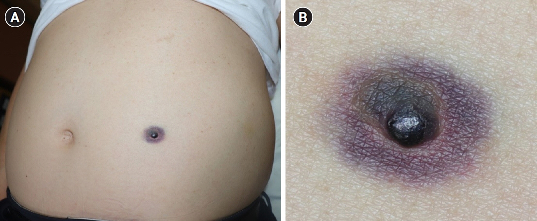
Fig. 1. (A, B) Blackish nodule on the abdomen surrounded by an ecchymotic targetoid halo.
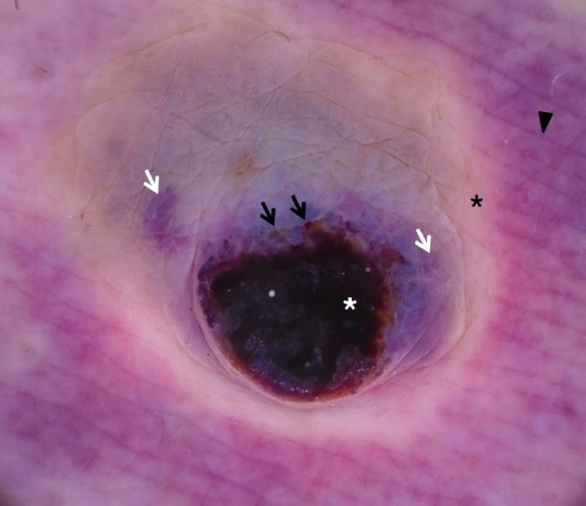
Fig. 2. Dermoscopy revealed brownish globules (black arrows) and violaceus globules (white arrows) intermingled with a structureless jet-black area (white asterisk) on the center. On the periphery, homogeneous milky red areas (black arrowhead) with an intermediate whitish circular ring (black asterisk) surrounding the central nodule were seen (×10).

Fig. 3. Histological findings. (A) Dome-shaped architecture was seen along with the extravasation of red blood cells throughout the lesion (hematoxylin-eosin [H&E] stain, ×20). (B) Well-circumscribed proliferation of dermal melanocytes arranged in a nest and vessel dilatation were noted (H&E stain, ×200). (C) On the periphery, only extravasation of red blood cells was seen (H&E stain, ×400).
Reference
-
References
1. Tomasini C, Broganelli P, Pippione M. Targetoid hemosiderotic nevus: a trauma-induced simulator of malignant melanoma. Dermatology. 2005; 210:200–5.2. Raucci M, Argenziano G, Zalaudek I, Lallas A, Longo C, Moscarella E. A worrisome sudden change: targetoid hemosiderotic nevus. J Am Acad Dermatol. 2014; 71:e5–6.
Article3. Patrizi A, Giacomini F, Savoia F, Misciali C, Neri I. Targetoid hemosiderotic naevus. J Eur Acad Dermatol Venereol. 2009; 23:493–4.
Article4. Zaballos P, Llambrich A, Del Pozo LJ, Landi C, Pizarro A, Vera A, et al. Dermoscopy of targetoid hemosiderotic hemangioma: a morphological study of 35 cases. Dermatology. 2015; 231:339–44.
Article5. Larre Borges A, Zalaudek I, Longo C, Dufrechou L, Argenziano G, Lallas A, et al. Melanocytic nevi with special features: clinical-dermoscopic and reflectance confocal microscopic-findings. J Eur Acad Dermatol Venereol. 2014; 28:833–45.
Article6. Oliveira A, Arzberger E, Massone C, Fink-Puches R, Zalaudek I, Hofmann-Wellenhof R. Dermoscopy, reflectance confocal microscopy and immunohistochemical analysis in melanocytic lesions with Meyerson’s phenomenon. Dermatology. 2014; 229:297–305.
Article
