EWSR1 rearranged primary renal myoepithelial carcinoma: a diagnostic conundrum
- Affiliations
-
- 1Department of Onco-Pathology, Mahamana Pandit Madan Mohan Malviya Cancer Centre, Varanasi, India
- KMID: 2545972
- DOI: http://doi.org/10.4132/jptm.2023.08.08
Abstract
- Primary renal myoepithelial carcinoma is an exceedingly rare neoplasm with an aggressive phenotype and Ewing sarcoma breakpoint region 1 (EWSR1) rearrangement in a small fraction of cases. In addition to its rarity, the diagnosis can be challenging for the pathologist due to morphologic heterogeneity, particularly on the biopsy specimen. At times, immunohistochemistry may be indecisive; therefore, molecular studies should be undertaken for clinching the diagnosis. We aim to illustrate a case of primary myoepithelial carcinoma of the kidney with EWSR1-rearrangement in a 67-year-old male patient who presented with right supraclavicular mass, which was clinically diagnosed as carcinoma of an unknown primary. An elaborate immunohistochemical work-up aided by fluorescent in-situ hybridization allowed us to reach a conclusive diagnosis. This unusual case report advocates that one should be aware of the histological mimickers and begin with broad differential diagnoses alongside sporadic ones and then narrow them down with appropriate ancillary studies.
Figure
-
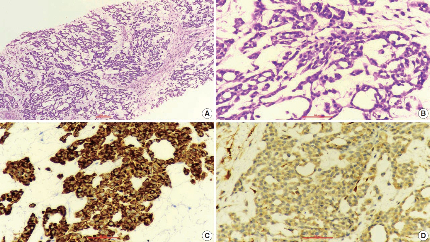
Fig. 1. The neoplastic cells are arranged in cords, trabeculae, tubules and scattered singly over a loose myxoid stroma (A). The neoplastic cells are oval to spindle-shaped and possess largely monomorphic nuclei, dispersed chromatin, inconspicuous nucleoli, and a scant to moderate amount of eosinophilic cytoplasm (B). The neoplastic cells are positive for AE1/AE3 (C) and smooth muscle actin (D).
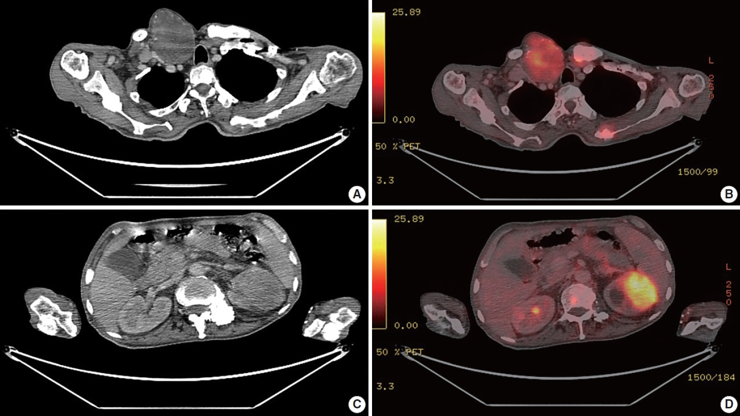
Fig. 2. Positron emission tomography and computed tomography (PET-CT) images of the hypermetabolic right supraclavicular mass (A, B). PET-CT images of the left renal lesion (C, D).
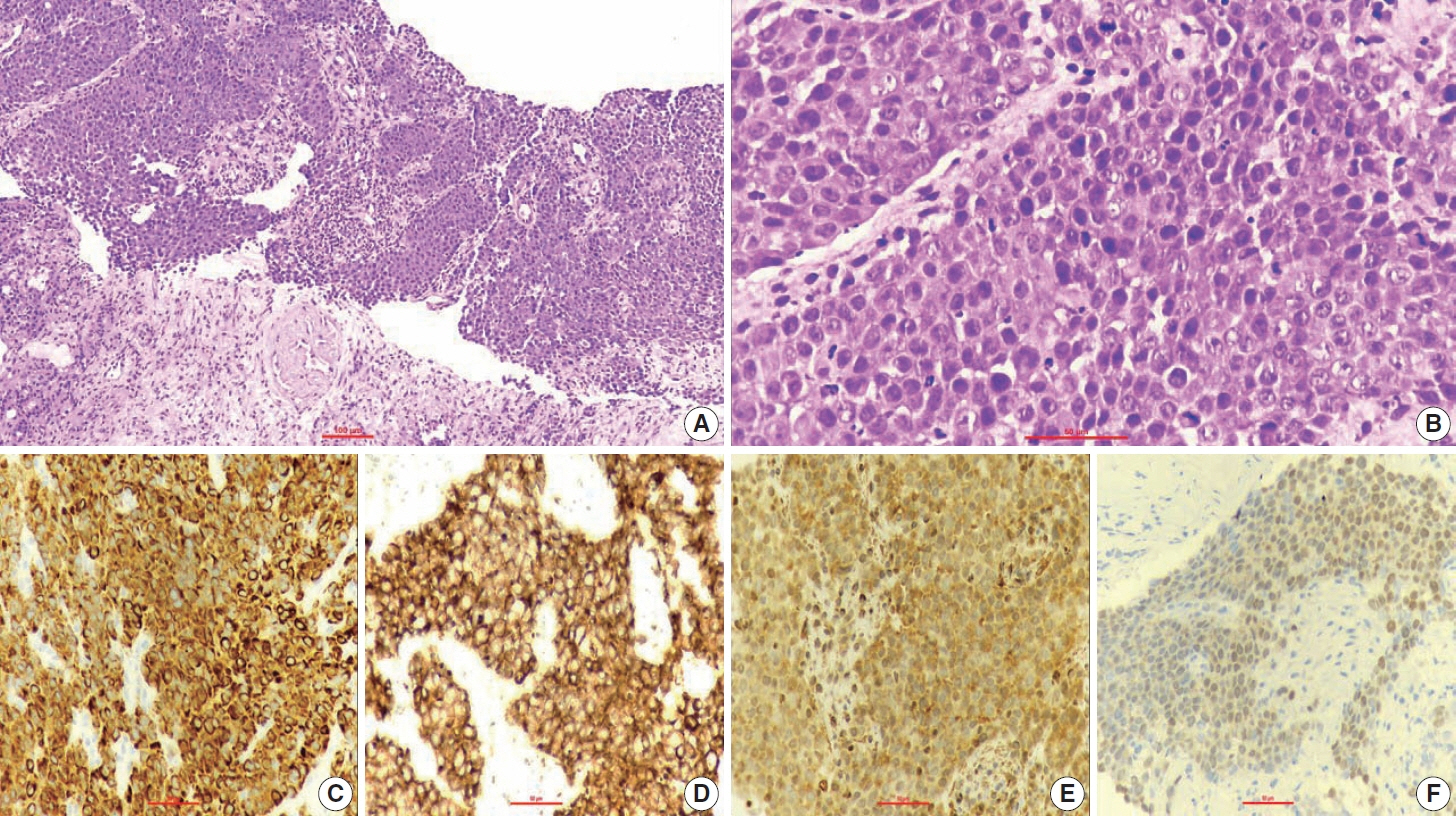
Fig. 3. The tumor cells are disposed in nests and sheets (A). The tumor cells are round, with a high nucleo-cytoplasmic ratio, fine to vesicular nuclei, prominent nucleoli, and scant cytoplasm (B). The tumor cells are positive for AE1/AE3 (C), CD10 (D), smooth muscle actin (E), and p63 (F).
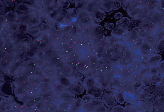
Fig. 4. Fluorescent in-situ hybridization using dual-color breakapart probe illustrating Ewing sarcoma breakpoint region 1 (EWSR1) gene rearrangement. The pink arrows indicate separated (split) red and green signals denoting a rearrangement involving the EWSR1 gene at 22q12. The white arrow points toward a normal fused signal, which is the site of the EWSR1 gene.
Reference
-
References
1. Jo VY. Soft tissue special issue: myoepithelial neoplasms of soft tissue: an updated review with emphasis on diagnostic considerations in the head and neck. Head Neck Pathol. 2020; 14:121–31.2. She YH, Chen YT, Huang YM, et al. Primary renal myoepithelial carcinoma with EWSR-1 gene rearrangement: case report and literature review. Acta Nephrol. 2021; 35:105–11.3. Cajaiba MM, Jennings LJ, Rohan SM, et al. Expanding the spectrum of renal tumors in children: primary renal myoepithelial carcinomas with a novel EWSR1-KLF15 fusion. Am J Surg Pathol. 2016; 40:386–94.4. Li Q, Mou Z, Yang K, Jiang H. A first case report of primary epithelial myoepithelial carcinoma-like renal tumor showing a perivascular pseudorosette-like pattern: description of morphologic, immunohistochemical, and genetic features. Medicine (Baltimore). 2019; 98:e17245.5. Yue D, Feng W, Ning C, Han LX, YaHong L. Myoepithelial carcinoma of the salivary gland: pathologic and CT imaging characteristics (report of 10 cases and literature review). Oral Surg Oral Med Oral Pathol Oral Radiol. 2017; 123:e182–7.6. Hiyama T, Kuno H, Sekiya K, Oda S, Kobayashi T. Imaging of malignant minor salivary gland tumors of the head and neck. Radiographics. 2021; 41:175–91.7. Hornick JL, Fletcher CD. Myoepithelial tumors of soft tissue: a clinicopathologic and immunohistochemical study of 101 cases with evaluation of prognostic parameters. Am J Surg Pathol. 2003; 27:1183–96.8. Antonescu CR, Zhang L, Chang NE, et al. EWSR1-POU5F1 fusion in soft tissue myoepithelial tumors: a molecular analysis of sixty-six cases, including soft tissue, bone, and visceral lesions, showing common involvement of the EWSR1 gene. Genes Chromosomes Cancer. 2010; 49:1114–24.9. Brandal P, Panagopoulos I, Bjerkehagen B, Heim S. t(19;22)(q13;q12) Translocation leading to the novel fusion gene EWSR1-ZNF444 in soft tissue myoepithelial carcinoma. Genes Chromosomes Cancer. 2009; 48:1051–6.10. Agaram NP, Chen HW, Zhang L, et al. EWSR1-PBX3: a novel gene fusion in myoepithelial tumors. Genes Chromosomes Cancer. 2015; 54:63–71.11. McConnell BB, Yang VW. Mammalian Kruppel-like factors in health and diseases. Physiol Rev. 2010; 90:1337–81.12. Uchida S, Tanaka Y, Ito H, et al. Transcriptional regulation of the CLC-K1 promoter by myc-associated zinc finger protein and kidney-enriched Kruppel-like factor, a novel zinc finger repressor. Mol Cell Biol. 2000; 20:7319–31.
- Full Text Links
-
- Actions
-
Cited
- CITED
-
- Close
- Share
-
- Similar articles
-
- Papillary Renal Cell Carcinoma in Transplanted Kidney and Xp11.2 Translocation/ Transcription Factor E3-Rearranged Renal Cell Carcinoma in the Native Kidney: A Case Report
- A Case of Myoepithelial Carcinoma Originated from Inferior Turbinate
- Epithelial-myoepithelial carcinoma arising in pleomorphic adenoma of palate
- Epithelial-Myoepithelial Carcinoma of Intercalated Duct of Parotid Gland
- A Case of Myoepithelial Carcinoma of Minor Salivary Gland
