Protective effects of red orange (Citrus sinensis [L.] Osbeck [Rutaceae]) extract against UVA-B radiation-induced photoaging in Skh:HR-2 mice
- Affiliations
-
- 1Technology Development Center, BTC Corporation, Ansan 15588, Korea
- 2Industry Coupled Cooperation Center for Bio Healthcare Materials, Hallym University, Chuncheon 24252, Korea
- KMID: 2545182
- DOI: http://doi.org/10.4162/nrp.2023.17.4.641
Abstract
- BACKGROUND/OBJECTIVES
The skin is the outermost organ of the human body and plays a protective role against external environmental damages, such as sunlight and pollution, which affect anti-oxidant defenses and skin inflammation, resulting in erythema or skin reddening, immunosuppression, and epidermal DNA damage.
MATERIALS/METHODS
The present study aimed to investigate the potential protective effects of red orange complex H extract (ROC) against ultraviolet (UV)-induced skin photoaging in Skh:HR-2 mice. ROC was orally administered at doses of 20, 40, and 80 mg/ kg/day for 13 weeks, along with UV irradiation of the mice for 10 weeks.
RESULTS
ROC improved UV-induced skin barrier parameters, including erythema, melanin production, transepidermal water loss, elasticity, and wrinkle formation. Notably, ROC inhibited the mRNA expression of pro-inflammatory cytokines (interleukin 6 and tumor necrosis factor α) and melanogenesis. In addition, ROC recovered the UV-induced decrease in the hyaluronic acid and collagen levels by enhancing genes expression. Furthermore, ROC significantly downregulated the protein and mRNA expression of matrix metalloproteinases responsible for collagen degradation. These protective effects of ROC against photoaging are associated with the suppression of UV-induced phosphorylation of c-Jun NH2-terminal kinase and activator protein 1 activation.
CONCLUSIONS
Altogether, our findings suggest that the oral administration of ROC exerts potential protective activities against photoaging in UV-irradiated hairless mice.
Figure
-
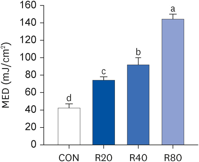
Fig. 1 Effect of ROC administration on the MED of the dorsal skin in Skh:HR-2 mice. ROC was administered via oral gavage for 13 weeks. The backs of mice were divided into 6 sections and irradiated with various UVA-B doses (30, 50, 70, 90, 120, and 150 mJ/cm2). After 24-h UVA-B irradiation, the MED was established. Each bar represents the mean ± SEM (n = 5).ROC, red orange complex H extract; MED, minimum erythema dose; UV, ultraviolet; CON, control group; R20, 20 mg/kg body weight/day red orange complex H extract-administered group; R40, 40 mg/kg body weight/day red orange complex H extract-administered group; R80, 80 mg/kg body weight/day red orange complex H extract-administered group.Means without the same letter differ significantly (P < 0.05).
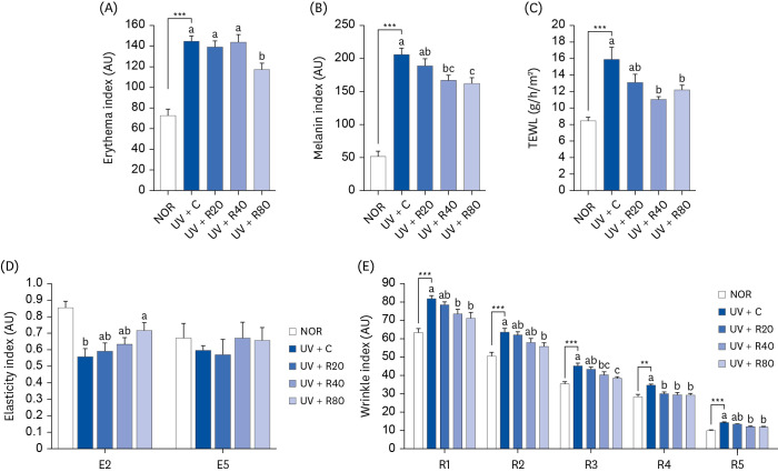
Fig. 2 Effect of ROC administration on skin index in UVA-B-irradiated Skh:HR-2 mice. ROC was administered via oral gavage for 13 weeks. After 3-week ROC administration, mice were exposed to UVA-B irradiation 3 times per week for 10 weeks. Various skin indices were estimated using an individual skin analyzer. (A) Erythema index; (B) Melanin index; (C) TEWL; (D) Elasticity index (E2, gross elasticity; E5, net elasticity); and (E) Wrinkle index (R1, total roughness; R2, average roughness; R3, maximum roughness; R4, smoothness depth; R5, arithmetic average roughness). Each bar represents the mean ± SEM (n = 10).ROC, red orange complex H extract; UV, ultraviolet; NOR, normal control group; UV + C, ultraviolet-irradiated control group; UV + R20, ultraviolet-irradiated and 20 mg/kg body weight/day red orange complex H extract administration group; UV + R40, ultraviolet-irradiated and 40 mg/kg body weight/day red orange complex H extract administration group; UV + R80, ultraviolet-irradiated and 80 mg/kg body weight/day red orange complex H extract administration group; TEWL, transepidermal water loss; AU, arbitrary unit.**P < 0.01 and ***P < 0.001 indicate statistically significant differences from the NOR group.Different letters indicate significant differences among the UV + C, UV + R20, UV + R40, and UV + R80 groups (P < 0.05).
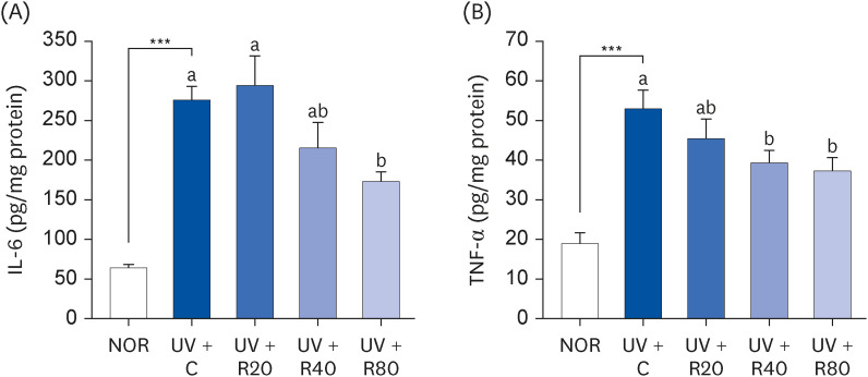
Fig. 3 Effect of ROC administration on IL-6 and TNF-α levels in the dorsal skin in UVA-B-irradiated Skh:HR-2 mice. ROC was administered via oral gavage for 13 weeks. After 3-week ROC administration, mice were exposed to UVA-B irradiation 3 times per week for ten weeks. Dorsal skin tissues were excised and homogenized. The levels of IL-6 (A) and TNF-α (B) in the skin homogenate were measured using enzyme-linked immunosorbent assay kits. Each bar represents the mean ± SEM (n = 10).ROC, red orange complex H extract; UV, ultraviolet; NOR, normal control group; UV + C, ultraviolet-irradiated control group; UV + R20, ultraviolet-irradiated and 20 mg/kg body weight/day red orange complex H extract administration group; UV + R40, ultraviolet-irradiated and 40 mg/kg body weight/day red orange complex H extract administration group; UV + R80, ultraviolet-irradiated and 80 mg/kg body weight/day red orange complex H extract administration group; IL-6, interleukin-6; TNF-α, tumor necrosis factor-α.***P < 0.001 significantly different from the NOR group.Different letters indicate significant differences among UV + C, UV + R20, UV + R40, and UV + R80 groups (P < 0.05).

Fig. 4 Effect of ROC administration on melanin content in the dorsal skin in UVA-B-irradiated Skh:HR-2 mice. ROC was administered via oral gavage for 13 weeks. After 3-week ROC administration, mice were exposed to UVA-B irradiation 3 times per week for ten weeks. (A) Dorsal skin sections were stained with Fontana-Masson’s silver stain. Representative images of staining are presented. Scale bar, 50 μm. (B) Melanin content in the dorsal skin. Each bar represents the mean ± SEM (n = 10).ROC, red orange complex H extract; UV, ultraviolet; NOR, normal control group; UV + C, ultraviolet-irradiated control group; UV + R20, ultraviolet-irradiated and 20 mg/kg body weight/day red orange complex H extract administration group; UV + R40, ultraviolet-irradiated and 40 mg/kg body weight/day red orange complex H extract administration group; UV + R80, ultraviolet-irradiated and 80 mg/kg body weight/day red orange complex H extract administration group.***P < 0.001 indicates statistically significant differences from the NOR group.Different letters indicate significant differences among UV + C, UV + R20, UV + R40, and UV + R80 groups (P < 0.05).
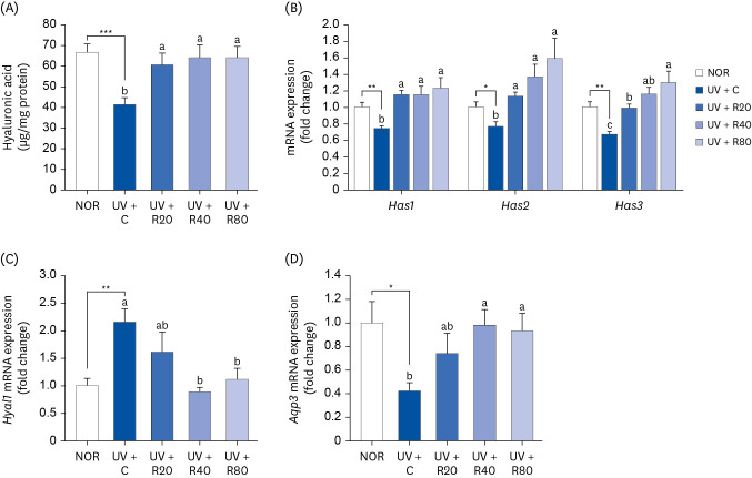
Fig. 5 Effect of ROC administration on HA synthesis in the dorsal skin in UVA-B-irradiated Skh:HR-2 mice. ROC was administered via oral gavage for 13 weeks. After 3-week ROC administration, mice were exposed to UVA-B irradiation 3 times per week for 10 weeks. (A) Dorsal skin tissues were excised and homogenized. HA levels in the skin homogenate were measured using enzyme-linked immunosorbent assay kits. (B-D) Total RNA in the dorsal skin tissues was extracted and reverse-transcribed, and real-time polymerase chain reaction was conducted. The expression of each mRNA was normalized to that of Gapdh and is represented relative to that of the NOR group. Each bar represents the mean ± SEM (n = 10).ROC, red orange complex H extract; HA, hyaluronic acid; UV, ultraviolet; NOR, normal control group; UV + C, UV-irradiated control group; UV + R20, UV-irradiated and 20 mg/kg body weight/day red orange complex H extract administration group; UV + R40, UV-irradiated and 40 mg/kg body weight/day red orange complex H extract administration group; UV + R80, UV-irradiated and 80 mg/kg body weight/day red orange complex H extract administration group; Has, hyaluronic acid synthase; Hyal, hyaluronidase; Aqp3, aquaporin 3; Gapdh, glyceraldehyde 3-phosphate dehydrogenase.*P < 0.05, **P < 0.01, and ***P < 0.001 indicate statistically significant differences from the NOR group.Different letters indicate significant differences among UV + C, UV + R20, UV + R40, and UV + R80 groups (P < 0.05).
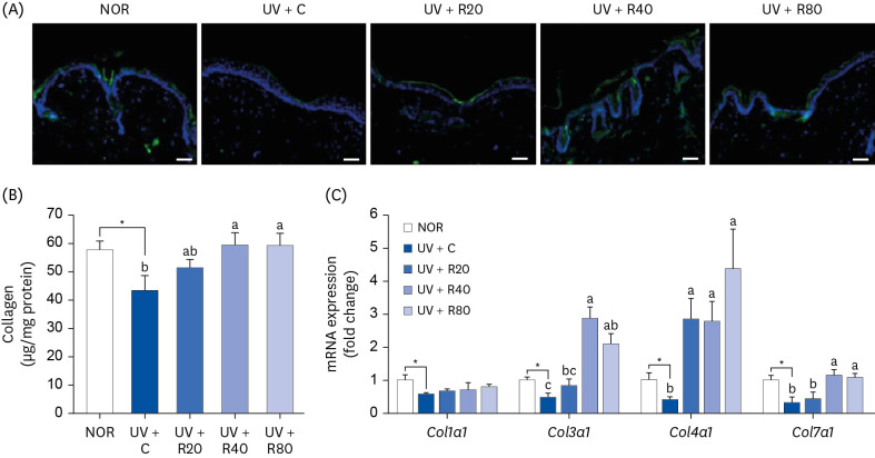
Fig. 6 Effect of ROC administration on collagen synthesis in the dorsal skin in UVA-B-irradiated Skh:HR-2 mice. ROC was administered via oral gavage for 13 weeks. After 3-week ROC administration, mice were exposed to UVA-B irradiation 3 times per week for ten weeks. (A) Dorsal skin sections were stained with collagen 1 antibody. Representative immunofluorescence staining images are displayed. Scale bar, 50 μm. (B) Dorsal skin tissues were excised and homogenized. The levels of collagen in skin homogenates were measured using hyaluronic acid enzyme-linked immunosorbent assay kits. (C) Total RNA in the dorsal skin tissues was extracted, reverse-transcribed, and subjected to real-time polymerase chain reaction. The expression of each mRNA was normalized to that of Gapdh and is represented relative to that of the NOR group. Each bar represents the mean ± SEM (n = 10).ROC, red orange complex H extract; UV, ultraviolet; NOR, normal control group; UV + C, ultraviolet-irradiated control group; UV + R20, ultraviolet-irradiated and 20 mg/kg body weight/day red orange complex H extract administration group; UV + R40, ultraviolet-irradiated and 40 mg/kg body weight/day red orange complex H extract administration group; UV + R80, ultraviolet-irradiated and 80 mg/kg body weight/day red orange complex H extract administration group; Col1a1, collagen type 1 alpha 1 chain; Col3a1, collagen type 3 alpha 1 chain; Col4a1, collagen type 4 alpha 1 chain; Col7a1, collagen type 7 alpha 1 chain; Gapdh, glyceraldehyde 3-phosphate dehydrogenase.*P < 0.05, significantly different from the NOR group.Different letters indicate significant differences among UV + C, UV + R20, UV + R40, and UV + R80 groups (P < 0.05).

Fig. 7 Effect of ROC administration on MMP-2 and MMP-13 expression in the dorsal skin in UVA-B-irradiated Skh:HR-2 mice. ROC was administered via oral gavage for 13 weeks. After 3-week ROC administration, mice were exposed to UVA-B irradiation 3 times per week for ten weeks. (A) Total lysates of dorsal skin tissues were prepared and analyzed using Western blotting with the indicated antibodies. Images of Western blots representative of 3 independent experiments are displayed. (B) Quantitative analysis of Western blot results. The protein abundance was normalized to β-actin and expressed relative to that of the NOR group. (C) Total RNA in the dorsal skin tissues was extracted, reverse-transcribed, and subjected to real-time polymerase chain reaction. The expression of each mRNA was normalized to that of Gapdh and is represented relative to that of the NOR group. Each bar represents the mean ± SEM (n = 10).ROC, red orange complex H extract; UV, ultraviolet; NOR, normal control group; UV + C, ultraviolet-irradiated control group; UV + R20, ultraviolet-irradiated and 20 mg/kg body weight/day ROC administration group; UV + R40, ultraviolet-irradiated and 40 mg/kg body weight/day ROC administration group; UV + R80, ultraviolet-irradiated and 80 mg/kg body weight/day ROC administration group; MMP, matrix metallopeptidase; Gapdh, glyceraldehyde 3-phosphate dehydrogenase.*P < 0.05, **P < 0.01, ***P < 0.001 significantly different from the NOR group.Different letters indicate significant differences among UV + C, UV + R20, UV + R40, and UV + R80 groups (P < 0.05).
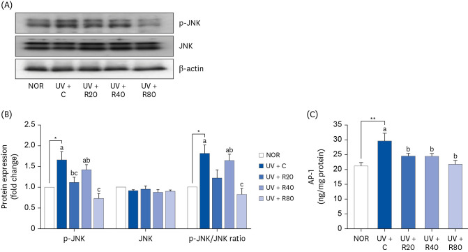
Fig. 8 Effect of ROC administration on JNK signaling pathway in the dorsal skin in UVA-B-irradiated Skh:HR-2 mice. ROC was administered via oral gavage for 13 weeks. After 3-week ROC administration, mice were exposed to UVA-B irradiation 3 times per week for ten weeks. (A) Total lysates of dorsal skin tissues were prepared and analyzed using Western blotting with the indicated antibodies. Images of Western blots representative of 3 independent experiments are displayed. (B) Quantitative analysis of Western blot results. The protein abundance was normalized to β-actin and expressed relative to that of the NOR group. (C) Nuclear protein from dorsal skin tissues was extracted, and the AP-1 content in the nuclear protein was measured. Each bar represents the mean ± SEM (n = 10).ROC, red orange complex H extract; UV, ultraviolet; NOR, normal control group; UV + C, ultraviolet-irradiated control group; UV + R20, ultraviolet-irradiated and 20 mg/kg body weight/day red orange complex H extract administration group; UV + R40, ultraviolet-irradiated and 40 mg/kg body weight/day red orange complex H extract administration group; UV + R80, ultraviolet-irradiated and 80 mg/kg body weight/day red orange complex H extract administration group; p-JNK, phospho c-Jun NH2-terminal kinase; JNK, c-Jun NH2-terminal kinase; AP-1, activator protein 1.*P < 0.05, **P < 0.01 statistically significant differences from the NOR group.Different letters indicate significant differences among UV + C, UV + R20, UV + R40, and UV + R80 groups (P < 0.05).

Fig. 9 Summary of the effects of the ROC on the photoaging.UV, ultraviolet; ROS, reactive oxygen species; ROC, red orange complex H extract; JNK, c-Jun NH2-terminal kinase; ERK1/2, extracellular signal-regulated kinase 1/2; AP-1, activator protein 1; MMP, matrix metalloproteinases; COL1a1, collagen type 1 alpha 1 chain; COL3a1, collagen type 3 alpha 1 chain; COL4a1, collagen type 4 alpha 1 chain; COL7a1, collagen type 7 alpha 1 chain; HYAL1, hyaluronidase 1; HAS, hyaluronic acid synthase; NF-κB, nuclear factor-κB; IL-6, interleukin 6; TNF-α, tumor necrosis factor α.
Reference
-
1. Parrado C, Mercado-Saenz S, Perez-Davo A, Gilaberte Y, Gonzalez S, Juarranz A. Environmental stressors on skin aging. Mechanistic insights. Front Pharmacol. 2019; 10:759. PMID: 31354480.
Article2. Gilchrest BA, Yaar M. Ageing and photoageing of the skin: observations at the cellular and molecular level. Br J Dermatol. 1992; 127(Suppl 41):25–30. PMID: 1390183.
Article3. Cavinato M, Waltenberger B, Baraldo G, Grade CVC, Stuppner H, Jansen-Dürr P. Plant extracts and natural compounds used against UVB-induced photoaging. Biogerontology. 2017; 18:499–516. PMID: 28702744.
Article4. Chiang HM, Chen HC, Lin TJ, Shih IC, Wen KC. Michelia alba extract attenuates UVB-induced expression of matrix metalloproteinases via MAP kinase pathway in human dermal fibroblasts. Food Chem Toxicol. 2012; 50:4260–4269. PMID: 22922035.
Article5. Hwang E, Park SY, Lee HJ, Lee TY, Sun ZW, Yi TH. Gallic acid regulates skin photoaging in UVB-exposed fibroblast and hairless mice. Phytother Res. 2014; 28:1778–1788. PMID: 25131997.
Article6. Gromkowska-Kępka KJ, Puścion-Jakubik A, Markiewicz-Żukowska R, Socha K. The impact of ultraviolet radiation on skin photoaging - review of in vitro studies. J Cosmet Dermatol. 2021; 20:3427–3431. PMID: 33655657.7. Matsumura Y, Ananthaswamy HN. Toxic effects of ultraviolet radiation on the skin. Toxicol Appl Pharmacol. 2004; 195:298–308. PMID: 15020192.
Article8. Shaulian E, Karin M. AP-1 as a regulator of cell life and death. Nat Cell Biol. 2002; 4:E131–E136. PMID: 11988758.
Article9. Geoffrey K, Mwangi AN, Maru SM. Sunscreen products: rationale for use, formulation development and regulatory considerations. Saudi Pharm J. 2019; 27:1009–1018. PMID: 31997908.
Article10. Parrado C, Philips N, Gilaberte Y, Juarranz A, González S. Oral photoprotection: effective agents and potential candidates. Front Med (Lausanne). 2018; 5:188. PMID: 29998107.
Article11. Cimino F, Cristani M, Saija A, Bonina FP, Virgili F. Protective effects of a red orange extract on UVB-induced damage in human keratinocytes. Biofactors. 2007; 30:129–138. PMID: 18356584.
Article12. Saija A, Tomaino A, Lo Cascio R, Rapisarda P, Dederen JC. In vitro antioxidant activity and in vivo photoprotective effect of a red orange extract. Int J Cosmet Sci. 1998; 20:331–342. PMID: 18505518.
Article13. Vitali F, Pennisi C, Tomaino A, Bonina F, De Pasquale A, Saija A, Tita B. Effect of a standardized extract of red orange juice on proliferation of human prostate cells in vitro . Fitoterapia. 2006; 77:151–155. PMID: 16530345.
Article14. Bonina FP, Puglia C, Frasca G, Cimino F, Trombetta D, Tringali G, Roccazzello A, Insiriello E, Rapisarda P, Saija A. Protective effects of a standardised red orange extract on air pollution-induced oxidative damage in traffic police officers. Nat Prod Res. 2008; 22:1544–1551. PMID: 19023818.
Article15. Balestra C, Cimino F, Theunissen S, Snoeck T, Provyn S, Canali R, Bonina A, Virgili F. A red orange extract modulates the vascular response to a recreational dive: a pilot study on the effect of anthocyanins on the physiological consequences of scuba diving. Nat Prod Res. 2016; 30:2101–2106. PMID: 26548425.
Article16. Tomasello B, Malfa GA, Acquaviva R, La Mantia A, Di Giacomo C. Phytocomplex of a standardized extract from red orange (Citrus sinensis L. Osbeck) against photoaging. Cells. 2022; 11:1447. PMID: 35563752.
Article17. Nobile V, Burioli A, Yu S, Zhifeng S, Cestone E, Insolia V, Zaccaria V, Malfa GA. Photoprotective and antiaging effects of a standardized red orange (Citrus sinensis (L.) Osbeck) extract in Asian and Caucasian subjects: a randomized, double-blind, controlled study. Nutrients. 2022; 14:2241. PMID: 35684041.
Article18. Peng LH, Liu S, Xu SY, Chen L, Shan YH, Wei W, Liang WQ, Gao JQ. Inhibitory effects of salidroside and paeonol on tyrosinase activity and melanin synthesis in mouse B16F10 melanoma cells and ultraviolet B-induced pigmentation in guinea pig skin. Phytomedicine. 2013; 20:1082–1087. PMID: 23746955.
Article19. Lee HS, Lim SM, Jung JI, Kim SM, Lee JK, Kim YH, Cha KM, Oh TK, Moon JM, Kim TY, et al. Gynostemma pentaphyllum extract ameliorates high-fat diet-induced obesity in C57BL/6N mice by upregulating SIRT1. Nutrients. 2019; 11:2475. PMID: 31618980.
Article20. Cho HJ, Kim WK, Kim EJ, Jung KC, Park S, Lee HS, Tyner AL, Park JH. Conjugated linoleic acid inhibits cell proliferation and ErbB3 signaling in HT-29 human colon cell line. Am J Physiol Gastrointest Liver Physiol. 2003; 284:G996–1005. PMID: 12571082.
Article21. US Food and Drug Administration. Guidance for Industry: Estimating the Maximum Safe Starting Dose in Initial Clinical Trials for Therapeutics in Adult Healthy Volunteers. Rockville (MD): US Food and Drug Administration;2005.22. Yoshizumi M, Nakamura T, Kato M, Ishioka T, Kozawa K, Wakamatsu K, Kimura H. Release of cytokines/chemokines and cell death in UVB-irradiated human keratinocytes, HaCaT. Cell Biol Int. 2008; 32:1405–1411. PMID: 18782623.
Article23. Duan X, Wu T, Liu T, Yang H, Ding X, Chen Y, Mu Y. Vicenin-2 ameliorates oxidative damage and photoaging via modulation of MAPKs and MMPs signaling in UVB radiation exposed human skin cells. J Photochem Photobiol B. 2019; 190:76–85. PMID: 30502588.
Article24. Pittayapruek P, Meephansan J, Prapapan O, Komine M, Ohtsuki M. Role of matrix metalloproteinases in photoaging and photocarcinogenesis. Int J Mol Sci. 2016; 17:868. PMID: 27271600.
Article25. Choi SI, Jung TD, Cho BY, Choi SH, Sim WS, Han X, Lee SJ, Kim YC, Lee OH. Anti-photoaging effect of fermented agricultural by-products on ultraviolet B-irradiated hairless mouse skin. Int J Mol Med. 2019; 44:559–568. PMID: 31198982.
Article26. Young AR. Acute effects of UVR on human eyes and skin. Prog Biophys Mol Biol. 2006; 92:80–85. PMID: 16600340.
Article27. Saewan N, Jimtaisong A. Natural products as photoprotection. J Cosmet Dermatol. 2015; 14:47–63. PMID: 25582033.
Article28. Yadav T, Mishra S, Das S, Aggarwal S, Rani V. Anticedants and natural prevention of environmental toxicants induced accelerated aging of skin. Environ Toxicol Pharmacol. 2015; 39:384–391. PMID: 25555260.
Article29. Grosso G, Galvano F, Mistretta A, Marventano S, Nolfo F, Calabrese G, Buscemi S, Drago F, Veronesi U, Scuderi A. Red orange: experimental models and epidemiological evidence of its benefits on human health. Oxid Med Cell Longev. 2013; 2013:157240. PMID: 23738032.
Article30. de Lima LP, de Paula BA. A review of the lipolytic effects and the reduction of abdominal fat from bioactive compounds and Moro orange extracts. Heliyon. 2021; 7:e07695. PMID: 34409177.
Article31. Ribeiro AP, Pereira AG, Todo MC, Fujimori AS, Dos Santos PP, Dantas D, Fernandes AA, Zanati SG, Hassimotto NM, Zornoff LA, et al. Pera orange (Citrus sinensis) and Moro orange (Citrus sinensis (L.) Osbeck) juices attenuate left ventricular dysfunction and oxidative stress and improve myocardial energy metabolism in acute doxorubicin-induced cardiotoxicity in rats. Nutrition. 2021; 91-92:111350. PMID: 34265580.32. Buscemi S, Rosafio G, Arcoleo G, Mattina A, Canino B, Montana M, Verga S, Rini G. Effects of red orange juice intake on endothelial function and inflammatory markers in adult subjects with increased cardiovascular risk. Am J Clin Nutr. 2012; 95:1089–1095. PMID: 22492368.
Article33. Magalhães ML, de Sousa RV, Miranda JR, Konig IF, Wouters F, Souza FR, Simão SD, da Silva LA, Nelson DL, das Graças CM. Effects of Moro orange juice (Citrus sinensis (L.) Osbeck) on some metabolic and morphological parameters in obese and diabetic rats. J Sci Food Agric. 2021; 101:1053–1064. PMID: 32767388.
Article34. Jiang SJ, Chu AW, Lu ZF, Pan MH, Che DF, Zhou XJ. Ultraviolet B-induced alterations of the skin barrier and epidermal calcium gradient. Exp Dermatol. 2007; 16:985–992. PMID: 18031457.
Article35. Haratake A, Uchida Y, Schmuth M, Tanno O, Yasuda R, Epstein JH, Elias PM, Holleran WM. UVB-induced alterations in permeability barrier function: roles for epidermal hyperproliferation and thymocyte-mediated response. J Invest Dermatol. 1997; 108:769–775. PMID: 9129231.
Article36. Mojumdar EH, Pham QD, Topgaard D, Sparr E. Skin hydration: interplay between molecular dynamics, structure and water uptake in the stratum corneum. Sci Rep. 2017; 7:15712. PMID: 29146971.
Article37. Dai G, Freudenberger T, Zipper P, Melchior A, Grether-Beck S, Rabausch B, de Groot J, Twarock S, Hanenberg H, Homey B, et al. Chronic ultraviolet B irradiation causes loss of hyaluronic acid from mouse dermis because of down-regulation of hyaluronic acid synthases. Am J Pathol. 2007; 171:1451–1461. PMID: 17982124.
Article38. Yun MS, Kim C, Hwang JK. Agastache rugosa Kuntze attenuates UVB-induced photoaging in hairless mice through the regulation of MAPK/AP-1 and TGF-β/Smad pathways. J Microbiol Biotechnol. 2019; 29:1349–1360. PMID: 31474086.
Article39. Razia S, Park H, Shin E, Shim KS, Cho E, Kim SY. Effects of Aloe vera flower extract and its active constituent isoorientin on skin moisturization via regulating involucrin expression: in vitro and molecular docking studies. Molecules. 2021; 26:2626. PMID: 33946287.
Article40. Lee HJ, Im AR, Kim SM, Kang HS, Lee JD, Chae S. The flavonoid hesperidin exerts anti-photoaging effect by downregulating matrix metalloproteinase (MMP)-9 expression via mitogen activated protein kinase (MAPK)-dependent signaling pathways. BMC Complement Altern Med. 2018; 18:39. PMID: 29382339.
Article41. Park JE, Pyun HB, Woo SW, Jeong JH, Hwang JK. The protective effect of Kaempferia parviflora extract on UVB-induced skin photoaging in hairless mice. Photodermatol Photoimmunol Photomed. 2014; 30:237–245. PMID: 24313661.
Article42. Han HS, Shin JS, Myung DB, Ahn HS, Lee SH, Kim HJ, Lee KT. Hydrangea serrata (Thunb.) Ser. extract attenuate UVB-induced photoaging through MAPK/AP-1 inactivation in human skin fibroblasts and hairless mice. Nutrients. 2019; 11:553. PMID: 30841605.
- Full Text Links
-
- Actions
-
Cited
- CITED
-
- Close
- Share
-
- Similar articles
-
- Protective effect of the standardized green tea seed extract on UVB-induced skin photoaging in hairless mice
- Protective effect of methanol extract from citrus press cakes prepared by far-infrared radiation drying on H2O2-mediated oxidative damage in Vero cells
- The Effects of UVB Irradiation and Sunscreen Agents on Photoaging of Hairless Albino Mouse Skin
- Ethanol Extract of Chaenomeles sinensis Inhibits the Development of Benign Prostatic Hyperplasia by Exhibiting Anti-oxidant and Anti-inflammatory Effects
- An Experimental Study on the Protective Effects of Ginseng Extract to Oxygen Toxicity
