A Case of Sinochoanal Polyp Originating From the Ethmoid Sinus
- Affiliations
-
- 1Department of Otolaryngology-Head and Neck Surgery, Hanyang University Hospital, Seoul, Republic of Korea
- 2Department of Pathology, Hanyang University Hospital, Seoul, Republic of Korea
- KMID: 2544622
- DOI: http://doi.org/10.18787/jr.2023.00020
Abstract
- A nasal polyp is a distinct mucosal pathology that obstructs the nasal cavity and paranasal sinuses, with various phenotypes and endotypes. Nasal polyps should be distinguished from inverted papillomas, squamous cell carcinomas, juvenile angiofibromas, lymphomas, and olfactory neuroblastomas. A choanal polyp is a solitary benign lesion that originates in the paranasal sinus and extends to the choana through the natural ostium of the sinus. Choanal polyps usually originate from the maxillary sinus; however, we recently experienced the case of 41-year old women with sinochoanal polyp originated from the ethmoid sinus. As choanal polyps can recur even after appropriate surgery, complete resection, including the surrounding mucosa at the site of origin, is required. Therefore, it is essential to consider anatomical differences in polypectomy. We recently diagnosed and successfully performed surgery on an ethmochoanal polyp; herein, we report our experience and present a literature review.
Keyword
Figure
-
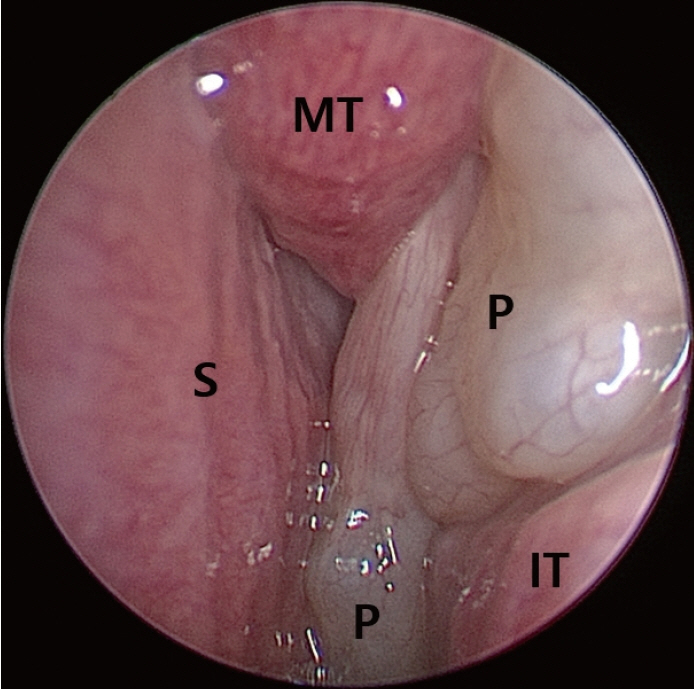
Fig. 1. Endoscopic findings of the ethmochoanal polyp. The pedunculated polyp (P) emerged from the left anterior ethmoid and extended to the middle meatus and the choana. MT, middle turbinate; IT, inferior turbinate; S, septum.

Fig. 2. Radiologic findings of the ethmochoanal polyp. Paranasal sinus computed tomography images (axial and coronal) of the ethmochoanal polyp (P). CT scans showed its origin (A), stalk (passage through the ethmoid sinus, B and D) and final extension to the middle meatus and choana (C and E).

Fig. 3. Gross findings of the ethmochoanal polyp. The ethmochoanal polyp, with a length of approximately 6.5 cm, showed a pedunculated structure. The stalk of its origin (O) was about 3.5 cm long, and the two branches of the polyp were each about 3 cm long.

Fig. 4. Pathologic findings of the ethmochoanal polyp. Representative images of an ethmochoanal polyp (A), a nasal polyp (B), and an antrochonal polyp (C) (H&E, magnification ×40 and ×400). The surfaces of the three polyps are lined by benign respiratory epithelium. The ethmochoanal polyp showed numerous dilated cystic structures lined by columnar epithelium and filled with mucus within the stroma. The stroma of the ethmochoanal polyp showed an edematous or loosely myxoid appearance without infiltration of mixed inflammatory cells. Compared to the ethmochoanal polyp, the nasal polyp and antrochonal polyp revealed prominent inflammatory cells composed of a mixture of lymphocytes, plasma cells, eosinophils, and neutrophils, without a dilated cystic structure in the stroma.
Reference
-
References
1. Kizil Y, Aydil U, Ceylan A, Uslu S, Baştürk V, İleri F. Analysis of choanal polyps. J Craniofac Surg. 2014; 25(3):1082–4.2. Lopatin A, Bykova V, Piskunov G. Choanal polyps: one entity, one surgical approach? Rhinology. 1997; 35(2):79–83.3. Chung SK, Chang BC, Dhong HJ. Surgical, radiologic, and histologic findings of the antrochoanal polyp. Am J Rhinol. 2002; 16(2):71–6.4. Stammberger H. Surgical treatment of nasal polyps: past, present, and future. Allergy. 1999; 54(Suppl 53):7–11.5. Min YG, Chung JW, Shin JS, Chi JG. Histologic structure of antrochoanal polyps. Acta Otolaryngol. 1995; 115(4):543–7.6. Cook PR, Davis WE, McDonald R, McKinsey JP. Antrochoanal polyposis: a review of 33 cases. Ear Nose Throat J. 1993; 72(6):401–11.7. Aydin O, Keskin G, Ustündağ E, Işeri M, Ozkarakaş H. Choanal polyps: an evaluation of 53 cases. Am J Rhinol. 2007; 21(2):164–8.8. Hong SK, Yoo YS, Shin YR, Chung SW. Choanal polyps originating from the ethmoid sinus: ethmochoanal polyps? Korean J Otorhinolaryngol-Head Neck Surg. 2002; 45(9):921–5.9. Yaman H, Yilmaz S, Karali E, Guclu E, Ozturk O. Evaluation and management of antrochoanal polyps. Clin Exp Otorhinolaryngol. 2010; 3(2):110–4.10. Bozzo C, Garrel R, Meloni F, Stomeo F, Crampette L. Endoscopic treatment of antrochoanal polyps. Eur Arch Otorhinolaryngol. 2007; 264(2):145–50.11. Suzuki S, Yasunaga H, Matsui H, Fushimi K, Kondo K, Yamasoba T. Complication rates after functional endoscopic sinus surgery: analysis of 50,734 Japanese patients. Laryngoscope. 2015; 125(8):1785–91.12. We J, Lee WH, Tan KL, Wee JH, Rhee CS, Lee CH, et al. Prevalence of nasal polyps and its risk factors: Korean National Health and Nutrition Examination Survey 2009-2011. Am J Rhinol Allergy. 2015; 29(1):e24–8.13. Fahy C, Jones NS. Nasal polyposis and facial pain. Clin Otolaryngol Allied Sci. 2001; 26(6):510–3.14. Youn EK, Chung EC, Lee YU. CT and MR evaluation of choanal polyps. J Korean Radiol Soc. 1998; 39(2):283–8.15. De Vuysere S, Hermans R, Marchal G. Sinochoanal polyp and its variant, the angiomatous polyp: MRI findings. Eur Radiol. 2001; 11(1):55–8.16. Diamantopoulos II, Jones NS, Lowe J. All nasal polyps need histological examination: an audit-based appraisal of clinical practice. J Laryngol Otol. 2000; 114(10):755–9.17. Kim SJ, Lee KH, Kim SW, Cho JS, Park YK, Shin SY. Changes in histological features of nasal polyps in a Korean population over a 17-year period. Otolaryngol Head Neck Surg. 2013; 149(3):431–7.18. Berg O, Carenfelt C, Silfverswärd C, Sobin A. Origin of the choanal polyp. Arch Otolaryngol Head Neck Surg. 1988; 114(11):1270–1.
- Full Text Links
-
- Actions
-
Cited
- CITED
-
- Close
- Share
-
- Similar articles
-
- Choanal Polyps Originating from the Ethmoid Sinus: Ethmochoanal Polyps?
- A Case of Ethmoid Mucocele
- A Case of Sinonasal Undifferentiated Carcinoma Originating from the Ethmoid Sinus
- A Case of Choanal Polyps Originating in the Both Sphenoid Sinuses
- Effect of Pediatric Endoscopic Sinus Surgery with Antrochoanal Polyps on Sinus Growth
