What’s new in hematopathology 2023: updates on mature T-cell neoplasms in the 5th edition of the WHO classification
- Affiliations
-
- 1Department of Translational Molecular Pathology, The University of Texas MD Anderson Cancer Center, Houston, TX, USA
- 2Department of Hematopathology, The University of Texas MD Anderson Cancer Center, Houston, TX, USA
- KMID: 2544333
- DOI: http://doi.org/10.4132/jptm.2023.06.15
Abstract
- The overview of the upcoming Blue Book of the 5th edition of the World Health Organization Classification of Hematolymphoid Tumors was published in Leukemia in June 2022. The updates on mature T-/NK-cell lymphomas and leukemias are organized in nine groups based on cell of origin, morphology, clinical scenario, and localization, and are highlighted in this newsletter.
Figure
-
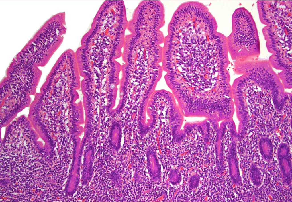
Fig. 1. Expansion of the lamina propria by small-sized and mature-appearing lymphocytes in indolent T-cell lymphoma of the gastrointestinal tract.
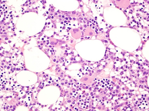
Fig. 2. Bone marrow core biopsy involved with HSTCL shows hypercellularity displaying small and hypolobated megakaryocytes, morphologically consistent with dysmegakaryopoiesis, raising the possibility of underlying myelodysplastic syndrome.

Fig. 3. CD3 immunostaining shows that the lymphocytes have a sinusoidal pattern characterized by clusters of lymphocytes in a cord-like pattern in HSTCL.
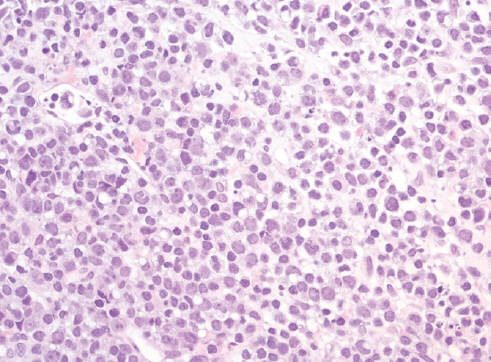
Fig. 4. Lymph node involved by ALK- ALCL shows diffuse replacement of normal architecture by large and anaplastic lymphocytes with no distinct nucleoli; some of them have a doughnut-like shape.
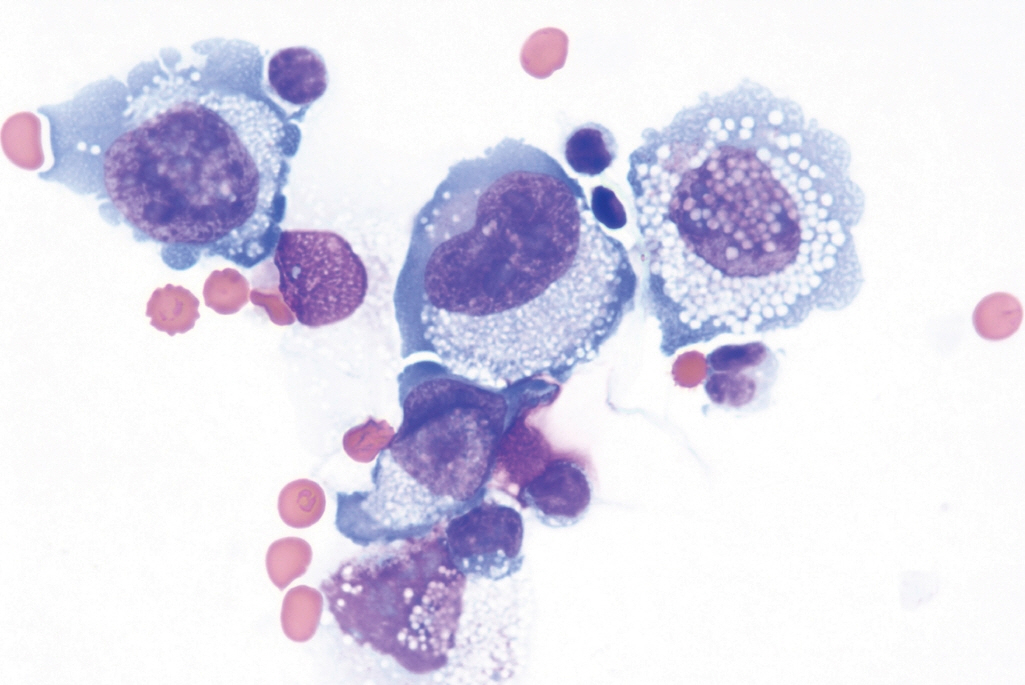
Fig. 5. Cytologic preparation of an effusion around a breast implant in a case of BIA-ALCL shows large and atypical cells with irregular nuclear contours, inconspicuous nuclei and abundant cytoplasm with small inclusions.
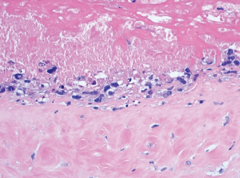
Fig. 6. Breast capsule with BIA-ALCL shows large and neoplastic cells confined to the luminal space in a necrotic background.
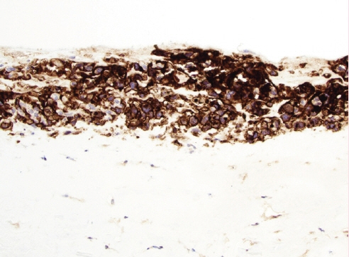
Fig. 7. CD30 immunostaining shows diffuse and strong cytoplasmic and nuclear positivity in the BIAALCL cells.

Fig. 8. nTFHL-AI with effacement of the nodal architecture and infiltration of the capsule and subcapsular sinus.

Fig. 9. Positivity for the checkpoint molecule PD-1 in scattered lymphoma cells supports a T follicular helper phenotype in a case of nTFHL-AI.
- Full Text Links
-
- Actions
-
Cited
- CITED
-
- Close
- Share
-
- Similar articles
-
- What’s new in kidney tumor pathology 2022: WHO 5th edition updates
- What’s new in neuropathology 2024: CNS WHO 5th edition updates
- What’s new in dermatopathology 2023: WHO 5th edition updates
- What’s new in genitourinary pathology 2023: WHO 5th edition updates for urinary tract, prostate, testis, and penis
- What’s new in soft tissue and bone pathology 2022–updates from the WHO classification 5th edition
