Loss of aquaporin-1 expression is associated with worse clinical outcomes in clear cell renal cell carcinoma: an immunohistochemical study
- Affiliations
-
- 1Department of Pathology, Seoul National University College of Medicine, Seoul, Korea
- 2Department of Pathology, Konkuk University Medical Center, Konkuk University School of Medicine, Seoul, Korea
- 3Department of Pathology, Yonsei University College of Medicine, Seoul, Korea
- 4Kidney Research Institute, Medical Research Center, Seoul National University College of Medicine, Seoul, Korea
- KMID: 2544330
- DOI: http://doi.org/10.4132/jptm.2023.06.17
Abstract
- Background
Aquaporin (AQP) expression has been investigated in various malignant neoplasms, and the overexpression of AQP is related to poor prognosis in some malignancies. However, the expression of AQP protein in clear cell renal cell carcinoma (ccRCC) has not been extensively investigated by immunohistochemistry with large sample size.
Methods
We evaluated the AQP expression in 827 ccRCC with immunohistochemical staining in tissue microarray blocks and classified the cases into two categories, high and low expression.
Results
High expression of aquaporin-1 (AQP1) was found in 320 cases (38.7%), but aquaporin-3 was not expressed in ccRCC. High AQP1 expression was significantly related to younger age, low TNM stage, low World Health Organization/International Society of Urologic Pathology nuclear grade, and absence of distant metastasis. Furthermore, high AQP1 expression was also significantly associated with longer overall survival (OS; p<.001) and progression-specific survival (PFS; p<.001) and was an independent predictor of OS and PFS in ccRCC.
Conclusions
Our study revealed the prognostic significance of AQP1 protein expression in ccRCC. These findings could be applied to predict the prognosis of ccRCC.
Figure
-
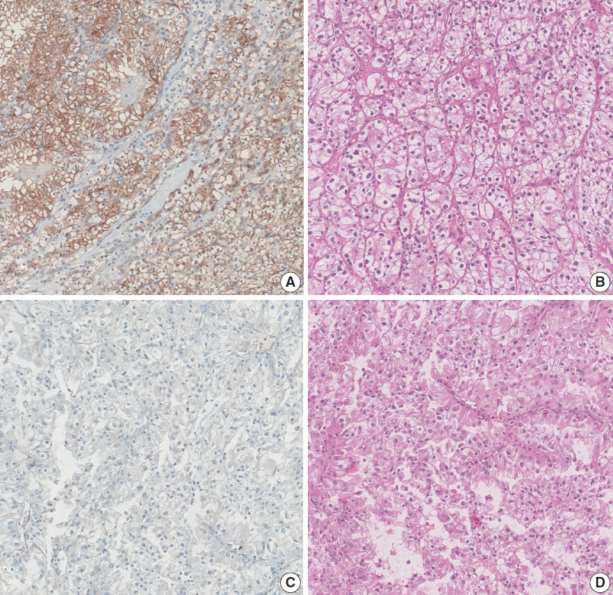
Fig. 1. Representative photographs of the expression levels of aquaporin 1 (AQP1) by immunohistochemistry and corresponding hematoxylin and eosin–stained slides. High expression of AQP1 (A) is associated with favorable histopathology and lower pathological stage (B), and low expression of AQP1 (C) is associated with unfavorable histological features and higher pathological stage (D).

Fig. 2. Representative photographs of the expression levels of aquaporin 3 (AQP3) by immunohistochemistry slides. In contrast to aquaporin 1, the staining intensity of AQP3 was too low in every clear cell renal cell carcinoma tissue (A) to evaluate and analyze statistically. Distal tubular epithelial cells, which normally express the AQP3 protein, are positive for AQP3 immunostaining (B).
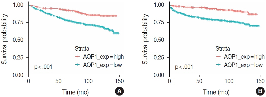
Fig. 3. Overall (A) and progression-free (B) survival curves showing Kaplan-Meier survival probability by time. The difference in overall and progression- free survival rates between higher and lower aquaporin 1 (AQP1) expression was statistically significant in both respects of survival.
Reference
-
References
1. Agre P, King LS, Yasui M, et al. Aquaporin water channels: from atomic structure to clinical medicine. J Physiol. 2002; 542:3–16.2. Chen J, Wang Z, Xu D, Liu Y, Gao Y. Aquaporin 3 promotes prostate cancer cell motility and invasion via extracellular signal-regulated kinase 1/2-mediated matrix metalloproteinase-3 secretion. Mol Med Rep. 2015; 11:2882–8.3. Kusayama M, Wada K, Nagata M, et al. Critical role of aquaporin 3 on growth of human esophageal and oral squamous cell carcinoma. Cancer Sci. 2011; 102:1128–36.4. Yang J, Zhang JN, Chen WL, et al. Effects of AQP5 gene silencing on proliferation, migration and apoptosis of human glioma cells through regulating EGFR/ERK/p38 MAPK signaling pathway. Oncotarget. 2017; 8:38444–55.5. Kang BW, Kim JG, Lee SJ, et al. Expression of aquaporin-1, aquaporin-3, and aquaporin-5 correlates with nodal metastasis in colon cancer. Oncology. 2015; 88:369–76.6. Moosavi MS, Elham Y. Aquaporins 1, 3 and 5 in different tumors, their expression, prognosis value and role as new therapeutic targets. Pathol Oncol Res. 2020; 26:615–25.7. Ticozzi-Valerio D, Raimondo F, Pitto M, et al. Differential expression of AQP1 in microdomain-enriched membranes of renal cell carcinoma. Proteomics Clin Appl. 2007; 1:588–97.8. Huang Y, Murakami T, Sano F, et al. Expression of aquaporin 1 in primary renal tumors: a prognostic indicator for clear-cell renal cell carcinoma. Eur Urol. 2009; 56:690–8.9. Otto W, Rubenwolf PC, Burger M, et al. Loss of aquaporin 3 protein expression constitutes an independent prognostic factor for progression-free survival: an immunohistochemical study on stage pT1 urothelial bladder cancer. BMC Cancer. 2012; 12:459.10. Bedford JJ, Leader JP, Walker RJ. Aquaporin expression in normal human kidney and in renal disease. J Am Soc Nephrol. 2003; 14:2581–7.11. Sato K, Miyamoto M, Takano M, Furuya K, Tsuda H. Different prognostic implications of aquaporin-1 and aquaporin-5 expression among different histological types of ovarian carcinoma. Pathol Oncol Res. 2020; 26:263–71.12. Zou W, Yang Z, Li D, Liu Z, Zou Q, Yuan Y. AQP1 and AQP3 expression are associated with severe symptoms and poor-prognosis of the pancreatic ductal adenocarcinoma. Appl Immunohistochem Mol Morphol. 2019; 27:40–7.13. Li C, Li X, Wu L, Jiang Z. Elevated AQP1 expression is associated with unfavorable oncologic outcome in patients with hilar cholangiocarcinoma. Technol Cancer Res Treat. 2017; 16:421–7.14. Xu WH, Shi SN, Xu Y, et al. Prognostic implications of aquaporin 9 expression in clear cell renal cell carcinoma. J Transl Med. 2019; 17:363.15. Dorward HS, Du A, Bruhn MA, et al. Pharmacological blockade of aquaporin-1 water channel by AqB013 restricts migration and invasiveness of colon cancer cells and prevents endothelial tube formation in vitro. J Exp Clin Cancer Res. 2016; 35:36.16. Jiang B, Li Z, Zhang W, et al. miR-874 Inhibits cell proliferation, migration and invasion through targeting aquaporin-3 in gastric cancer. J Gastroenterol. 2014; 49:1011–25.
- Full Text Links
-
- Actions
-
Cited
- CITED
-
- Close
- Share
-
- Similar articles
-
- Prognostic Significance of E-cadherin Expression in Renal Cell Carcinoma
- Differentiation of Chromophobe Renal Cell Carcinoma and Clear Cell Renal Cell Carcinoma by Using Helical CT
- Loss of Nuclear BAP1 Expression Is Associated with High WHO/ISUP Grade in Clear Cell Renal Cell Carcinoma
- The Expressions of Cytokeratin 16, Involucrin and PCNA in Clear Cell Acanthoma on Areola
- A Case of Metastatic Renal Cell Carcinoma to the Gallbladder
