Intraindividual Comparison of MRIs with Extracellular and Hepatobiliary Contrast Agents for the Noninvasive Diagnosis of Hepatocellular Carcinoma Using the Korean Liver Cancer Association–National Cancer Center 2022 Criteria
- Affiliations
-
- 1Department of Radiology and Research Institute of Radiological Science, Severance Hospital, Yonsei University College of Medicine, Seoul, Korea
- 2Department of Surgery, Severance Hospital, Yonsei University College of Medicine, Seoul, Korea
- 3Department of Internal Medicine, Severance Hospital, Yonsei University College of Medicine, Seoul, Korea
- KMID: 2544174
- DOI: http://doi.org/10.4143/crt.2022.1645
Abstract
- Purpose
The aim of the present study was to evaluate the per-lesion sensitivity and specificity of the Korean Liver Cancer Association–National Cancer Center (KLCA-NCC) 2022 criteria for the noninvasive diagnosis of hepatocellular carcinoma (HCC), with intraindividual comparison of the diagnostic performance of magnetic resonance imaging with extracellular agents (ECA-MRI) and hepatobiliary agents (HBA-MRI).
Materials and Methods
Patients at high risk for HCC who were referred to a tertiary academic institution for hepatic lesions with size ≥ 10 mm between July 2019 and June 2022 were enrolled. A total of 91 patients (mean age, 58.1 years; 76 men and 15 women) with 118 lesions who underwent both ECA-MRI and HBA-MRI were eligible for final analysis. The per-lesion sensitivities and specificities of the KLCA-NCC 2022 criteria using ECA-MRI and HBA-MRI were compared using McNemar’s test.
Results
The 118 lesions were 93 HCCs, 4 non-HCC malignancies, and 21 benign lesions. On HBA-MRI, the “definite” HCC category showed significantly higher sensitivity than ECA-MRI (78.5% vs. 58.1%, p < 0.001), with identical specificity (92.0% vs. 92.0%, p > 0.999). For “probable” or “definite” HCC categories, there were no differences in the sensitivity (84.9% vs. 84.9%, p > 0.999) and specificity (84.0% vs. 84.0%, p > 0.999) between ECA-MRI and HBA-MRI.
Conclusion
The “definite” HCC category of the KLCA-NCC 2022 criteria showed higher sensitivity in diagnosing HCC on HBA-MRI compared with ECA-MRI, without compromising specificity. There were no significant differences in the sensitivity and specificity of “probable” or “definite” HCC categories according to ECA-MRI and HBA-MRI.
Figure
-
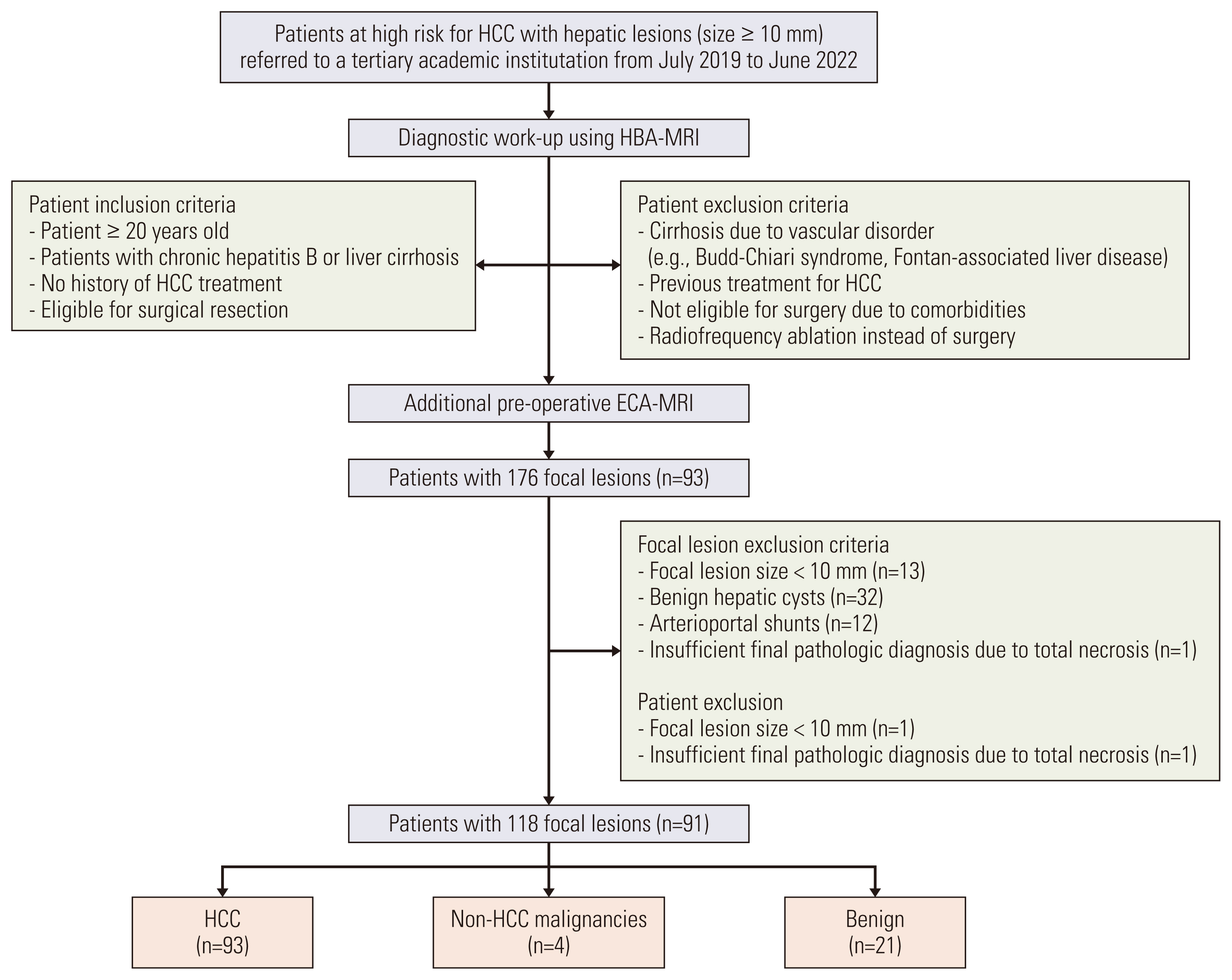
Fig. 1 Flowchart of the inclusion and exclusion criteria of the study population. ECA-MRI, magnetic resonance imaging with an extracellular agent; HBA-MRI, magnetic resonance imaging with a hepatobiliary agent; HCC, hepatocellular carcinoma.
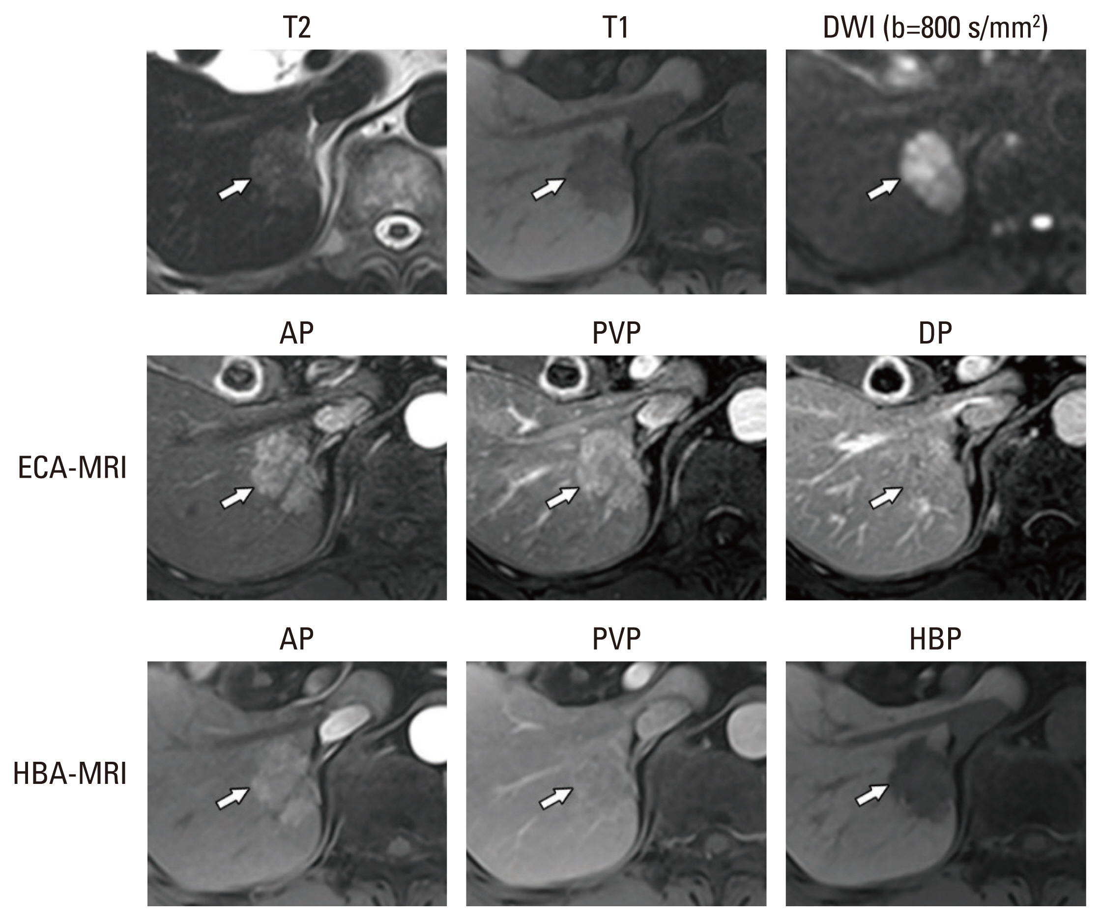
Fig. 2 Categorization of a pathologically confirmed HCC on ECA-MRI and HBA-MRI according to the KLCA-NCC 2022 criteria. A 3.2-cm sized mass in the right posterior section of the liver (arrows) shows moderate T2 hyperintensity and T1 hypointensity. The lesion did not show targetoid appearance on DWI nor enhanced sequences. On ECA-MRI, the mass showed APHE without demonstrable washout appearance during the PVP or DP, and was categorized as “probable” HCC. On HBA-MRI, the mass showed APHE without washout appearance on PVP, but with washout appearance on HBP, and was categorized as “definite” HCC. AP, arterial phase; APHE, arterial phase hyperenhancement; DP, delayed phase; DWI, diffusion-weighted images; ECA-MRI, magnetic resonance imaging with an extracellular agent; HBA-MRI, magnetic resonance imaging with a hepatobiliary agent; HBP, hepatobiliary phase; HCC, hepatocellular carcinoma; KLCA-NCC, Korean Liver Cancer Association-National Cancer Center; PVP, portal venous phase.
Reference
-
References
1. Marrero JA, Kulik LM, Sirlin CB, Zhu AX, Finn RS, Abecassis MM, et al. Diagnosis, staging, and management of hepatocellular carcinoma: 2018 practice guidance by the American Association for the Study of Liver Diseases. Clin Liver Dis. 2019; 13:1.
Article2. Asrani SK, Devarbhavi H, Eaton J, Kamath PS. Burden of liver diseases in the world. J Hepatol. 2019; 70:151–71.
Article3. Omata M, Cheng AL, Kokudo N, Kudo M, Lee JM, Jia J, et al. Asia-Pacific clinical practice guidelines on the management of hepatocellular carcinoma: a 2017 update. Hepatol Int. 2017; 11:317–70.
Article4. European Association for the Study of the Liver. EASL Clinical Practice Guidelines: management of hepatocellular carcinoma. J Hepatol. 2018; 69:182–236.5. American College of Radiology. Committee on LI-RADS® (Liver). Liver Imaging Reporting and Data System (LI-RADS) version 2018 Core [Internet]. Reston, VA: American College of Radiology;2018. [cited 2022 Oct 1]. Available from: https://www.acr.org/-/media/ACR/Files/RADS/LI-RADS/LI-RADS-2018-Core.pdf?la=en.6. Korean Liver Cancer Association (KLCA); National Cancer Center (NCC). 2018 Korean Liver Cancer Association-National Cancer Center Korea practice guidelines for the management of hepatocellular carcinoma. Korean J Radiol. 2019; 20:1042–113.7. Kim TH, Kim SY, Tang A, Lee JM. Comparison of international guidelines for noninvasive diagnosis of hepatocellular carcinoma: 2018 update. Clin Mol Hepatol. 2019; 25:245–63.
Article8. Korean Liver Cancer Association (KLCA); National Cancer Center. 2022 KLCA-NCC Korea practice guidelines for the management of hepatocellular carcinoma. Clin Mol Hepatol. 2022; 28:583–705.9. Kim TK, Lee KH, Jang HJ, Haider MA, Jacks LM, Menezes RJ, et al. Analysis of gadobenate dimeglumine-enhanced MR findings for characterizing small (1–2-cm) hepatic nodules in patients at high risk for hepatocellular carcinoma. Radiology. 2011; 259:730–8.
Article10. Tang A, Bashir MR, Corwin MT, Cruite I, Dietrich CF, Do RK, et al. Evidence supporting LI-RADS major features for CT- and MR imaging-based diagnosis of hepatocellular carcinoma: a systematic review. Radiology. 2018; 286:29–48.11. Cohen J. A coefficient of agreement for nominal scales. Educ Psychol Meas. 1960; 20:37–46.
Article12. Byun J, Choi SH, Byun JH, Lee SJ, Kim SY, Won HJ, et al. Comparison of the diagnostic performance of imaging criteria for HCCs ≤ 3.0 cm on gadoxetate disodium-enhanced MRI. Hepatol Int. 2020; 14:534–43.13. Jeon SK, Lee JM, Joo I, Yoo J, Park JY. Comparison of guidelines for diagnosis of hepatocellular carcinoma using gadoxetic acid-enhanced MRI in transplantation candidates. Eur Radiol. 2020; 30:4762–71.
Article14. Lee S, Kim MJ. Validation of the Korean Liver Cancer Association-National Cancer Center 2018 criteria for the noninvasive diagnosis of hepatocellular carcinoma using magnetic resonance imaging. J Liver Cancer. 2020; 20:120–7.
Article15. Lee S, Kim SS, Chang DR, Kim H, Kim MJ. Comparison of LI-RADS 2018 and KLCA-NCC 2018 for noninvasive diagnosis of hepatocellular carcinoma using magnetic resonance imaging. Clin Mol Hepatol. 2020; 26:340–51.
Article16. Kim DH, Kim B, Youn SY, Kim H, Choi JI. Diagnostic performance of KLCA-NCC 2018 criteria for hepatocellular carcinoma using magnetic resonance imaging: a systematic review and meta-analysis. Diagnostics (Basel). 2021; 11:1763.
Article17. Hwang SH, Park MS, Park S, Lim JS, Kim SU, Park YN. Comparison of the current guidelines for diagnosing hepatocellular carcinoma using gadoxetic acid-enhanced magnetic resonance imaging. Eur Radiol. 2021; 31:4492–503.
Article18. Park SH, Shim YS, Kim B, Kim SY, Kim YS, Huh J, et al. Retrospective analysis of current guidelines for hepatocellular carcinoma diagnosis on gadoxetic acid-enhanced MRI in at-risk patients. Eur Radiol. 2021; 31:4751–63.
Article19. Lee SM, Lee JM, Ahn SJ, Kang HJ, Yang HK, Yoon JH. Diagnostic performance of 2018 KLCA-NCC practice guideline for hpatocellular carcinoma on gadoxetic acid-enhanced MRI in patients with chronic hepatitis B or cirrhosis: comparison with LI-RADS version 2018. Korean J Radiol. 2021; 22:1066–76.
Article20. Joo I, Lee JM, Lee DH, Jeon JH, Han JK, Choi BI. Noninvasive diagnosis of hepatocellular carcinoma on gadoxetic acid-enhanced MRI: can hypointensity on the hepatobiliary phase be used as an alternative to washout? Eur Radiol. 2015; 25:2859–68.21. Choi MH, Choi JI, Lee YJ, Park MY, Rha SE, Lall C. MRI of small hepatocellular carcinoma: typical features are less frequent below a size cutoff of 1.5 cm. AJR Am J Roentgenol. 2017; 208:544–51.
Article22. Yoon J, Hwang JA, Lee S, Lee JE, Ha SY, Park YN. Clinicopathologic and MRI features of combined hepatocellular-cholangiocarcinoma in patients with or without cirrhosis. Liver Int. 2021; 41:1641–51.
- Full Text Links
-
- Actions
-
Cited
- CITED
-
- Close
- Share
-
- Similar articles
-
- Emerging Role of Hepatobiliary Magnetic Resonance Contrast Media and Contrast-Enhanced Ultrasound for Noninvasive Diagnosis of Hepatocellular Carcinoma: Emphasis on Recent Updates in Major Guidelines
- Sonazoid-enhanced ultrasonography for noninvasive imaging diagnosis of hepatocellular carcinoma: special emphasis on the 2022 KLCA-NCC guideline
- Diagnostic performance of the 2022 KLCA-NCC criteria for hepatocellular carcinoma on magnetic resonance imaging with extracellular contrast and hepatobiliary agents: comparison with the 2018 KLCA-NCC criteria
- Comparison of international guidelines for noninvasive diagnosis of hepatocellular carcinoma: 2018 update
- Validation of the Korean Liver Cancer Association-National Cancer Center 2018 Criteria for the Noninvasive Diagnosis of Hepatocellular Carcinoma Using Magnetic Resonance Imaging
