Degenerated lumbricals in the feet of adult human cadavers: case series
- Affiliations
-
- 1Department of Anatomy, All India Institute of Medical Sciences, Jodhpur, India
- 2Department of Anatomy, Mahatma Gandhi University of Medical College and Hospital, Jaipur, India
- 3Department of Anatomy, All India Institute of Medical Sciences, New Delhi, India
- KMID: 2544094
- DOI: http://doi.org/10.5115/acb.22.225
Abstract
- In the foot, the lumbricals flex the metatarsophalangeal joints and extend the interphalangeal joints. The lumbricals are known to be affected in neuropathies. It is not known whether they may degenerate in normal individuals. Here, we report our findings of isolated degenerated lumbricals in seemingly normal feet of two cadavers. We explored lumbricals in 20 male and 8 female cadavers that were 60–80 years of age at the time of death. As part of routine dissection, we exposed the tendons of the flexor digitorum longus and the lumbricals. From the degenerated lumbricals, we took some tissue for paraffin-embedding, sectioning, and staining by hematoxylin and eosin, and Masson’s trichrome technique. Of the 224 lumbricals studied, we found four apparently degenerated lumbricals in two male cadavers. In the first, the 2nd and 4th lumbricals in the left foot and the 2nd in the right foot were degenerated. In the second, the right 4th lumbrical was degenerated. Microscopically, the degenerated tissue was made of bundles of collagen. The lumbricals may have degenerated due to compression of their nerve supply. We cannot comment on whether the functionality of the feet were affected by these isolated degeneration of the lumbricals.
Keyword
Figure
-
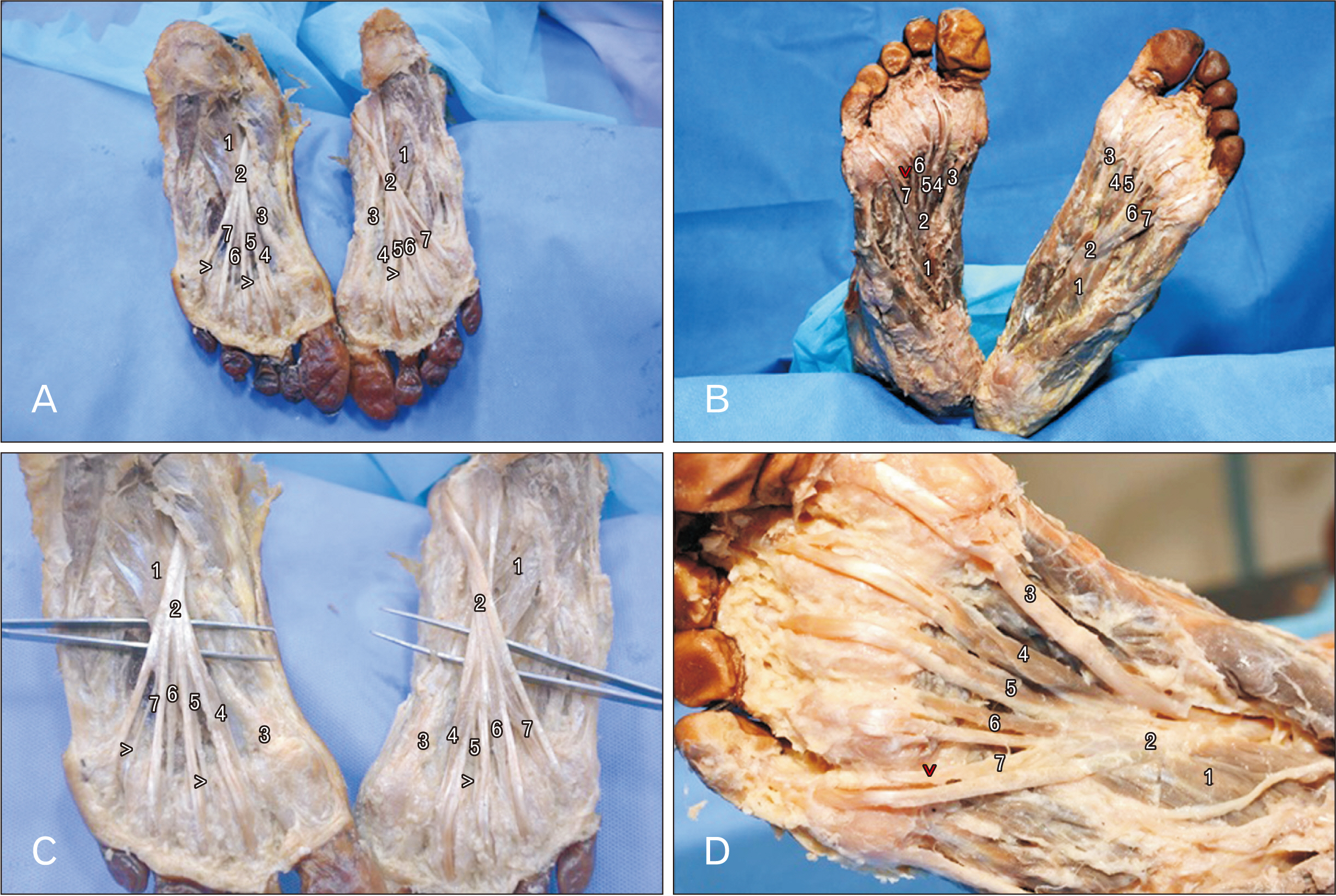
Fig. 1 Photographs of lumbricals of the foot showing: (A) degenerated 2nd and 4th lumbricals in the left and 2nd lumbrical in the right foot; (B) enlarged view of (A) showing degenerated 2nd and 4th lumbricals in the left and 2nd lumbrical in the right foot; (C) the degenerated fourth lumbrical of the right foot and normal lumbricals in the left foot; and (D) enlarged view of the right foot of (C) showing degenerated fourth lumbrical. 1, flexor accessorius; 2, tendon of flexor digitorum longus; 3, tendon of flexor hallucis longus; 4, first lumbrical; 5, second lumbrical; 6, third lumbrical; 7, fourth lumbrical; >, degenerated lumbrical.
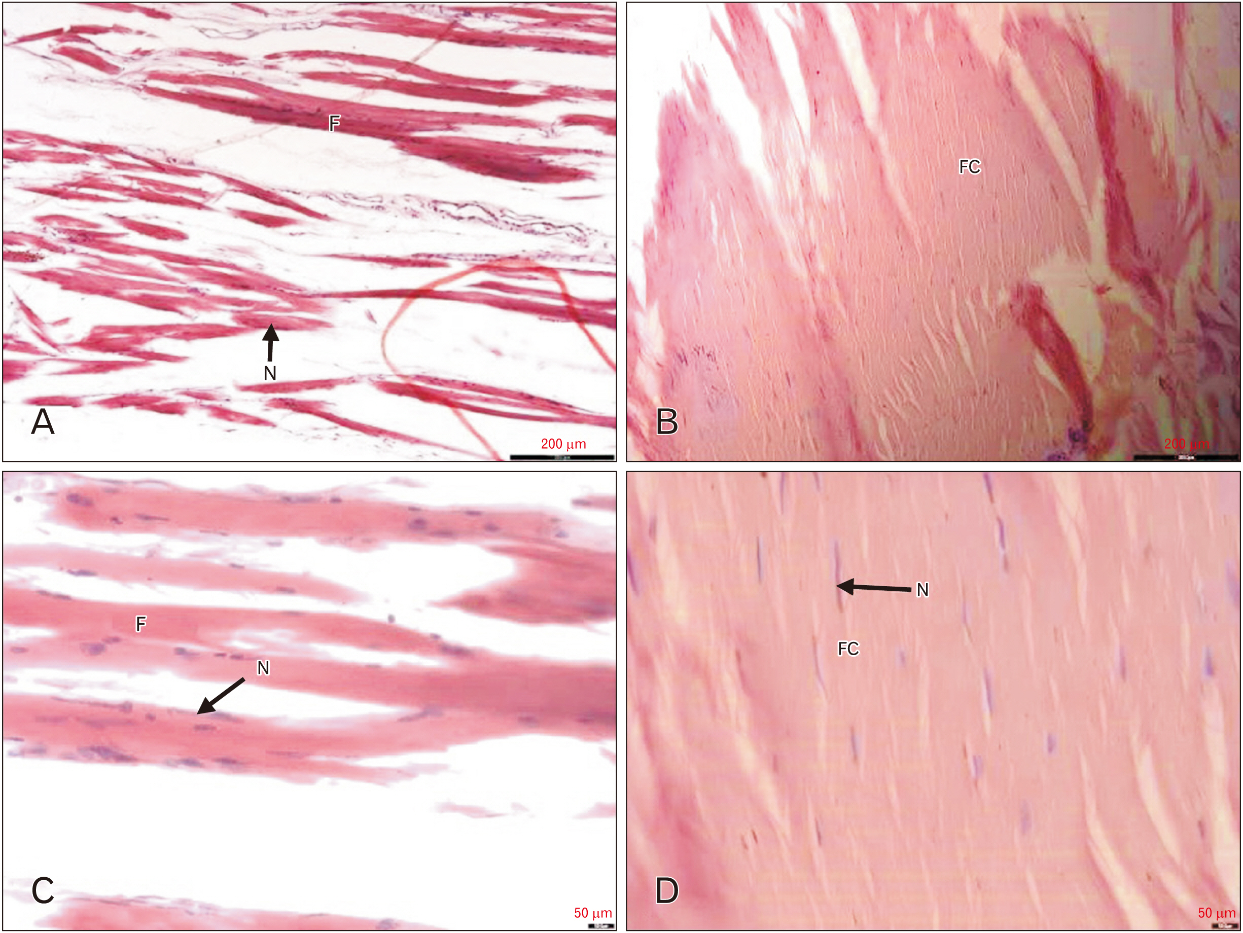
Fig. 2 The photomicrographs of longitudinally sectioned lumbrical muscles, stained with hematoxylin and eosin, showing: (A) normal lumbrical muscle at low power (10×); (B) normal lumbrical muscle at high power (40×); (C) degeneration of lumbrical muscle at low power (10×); and (D) degeneration of lumbrical muscle at high power (40×). Scale bars=200 µm (A, C); scale bars=50 µm (B, D). F, muscle fascicle; N, nucleus; FC, fascicle of collagen fibers.
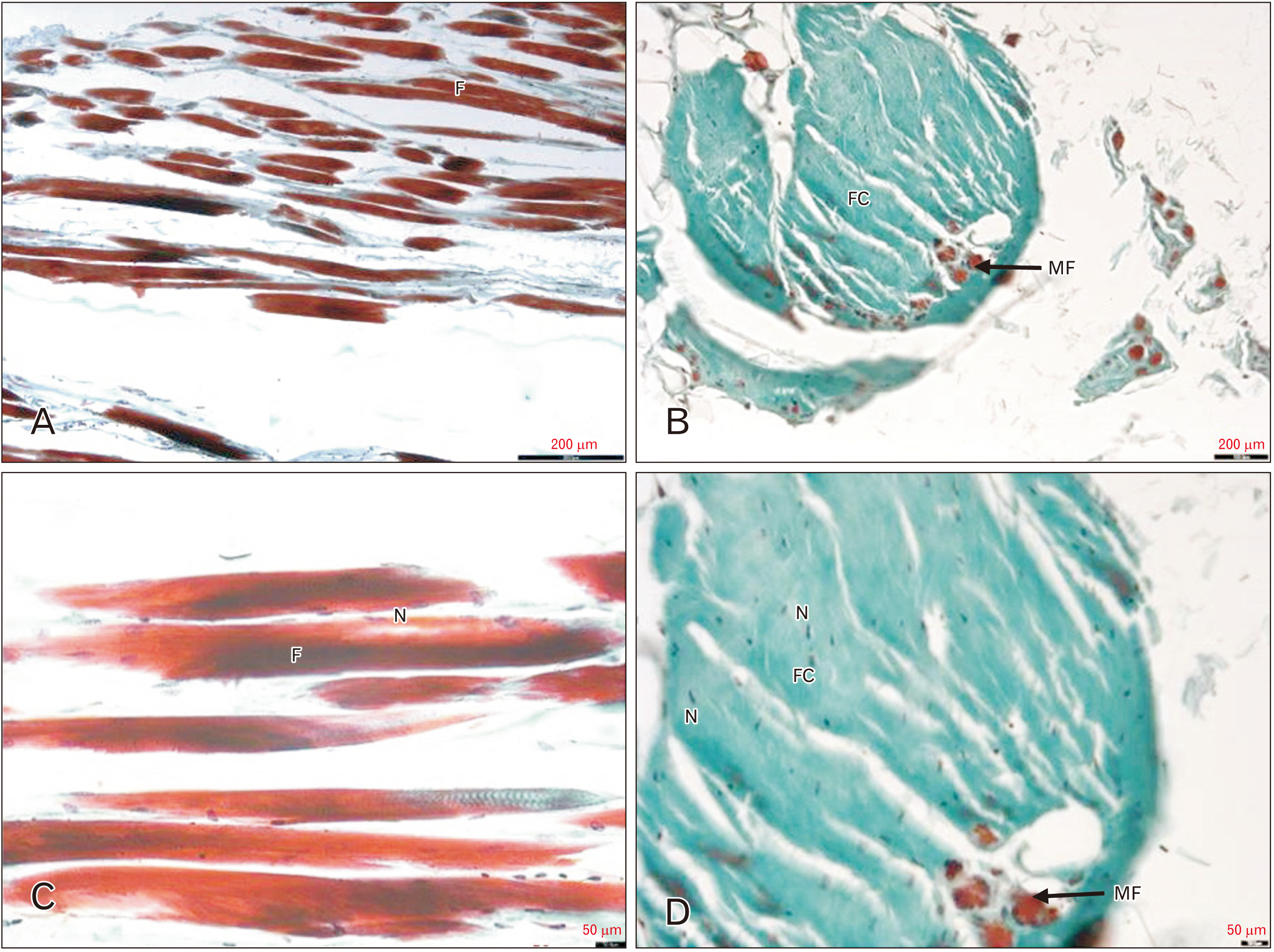
Fig. 3 The photomicrographs of longitudinally sectioned lumbrical muscles, stained with Masson’s trichrome technique, showing: (A) normal lumbrical muscle at low power (10×); (B) normal lumbrical muscle at high power (40×); (C) degeneration of lumbrical muscle at low power (10×); and (D) degeneration of lumbrical muscle at high power (40×). Scale bars=200 µm (A, C), scale bars=50 µm (B, D). F, muscle fascicle ; N, nucleus; FC, fascicle of collagen fibres; MF, muscle fibres.
Reference
-
References
1. Standring S. 2021. Gray's anatomy: the anatomical basis of clinical practice. 42nd ed. Elsevier;p. 979. p. 1458. p. 1462.2. Malhotra A, Kulesza R. 2022; A case report of an accessory flexor digitorum profundus indicis contributing the first lumbrical. Anat Cell Biol. 55:390–3. DOI: 10.5115/acb.22.014. PMID: 35768340. PMCID: PMC9519769.
Article3. Koshi R. 2017. Cunningham's manual of practical anatomy. Volume 1, Upper and lower limbs. 16th ed. Oxford University Press;p. 248–52. DOI: 10.5962/bhl.title.43984.4. Hur MS, Kim JH, Gil YC, Kim HJ, Lee KS. 2015; New insights into the origin of the lumbrical muscles of the foot: tendinous slip of the flexor hallucis longus muscle. Surg Radiol Anat. 37:1161–7. DOI: 10.1007/s00276-015-1488-6. PMID: 25963118.
Article5. Sinnatamby CS. 2011. Apr. 8. Last's anatomy: regional and applied. 12th ed. p. 163.6. Shawn Allen DC, Dabco IFW, Robert Lardner PT. 2008. The importance of the lumbricals [Internet]. Dynamic Chiropractic;Available from: https://www.dynamicchiropractic.com/mpacms/dc/article.php?id=53168. cited 2022 Aug 22.7. Fiolkowski P, Brunt D, Bishop M, Woo R, Horodyski M. 2003; Intrinsic pedal musculature support of the medial longitudinal arch: an electromyography study. J Foot Ankle Surg. 42:327–33. DOI: 10.1053/j.jfas.2003.10.003. PMID: 14688773.
Article8. Okamura K, Kanai S, Oki S, Tanaka S, Hirata N, Sakamura Y, Idemoto N, Wada H, Otsuka A. 2017; Does the weakening of intrinsic foot muscles cause the decrease of medial longitudinal arch height? J Phys Ther Sci. 29:1001–5. DOI: 10.1589/jpts.29.1001. PMID: 28626309. PMCID: PMC5468184.
Article9. Kalin PJ, Hirsch BE. 1987; The origins and function of the interosseous muscles of the foot. J Anat. 152:83–91. DOI: 10.53347/rid-85312. PMID: 3654378. PMCID: PMC1261748.10. Chaney DM, Lee MS, Khan MA, Krueger WA, Mandracchia VJ, Yoho RM. 1996; Study of ten anatomical variants of the foot and ankle. J Am Podiatr Med Assoc. 86:532–7. DOI: 10.7547/87507315-86-11-532. PMID: 8961655.
Article11. Drury RAB, Wallington EA. 1980. Carleton's histological technique. 5th ed. Oxford University Press;p. 57–149. p. 183–5. DOI: 10.5962/bhl.title.23578.12. Jackman RW, Kandarian SC. 2004; The molecular basis of skeletal muscle atrophy. Am J Physiol Cell Physiol. 287:C834–43. DOI: 10.1152/ajpcell.00579.2003. PMID: 15355854.
Article13. Peri G. 1991; The "critical zones" of entrapment of the nerves of the lower limb. Surg Radiol Anat. 13:139–43. DOI: 10.1007/BF01623889. PMID: 1925916.
Article14. Schon LC, Baxter DE. 1990; Neuropathies of the foot and ankle in athletes. Clin Sports Med. 9:489–509. DOI: 10.1016/S0278-5919(20)30743-2. PMID: 2183956.
Article15. Bejjani FJ, Jahss MH. 1986; Le Double's study of muscle variations of the human body. Part II: muscle variations of the foot. Foot Ankle. 6:157–76. DOI: 10.1177/107110078600600401. PMID: 3514399.
Article
- Full Text Links
-
- Actions
-
Cited
- CITED
-
- Close
- Share
-
- Similar articles
-
- Variations of the Lumbrical Muscles in Hands of Koreans
- Comparison between Suture-Button Technique with Syndesmotic Repair and Screw Fixation Technique for Complete Ankle Syndesmotic Injury: Biomechanical Cadaveric Study
- The use of the dorsal metacarpal artery for reconstruction of distal dorsal finger defects: an anatomic study and clinical experience
- Morphological Study of the Fibularis Brevis Tendon and Plantar Aponeurosis Lateral Band Insertion on the Fifth Metatarsal Base in Korean Cadavers
- Profiles, tissue, and microbial integrity of cadavers used in medical faculties in South-western Uganda: implication in anatomical education
