Anatomical study of divergences in facial artery endings
- Affiliations
-
- 1Department of Basic Medical Sciences, College of Medicine, King Saud Bin Abdul Aziz University for Health Sciences, (KSAU-HS), King Abdullah International Medical Research Center (KAIMRC), National Guard Health Affair (NGHA), King Abdul-Aziz Medical City, Jeddah, Saudi Arabia
- KMID: 2544083
- DOI: http://doi.org/10.5115/acb.22.262
Abstract
- Despite several studies focusing on the facial arteries variable courses, the findings have significantly differed. The divergent findings have made it increasingly challenging to establish consistent correlations. Thus, as a vital artery, the facial artery is prone to numerous variations, which makes the identification of the variations vital to clinical practice, particularly for the orofacial and rhinoplastic surgery, and the increasingly selective chemotherapy procedures. The present research uses angiography images for analysis in studying the bilateral facial artery variations noted in patients undergoing carotid angiography for the evaluation of congenital anomalies, cerebral vascular malformations, and intra-arterial procedures. Conventional angiography was used, as it is a vital assessment tool that helps in the assessment of variations in the facial arteries and is suitable in evaluating smaller vascular anatomy, due to the perfect spatial resolution and portrayal of vascular anatomy. Thus, rather than normal ending of the facial artery as an angular artery, the study disclosed that in certain instances, the artery termination took the form of a superior labial artery with a small lateral nasal artery branch located closer to the midline compared to the normal cases. Also, the study has disclosed a conspicuous pre-masseteric branch with small branches originating from the infraorbital artery and providing potential compensation for the facial artery’s shortness. Regardless of the infrequency of such variations, it is vital that they are considered during the performance of any facial surgical procedure.
Keyword
Figure
-

Fig. 1 mages showing the distinct categories of facial arteries based on the arteries final branching. Adapted from Niemann et al. [11], and Meegalla et al. [12], with the permission of the original authors.
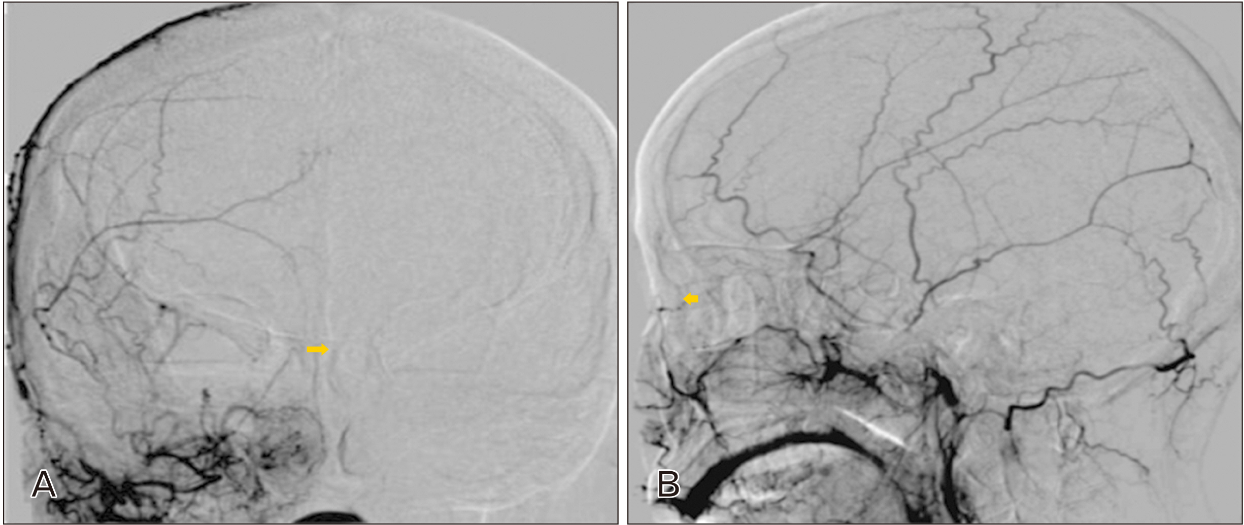
Fig. 2 Type 1A Facial artery and external carotid artery angiography of a cerebral infarction patient (female) who is 44 years old, with an angular branching of the facial artery observed without supratrochlear or a duplex branch. (A) Anteroposterior view (yellow arrow). (B) Lateral view (yellow arrow).
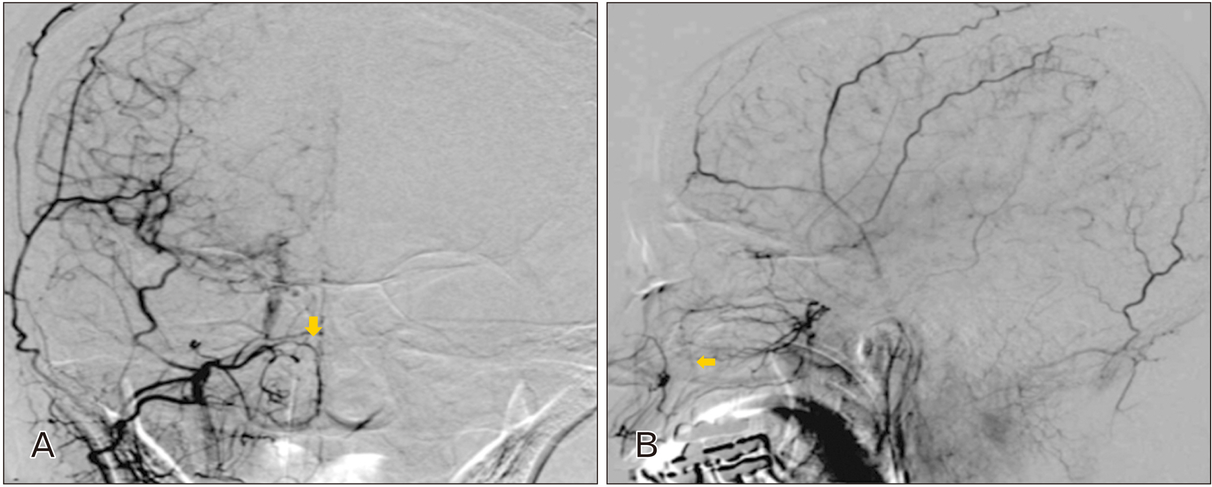
Fig. 3 Type 2 facial artery variation and external carotid artery angiography of a female moyamoya disease patient aged 40 years. (A) Anteroposterior view (yellow arrow). (B) Lateral view (yellow arrow).
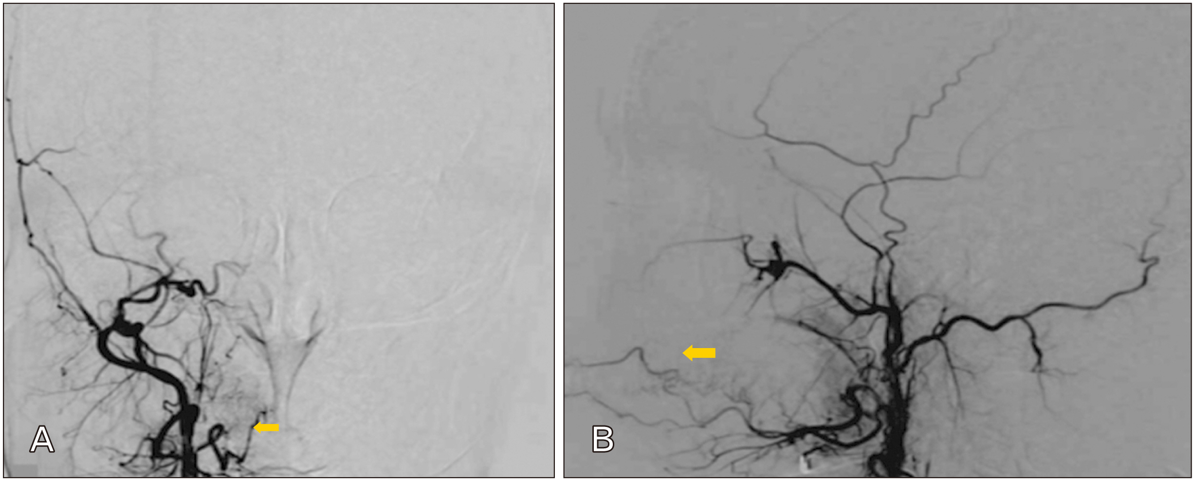
Fig. 4 Type 3 Facial artery variation and am external carotid angiography of a 41 year old female cavernous fistula patient. For this patient, the facial artery’s superior labial branch, indicated using a yellow arrow, marks the end branch. (A) Anteroposterior view. (B) Lateral view.
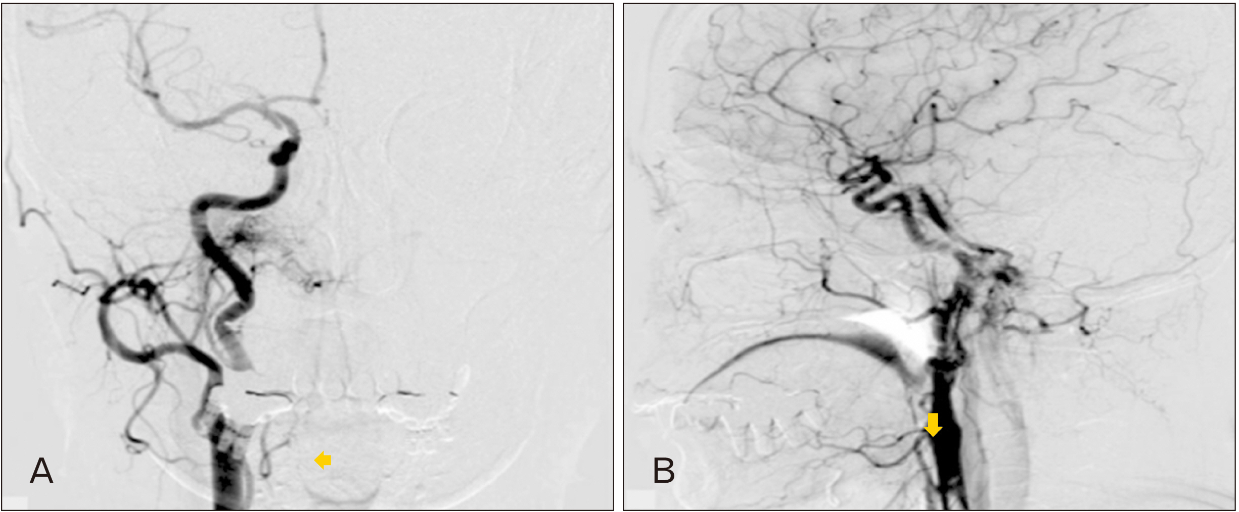
Fig. 5 Type 4 facial artery variation and external carotid artery angiography of a dural arteriovenous fistula patient aged 57 years old. For this patient, only the facial artery’s inferior labial artery branches, indicated by the yellow arrow, can be observed, devoid of other branches. (A) Anteroposterior view. (B) Lateral view.
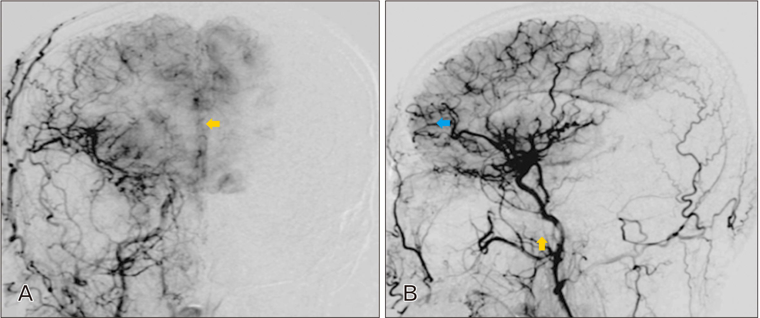
Fig. 6 Type 1C Facial artery variation and external carotid artery angiography of a female spontaneous intracranial haemorrhage patient aged 65 years. Blue arrows indicate the angular artery’s duplex, which has been separated from the facial artery’s lateral nasal branch indicated using the yellow arrows. (A) Anteroposterior view. (B) Lateral view.
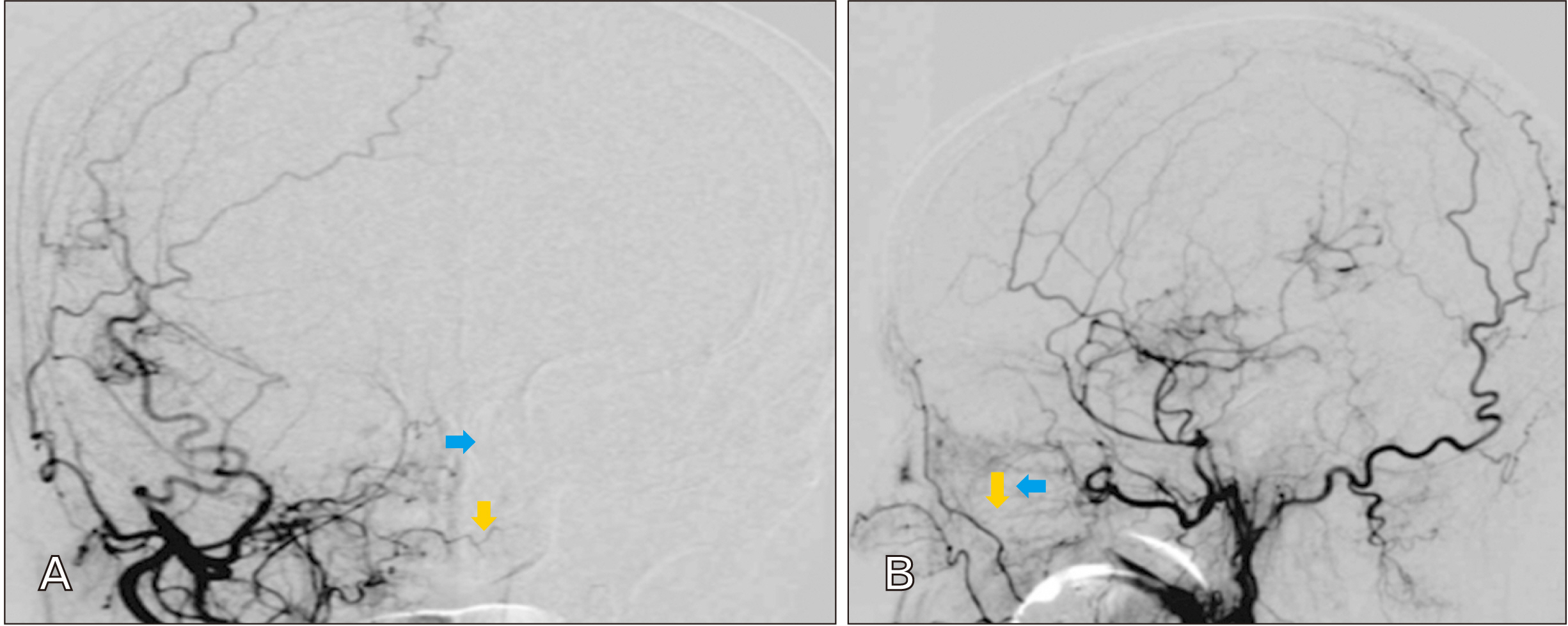
Fig. 7 Type 2 facial artery variation and external carotid artery angiography photo of a moyamoya disease patient aged 70 years, and with maxillary artery (yellow arrow) indicating an infraorbital branching that has extended to the angular artery territory. (A) Anteroposterior view. (B) Lateral view.
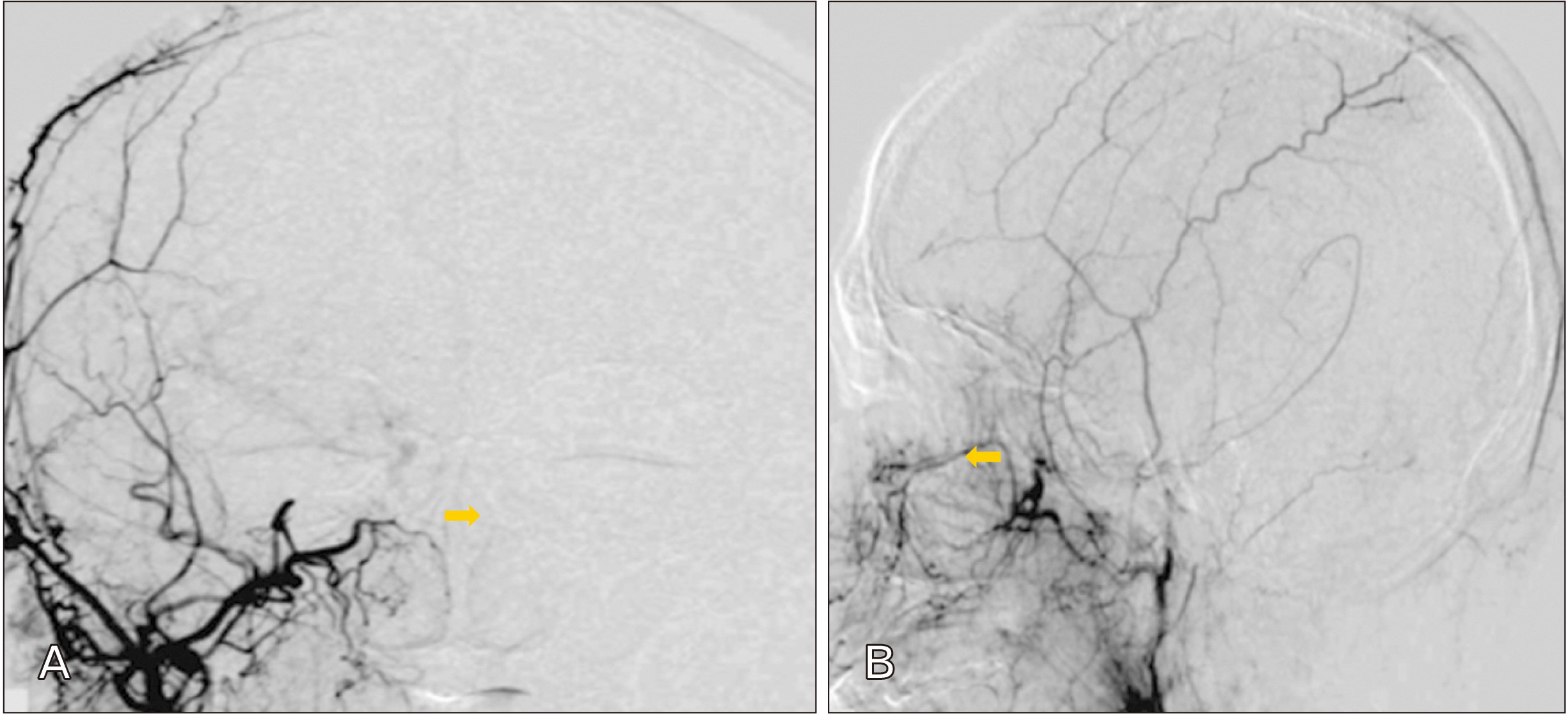
Fig. 8 Type 2 facial artery variation and external carotid artery angiography of a male cerebral infarction patient aged 55 years old. The blue arrow indicates the angular artery’s Supratrochlear branch extended to the frontal bone, and is categorized as Type 1B, even as the infraorbital artery, indicated by the yellow arrow ends prior to reaching the angular artery’s territory and has been categorized as Type 2. (A) Anteroposterior view. (B) Lateral view.
Reference
-
References
1. Braz A, Humphrey S, Weinkle S, Yee GJ, Remington BK, Lorenc ZP, Yoelin S, Waldorf HA, Azizzadeh B, Butterwick KJ, de Maio M, Sadick N, Trevidic P, Criollo-Lamilla G, Garcia P. 2015; Lower face: clinical anatomy and regional approaches with injectable fillers. Plast Reconstr Surg. 136(5 Suppl):235S–257S. DOI: 10.1097/PRS.0000000000001836. PMID: 26441104.2. Funt D, Pavicic T. 2013; Dermal fillers in aesthetics: an overview of adverse events and treatment approaches. Clin Cosmet Investig Dermatol. 6:295–316. DOI: 10.2147/CCID.S50546. PMID: 24363560. PMCID: PMC3865975.3. Pilsl U, Anderhuber F, Neugebauer S. 2016; The facial artery-the main blood vessel for the anterior face? Dermatol Surg. 42:203–8. DOI: 10.1097/DSS.0000000000000599. PMID: 26818208.
Article4. Lohn JW, Penn JW, Norton J, Butler PE. 2011; The course and variation of the facial artery and vein: implications for facial transplantation and facial surgery. Ann Plast Surg. 67:184–8. DOI: 10.1097/SAP.0b013e31822484ae. PMID: 21712695.5. Furukawa M, Mathes DW, Anzai Y. 2013; Evaluation of the facial artery on computed tomographic angiography using 64-slice multidetector computed tomography: implications for facial reconstruction in plastic surgery. Plast Reconstr Surg. 131:526–35. DOI: 10.1097/PRS.0b013e31827c6f18. PMID: 23446566.6. Koh KS, Kim HJ, Oh CS, Chung IH. 2003; Branching patterns and symmetry of the course of the facial artery in Koreans. Int J Oral Maxillofac Surg. 32:414–8. DOI: 10.1054/ijom.2002.0372. PMID: 14505627.
Article7. Lasjaunias P, Vignaud J, Hasso AN. 1975; Maxillary artery blood supply to the orbit: normal and pathological aspects. Neuroradiology. 9:87–97. DOI: 10.1007/BF00341426.8. Lee JG, Yang HM, Choi YJ, Favero V, Kim YS, Hu KS, Kim HJ. 2015; Facial arterial depth and relationship with the facial musculature layer. Plast Reconstr Surg. 135:437–44. DOI: 10.1097/PRS.0000000000000991. PMID: 25626791.
Article9. Desai H, Yu H, Ohana E, Gunnell ET, Kim J, Isaacson A. 2018; Comparative analysis of cone-beam CT angiogram and conventional CT angiogram for prostatic artery identification prior to embolization. J Vasc Interv Radiol. 29:229–32. DOI: 10.1016/j.jvir.2017.09.020. PMID: 29414195.10. Park JH, Chung JW, Lee KW, Park YB, Han MC. 1997; CT angiography of Takayasu arteritis: comparison with conventional angiography. J Vasc Interv Radiol. 8:393–400. DOI: 10.1016/S1051-0443(97)70579-7. PMID: 9152912.
Article11. Niemann K, Lazarus L, Rennie C. 2019; An anatomical study of the facial artery. Int J Morphol. 37:1310–5. DOI: 10.4067/S0717-95022019000401310.
Article12. Meegalla N, Sood G, Nessel TA, Downs BW. 2022. Anatomy, head and neck, facial arteries [Internet]. StatPearls;Available from: https://europepmc.org/article/nbk/nbk536932. cited 2022 Feb 13.13. Cho KH, Dalla Pozza E, Toth G, Bassiri Gharb B, Zins JE. 2019; Pathophysiology study of filler-induced blindness. Aesthet Surg J. 39:96–106. DOI: 10.1093/asj/sjy141. PMID: 29873688.
Article14. Thanasarnaksorn W, Cotofana S, Rudolph C, Kraisak P, Chanasumon N, Suwanchinda A. 2018; Severe vision loss caused by cosmetic filler augmentation: case series with review of cause and therapy. J Cosmet Dermatol. 17:712–8. DOI: 10.1111/jocd.12705. PMID: 30006992.
Article15. Marx C, Kumar P, Reddy S, Vollala VR. 2008; Bilateral variation of facial artery: a case report. Rom J Morphol Embryol. 49:399–401. PMID: 18758647.16. Kumar A, Elumalai G, Thangamani M, Palayathan N, Singh MK. 2014; A rare variation in facial artery and its implications in facial surgery: case report. J Surg. 2:68–71. DOI: 10.11648/j.js.20140205.13.
Article17. Yonenaga K, Tohnai I, Mitsudo K, Mori Y, Saijo H, Iwai T, Yonehara Y, Ota Y, Torigoe K, Takato T. 2011; Anatomical study of the external carotid artery and its branches for administration of superselective intra-arterial chemotherapy via the superficial temporal artery. Int J Clin Oncol. 16:654–9. DOI: 10.1007/s10147-011-0238-y. PMID: 21537883.
Article18. Loukas M, Hullett J, Louis RG Jr, Kapos T, Knight J, Nagy R, Marycz D. 2006; A detailed observation of variations of the facial artery, with emphasis on the superior labial artery. Surg Radiol Anat. 28:316–24. DOI: 10.1007/s00276-006-0093-0. PMID: 16547605.
Article19. Mitz V, Ricbourg B, Lassau JP. 1973; Facial branches of the facial artery in adults. Typology, variations and respective cutaneous areas. Ann Chir Plast. 18:339–50. French. PMID: 4775922.20. Niranjan NS. 1988; An anatomical study of the facial artery. Ann Plast Surg. 21:14–22. DOI: 10.1097/00000637-198807000-00003. PMID: 3421650.
Article21. Wang D, Xiong S, Zeng N, Wu Y. 2022; Facial arterial variations in Asians: a study on computed tomographic angiography. Aesthet Surg J. 42:527–34. DOI: 10.1093/asj/sjab380. PMID: 34724046.
Article22. Kim JH, Kang NH, Lee IH, Seo YJ, Yang HJ, Song SH, Oh SH. 2011; Preoperative evaluation of the facial artery using facial angio computed tomography. Arch Plast Surg. 38:719–24.23. Koziej M, Trybus M, Hołda M, Polak J, Wnuk J, Brzegowy P, Popiela T, Walocha J, Chrapusta A. 2019; Anatomical map of the facial artery for facial reconstruction and aesthetic procedures. Aesthet Surg J. 39:1151–62. DOI: 10.1093/asj/sjz028. PMID: 30721996.
Article24. Kaatee R, Beek FJ, de Lange EE, van Leeuwen MS, Smits HF, van der Ven PJ, Beutler JJ, Mali WP. 1997; Renal artery stenosis: detection and quantification with spiral CT angiography versus optimized digital subtraction angiography. Radiology. 205:121–7. DOI: 10.1148/radiology.205.1.9314973. PMID: 9314973.
Article25. Perrini P, Cardia A, Fraser K, Lanzino G. 2007; A microsurgical study of the anatomy and course of the ophthalmic artery and its possibly dangerous anastomoses. J Neurosurg. 106:142–50. DOI: 10.3171/jns.2007.106.1.142. PMID: 17236500.
Article26. Lee JS, Kim JY, Jung C, Woo SJ. 2020; Iatrogenic ophthalmic artery occlusion and retinal artery occlusion. Prog Retin Eye Res. 78:100848. DOI: 10.1016/j.preteyeres.2020.100848. PMID: 32165219.
Article27. Sito G, Manzoni V, Sommariva R. 2019; Vascular complications after facial filler injection: a literature review and meta-analysis. J Clin Aesthet Dermatol. 12:E65–72. PMID: 31360292. PMCID: PMC6624005.
- Full Text Links
-
- Actions
-
Cited
- CITED
-
- Close
- Share
-
- Similar articles
-
- Preoperative Evaluation of the Facial Artery Using Facial Angio Computed Tomography
- The branching patterns and termination points of the facial artery: a cadaveric anatomical study
- A Case Report of Posttraumatic Pseudoaneurysm of the Superficial Temporal Artery
- Effectiveness of Vascular Network Including Angular Artery in Inner Canthal Region for Mid-facial Reconstruction
- Preservation of the Facial Artery during Submandibular Gland Resection: Preoperative Sonographic Assessment of Facial Artery
