The sphenopalatine vein: anatomical study of a rarely described structure
- Iwanaga J
 1,2,3,8
 - Pineda E4
- Miyamoto Y5
- Wysiadecki G6
- Anadkat S8
- Tubbs RS1,2,7,8,9,
- Affiliations
-
- 1Department of Neurosurgery, Tulane Center for Clinical Neurosciences, Tulane University School of Medicine, New Orleans, LA, USA
- 2Department of Neurology, Tulane Center for Clinical Neurosciences, Tulane University School of Medicine, New Orleans, LA, USA
- 3Department of Oral and Maxillofacial Anatomy, Graduate School of Medical and Dental Sciences, Tokyo Medical and Dental University, Tokyo, Japan
- 4Tulane University School of Medicine, New Orleans, LA, USA
- 5Department of Otorhinolaryngology, Graduate School of Medical Sciences, Kyusyu University, Fukuoka, Japan
- 6Department of Normal and Clinical Anatomy, Chair of Anatomy and Histology, Medical University of Lodz, Lodz, Poland
- 7Department of Anatomical Sciences, St. George’s University, St. George’s, Grenada, West Indies, USA
- 8Department of Structural & Cellular Biology, Tulane University School of Medicine, New Orleans, LA, USA
- 9Department of Surgery, Tulane University School of Medicine, New Orleans, LA, USA
- 10Department of Neurosurgery and Ochsner Neuroscience Institute, Ochsner Health System, New Orleans, LA, USA
- 11University of Queensland, Brisbane, Australia
- KMID: 2544081
- DOI: http://doi.org/10.5115/acb.22.231
Abstract
- Although in counterpart, the sphenopalatine artery (SPA), has been well described in the medical literature, the sphenopalatine vein (SPV) has received scant attention. Therefore, the present anatomical study was performed. Additionally, we discuss the variations, embryology, and clinical significance of the SPV. Adult cadaveric specimens underwent dissection of the SPV. In addition, some specimens were submitted for histological analysis of this structure. The SPV was found to drain from the sphenoidal sinus and nasal septum. Small tributaries traveled through the nasal septum with the posterior septal branches of the SPA and nasopalatine nerve. The SPA and SPV were found to travel through the sphenopalatine foramen and another tributary was found to perforate the medial plate of the pterygoid process and to connect to the pterygoid venous plexus which traveled lateral to the medial plate of the pterygoid process. The vein traveled through the posterior part of the lateral wall of the nasal cavity with the posterior lateral nasal branches of the SPA and the lateral superior posterior nasal branches of the maxillary nerve. To our knowledge, this is the first anatomical study on the SPV in humans. Data on the SPV provides an improved anatomical understanding of the vascular network of the nasal cavity. Developing a more complete picture of the nasal cavity and its venous supply might help surgeons and clinicians better manage clinical entities such as posterior epistaxis, cavernous sinus infections, and perform endoscopic surgery with fewer complications.
Figure
-
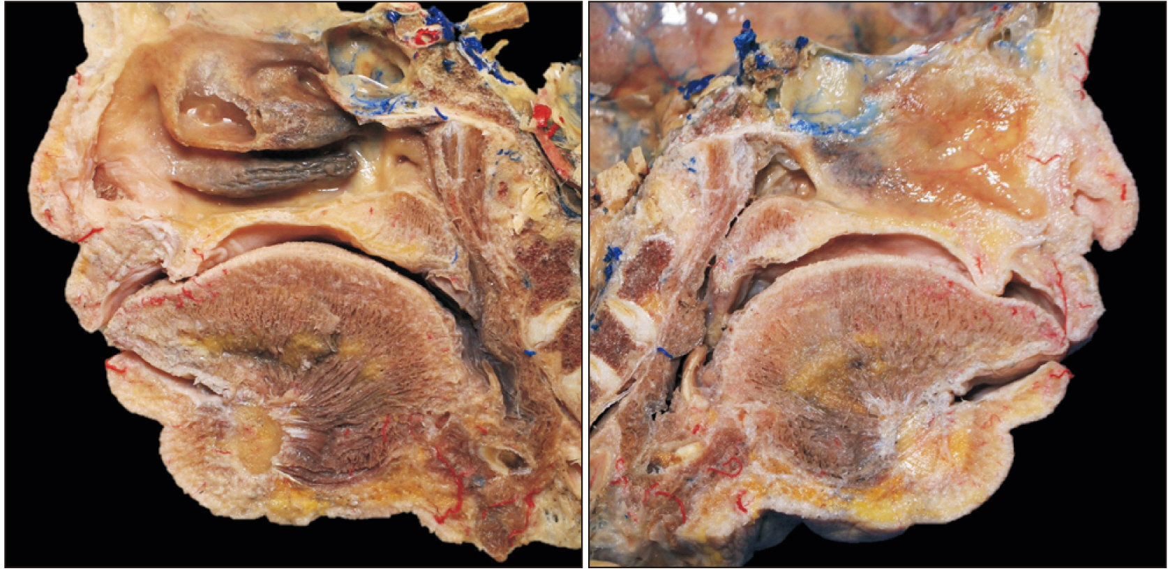
Fig. 1 Cadaveric head bisected using a bone saw and separated into two parts, one with the nasal septum (the right image) and the other without (the left image).

Fig. 2 The nasal septum and adjacent palate harvested from the midline (the left image). The additional coronal section was made for histological analysis (the right image).
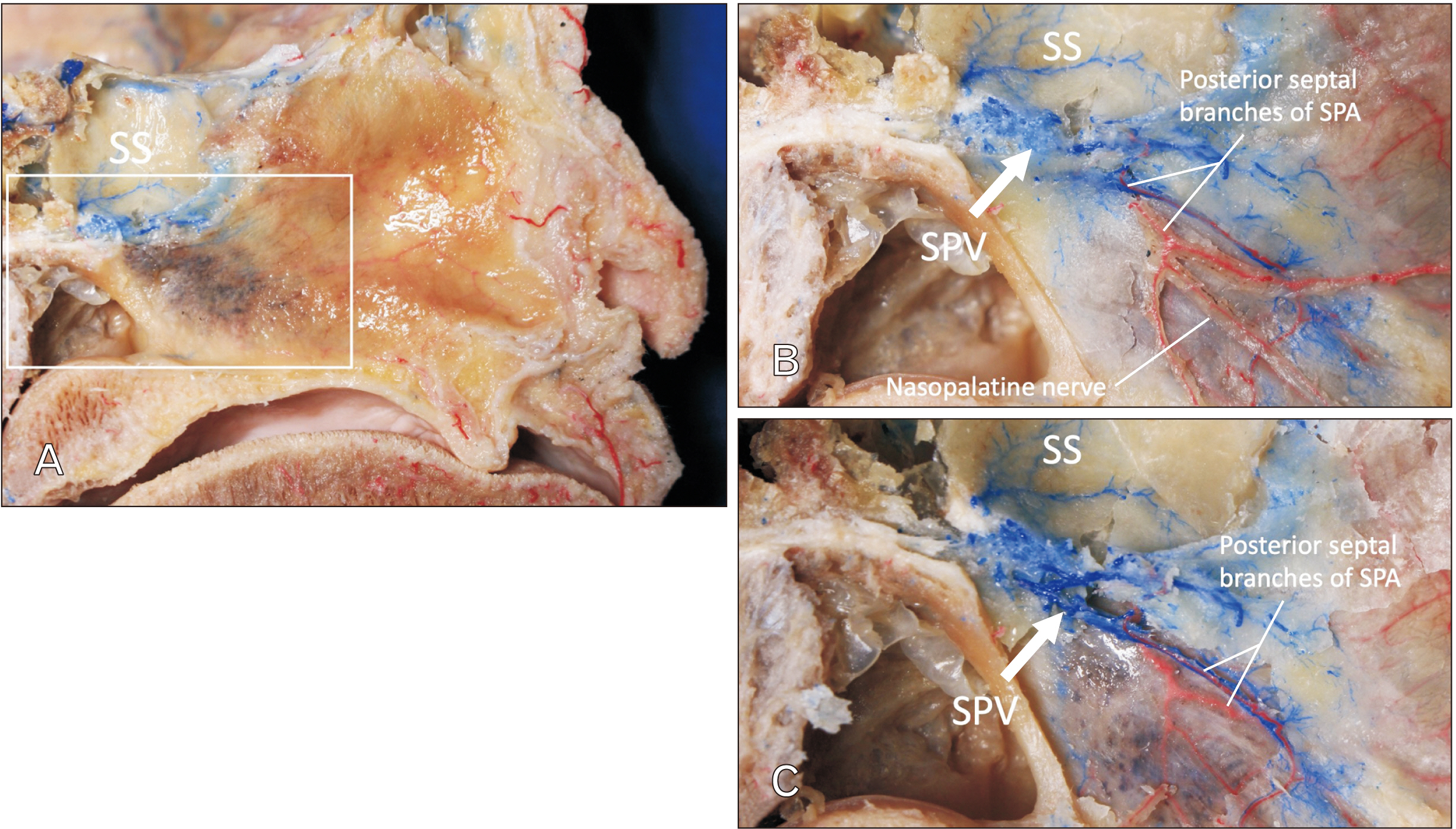
Fig. 3 Posterior part of the nasal septum (midsagittally hemisected head). (A) Specimen prior to dissection. (B) Following removal of the mucosa of the right side of the nasal septum. SPV drains from the SS and nasal septum. (C) Following careful bony removal around the SPV, note that the SPV courses with the posterior septal branches of the SPA. SS, sphenoidal sinus; SPV, sphenopalatine vein; SPA, sphenopalatine artery.

Fig. 4 Posterior part of the lateral wall of the nasal cavity. (A) Specimen prior to dissection. (B) Following removal of the mucosa. Note the SPA and vein go through the sphenopalatine foramen (white arrow) and another tributary (yellow arrow) perforates the MP to connect with the SPV which runs with the posterior lateral nasal branches of SPA. ET, eustachian tube; INC, inferior nasal concha; MNC, middle nasal concha; MP, medial plate of pterygoid process; LSPN, lateral superior posterior nasal branches of the maxillary nerve; SS, sphenoidal sinus; SPV, sphenopalatine vein; SPA, sphenopalatine artery.

Fig. 5 Masson-trichrome stain of the NS. The right image is a magnified image of dotted rectangular area of the left one. Note the posterior septal branches of the SPA and SPV are shown with close proximity. The perpendicular plate of the ethmoid bone is shown with arrowheads. NS, nasal septum; SPV, sphenopalatine vein; SPA, sphenopalatine artery. White scale bar: 5 mm, black scale bar: 500 μm.
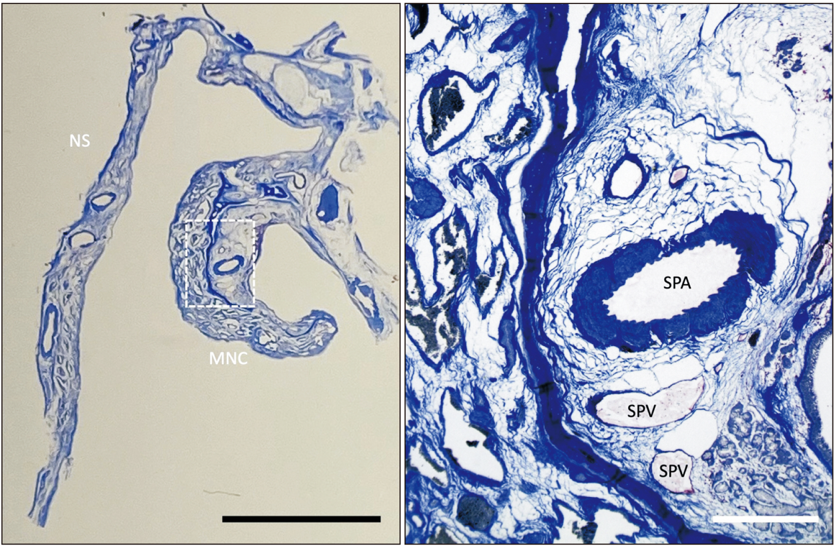
Fig. 6 Masson-trichrome stain of the NS and MNC. The right image is a magnified image of dotted rectangular area of the left one. Note that the posterior lateral nasal branches of the SPA and SPV are shown in close proximity. NS, nasal septum; MNC, middle nasal concha; SPV, sphenopalatine vein; SPA, sphenopalatine artery. Black scale bar: 5 mm, white scale bar: 500 μm.
Reference
-
References
1. Morosanu CO, Humphreys C, Egerton S, Tierney CM. 2022; Woodruff's plexus-arterial or venous? Surg Radiol Anat. 44:169–81. DOI: 10.1007/s00276-021-02852-0. PMID: 34714375.
Article2. Iwanaga J, Singh V, Takeda S, Ogeng'o J, Kim HJ, Moryś J, Ravi KS, Ribatti D, Trainor PA, Sañudo JR, Apaydin N, Sharma A, Smith HF, Walocha JA, Hegazy AMS, Duparc F, Paulsen F, Del Sol M, Adds P, Louryan S, Fazan VPS, Boddeti RK, Tubbs RS. 2022; Standardized statement for the ethical use of human cadaveric tissues in anatomy research papers: recommendations from Anatomical Journal Editors-in-Chief. Clin Anat. 35:526–8. DOI: 10.1002/ca.23849. PMID: 35218594.3. MacArthur FJ, McGarry GW. 2017; The arterial supply of the nasal cavity. Eur Arch Otorhinolaryngol. 274:809–15. DOI: 10.1007/s00405-016-4281-1. PMID: 27568352.
Article4. Hauman CH, Chandler NP, Tong DC. 2002; Endodontic implications of the maxillary sinus: a review. Int Endod J. 35:127–41. DOI: 10.1046/j.0143-2885.2001.00524.x. PMID: 11843967.
Article5. Souza SS, Raggio BS. 2022. Anatomy, head and neck, sphenopalatine foramen [Internet]. StatPearls;Available from: https://www.ncbi.nlm.nih.gov/books/NBK549911/. cited 2022 Jun 8.6. Krulewitz NA, Fix ML. 2019; Epistaxis. Emerg Med Clin North Am. 37:29–39. DOI: 10.1016/j.emc.2018.09.005. PMID: 30454778.
Article7. Seikaly H. 2021; Epistaxis. N Engl J Med. 384:944–51. DOI: 10.1056/NEJMcp2019344. PMID: 33704939.
Article8. Munawar K, Nayak G, Fatterpekar GM, Sen C, Zagzag D, Zan E, Hagiwara M. 2020; Cavernous sinus lesions. Clin Imaging. 68:71–89. DOI: 10.1016/j.clinimag.2020.06.029. PMID: 32574933.
Article9. Lung MA. 1995; The role of the autonomic nerves in the control of nasal circulation. Biol Signals. 4:179–85. DOI: 10.1159/000109439. PMID: 8750945.
Article10. Malm L. 1973; Stimulation of sympathetic nerve fibres to the nose in cats. Acta Otolaryngol. 75:519–26. DOI: 10.3109/00016487309139783. PMID: 4146975.
Article11. Eccles R, Wilson H. 1974; The autonomic innervation of the nasal blood vessels of the cat. J Physiol. 238:549–60. DOI: 10.1113/jphysiol.1974.sp010542. PMID: 4854660. PMCID: PMC1330901.
Article12. Iwanaga J, Wilson C, Simonds E, Vetter M, Schmidt C, Yilmaz E, Choi PJ, Oskouian RJ, Tubbs RS. 2018; Clinical anatomy of blockade of the pterygopalatine ganglion: literature review and pictorial tour using cadaveric images. Kurume Med J. 65:1–5. DOI: 10.2739/kurumemedj.MS651001. PMID: 30158355.
Article13. Jackson RT, Rooker DW. 1971; Stimulation and section of the vidian nerve in relation to autonomic control of the nasal vasculature. Laryngoscope. 81:565–9. DOI: 10.1288/00005537-197104000-00007. PMID: 5553124.
Article14. Lung MA, Wang JC. 1987; Arterial supply, venous drainage and collateral circulation in the nose of the anaesthetized dog. J Physiol. 391:57–70. DOI: 10.1113/jphysiol.1987.sp016725. PMID: 3443958. PMCID: PMC1192201.
Article15. Iwanaga J, Singh V, Ohtsuka A, Hwang Y, Kim HJ, Moryś J, Ravi KS, Ribatti D, Trainor PA, Sañudo JR, Apaydin N, Şengül G, Albertine KH, Walocha JA, Loukas M, Duparc F, Paulsen F, Del Sol M, Adds P, Hegazy A, Tubbs RS. 2021; Acknowledging the use of human cadaveric tissues in research papers: recommendations from Anatomical Journal editors. Clin Anat. 34:2–4. DOI: 10.1002/ca.23671. PMID: 32808702.
Article
- Full Text Links
-
- Actions
-
Cited
- CITED
-
- Close
- Share
-
- Similar articles
-
- The Morphology and Anatomical Location of the Sphenopalatine Foramen
- Trans-Inferior Turbinate Approach for Endoscopic Sphenopalatine Artery Ligation
- Stereotactic Sphenopalatine Ganglionotomy Using Radiofrequency Thermocoagulation: Case reports
- Pulsed Radiofrequency of the Sphenopalatine Ganglion for Treatment of a Cluster Headache: A case report
- Transposition of Femoral Artery and Vein in Fossa Ovalis Encountered during Varicose Vein Surgery: 3 cases report
