Acute Crit Care.
2023 May;38(2):244-248. 10.4266/acc.2021.01102.
What should an intensivist know about pneumocephalus and tension pneumocephalus?
- Affiliations
-
- 1Department of Surgical Intensive Care Medicine, Rashid Hospital, Dubai, UAE
- 2Dubai Medical College, Dubai, UAE
- KMID: 2543647
- DOI: http://doi.org/10.4266/acc.2021.01102
Abstract
- Collection of air in the cranial cavity is called pneumocephalus. Although simple pneumocephalus is a benign condition, accompanying increased intracranial pressure can produce a life-threatening condition comparable to tension pneumothorax, which is termed tension pneumocephalus. We report a case of tension pneumocephalus after drainage of a cerebrospinal fluid hygroma. The tension pneumocephalus was treated with decompression craniotomy, but the patient later died due to the complications related to critical care. Traumatic brain injury and neurosurgical intervention are the most common causes of pneumocephalus. Pneumocephalus and tension pneumocephalus are neurosurgical emergencies, and anesthetics and intensive care management like the use of nitrous oxide during anesthesia and positive pressure ventilation have important implications in their development and progress. Clinically, patients can present with various nonspecific neurological manifestations that are indistinguishable from a those of a primary neurological condition. If the diagnosis is questionable, patients should be investigated using computed tomography of the brain. Immediate neurosurgical consultation with decompression is the treatment of choice.
Keyword
Figure
-

Figure 1. Computed tomography of the brain showing cerebrospinal hygroma left side more than right side at cerebral convexities.
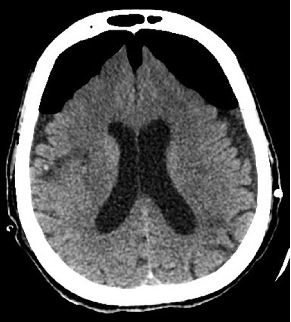
Figure 2. Computed tomography of the brain showing classical “Mount Fuji” sign indicating the development of tension pneumocephalus.
Reference
-
1. Harvey JJ, Harvey SC, Belli A. Tension pneumocephalus: the neurosurgical emergency equivalent of tension pneumothorax. BJR Case Rep. 2016; 2:20150127.2. Wolff E. Luftansammlung im rechten seitenventrikel des gehirns (pneumozephalus). Münch Med Wochenschr. 1914; 61:899.3. Kessler LA, Stern WZ. The ventriculopleural shunt procedure for hydrocephalus: case report of an unusual complication. J Pediatr. 1962; 60:418–20.4. Lee SH, Koh JS, Bang JS, Kim MC. Extensive tension pneumocephalus caused by spinal tapping in a patient with Basal skull fracture and pneumothorax. J Korean Neurosurg Soc. 2009; 45:318–21.
Article5. Sweni S, Senthilkumaran S, Balamurugan N, Thirumalaikolundusubramanian P. Tension pneumocephalus: a case report with review of literature. Emerg Radiol. 2013; 20:573–8.
Article6. Chang Y, Kim TG, Chung SY. High-flow nasal cannula-induced tension pneumocephalus. Indian J Crit Care Med. 2020; 24:592–5.
Article7. Clement AR, Palaniappan D, Panigrahi RK. Tension pneumocephalus. Anesthesiology. 2017; 127:710.
Article8. Pulickal GG, Sitoh YY, Ng WH. Tension pneumocephalus. Singapore Med J. 2014; 55:e46–8.
Article9. Michel SJ. The Mount Fuji sign. Radiology. 2004; 232:449–50.
Article10. Heidari SF. A patient with massive pneumocephalus without sign of tension pneumocephalus. J Intensive Crit Care. 2016; 2:3.
Article11. Arbit E, Shah J, Bedford R, Carlon G. Tension pneumocephalus: treatment with controlled decompression via a closed water-seal drainage system: case report. J Neurosurg. 1991; 74:139–42.12. Das JM, Bajaj J. Pneumocephalus [Internet]. StatPearls Publishing;2021. [cited 2021 Sep 11]. Available from: https://www.ncbi.nlm.nih.gov/books/NBK535412.13. Amato-Watkins A, Rao VM, Leach P. Air travel after intracranial surgery: a survey of advice given to patients by consultant neurosurgeons in the UK. Br J Neurosurg. 2013; 27:9–11.
Article
- Full Text Links
-
- Actions
-
Cited
- CITED
-
- Close
- Share
-
- Similar articles
-
- A Case of Intracerebral Tension Pneumocephalus
- Subdural Tension Pneumocephalus Follwing Surgery
- Tension Pneumocephalus after Transsphenoidal Surgery: Report of Two Cases
- A Case of Subdural Tension Pneumocephalus after Operation
- Communicating Hydrocephalus Onset Following a Traumatic Tension Pneumocephalus
