An interesting case of survival to multiple ruptures of aneurysms, with persistent trigeminal artery, cranial nerve deficit, and evolutionary exposure of neurovascular treatment
- Affiliations
-
- 1Department of Neurosurgery, Complejo Hospitalario Dr. Arnulfo Arias Madrid, Transístmica, Panamá
- 2General Medicine, University of Panama, Transístmica, Panamá
- KMID: 2543515
- DOI: http://doi.org/10.7461/jcen.2022.E2022.06.009
Abstract
- Subarachnoid hemorrhage secondary to rupture of an aneurysm is a severe condition, associated with a high rate of morbidity and mortality. There are few cases in the literature of rupture of an aneurysm of the persistent trigeminal artery. This is the case of a 62-year-old female who has suffered multiple ruptures of aneurysms, in different decades of her life, with the development of de novo aneurysm, been this the presented case, a rupture of aneurysm of the persistent trigeminal artery. This patient has survival to these conditions and remain without important morbidity. The case manifested with a clinical picture of third and seventh cranial nerve deficit, which this last one, there are not previous publications of cases with this deficit. This aneurysm was embolized with coils, and the postoperative condition was satisfactory, been discharged at 4 postoperative days.
Keyword
Figure
-
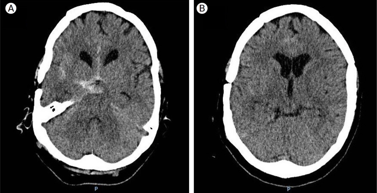
Fig. 1. Head computed tomography (CT) scan. (A) Subarachnoid hemorrhage is observed at the level of the suprasellar cistern. (B) There is no evidence of drainage of blood to the ventricular system.
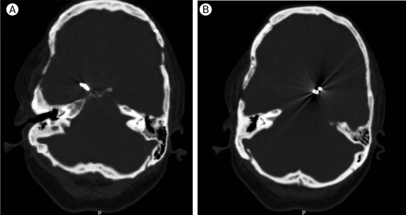
Fig. 2. Head computed tomography (CT) scan (bone window). (A) A foreign object is observed which seems to represent a vascular spring clip on the right side. (B) Another foreign body appears suggestive of a malleable vascular clip on the left side.
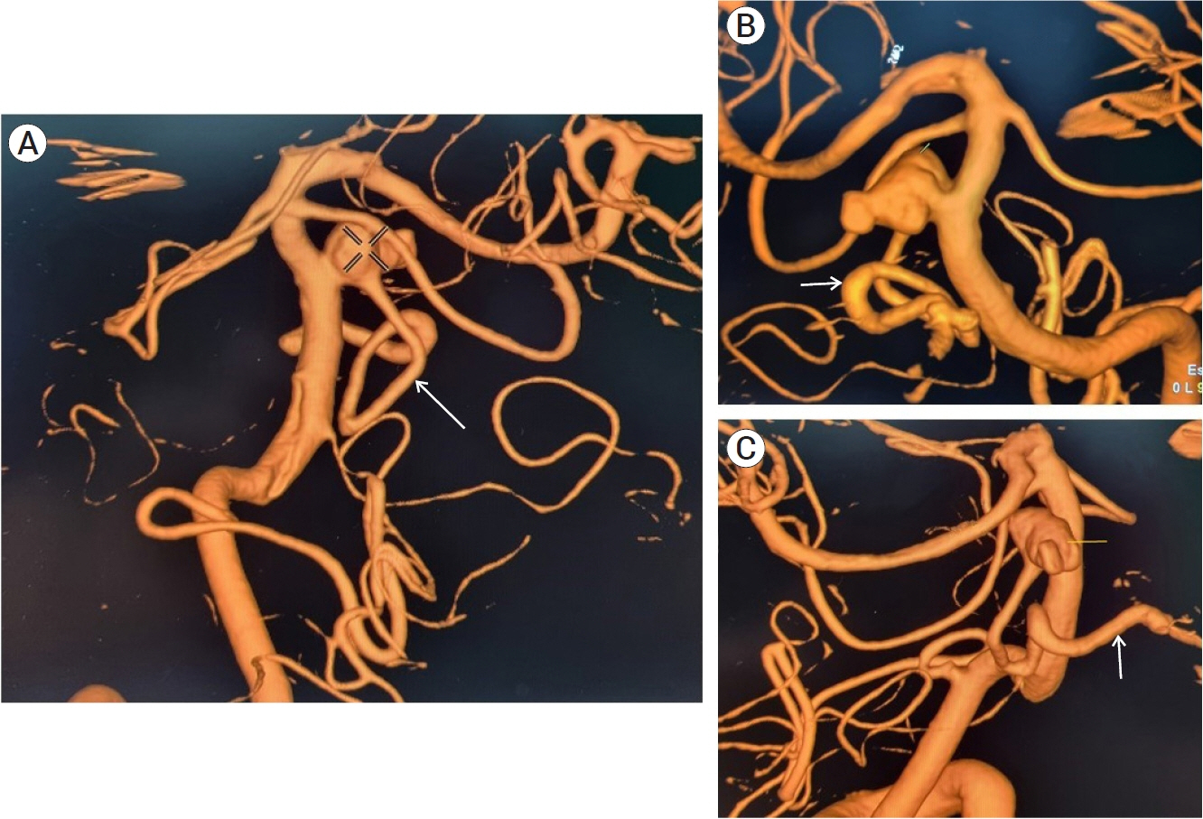
Fig. 3. Cerebral angiography (3D reconstruction). Image (A) shows the panoramic view, with a cross indicating the aneurysm with dimensions of 7.2×8.5 mm and a neck of 4 mm. (B) Anteroposterior view (note the relationship of the aneurysm with the anatomical exit of the third cranial nerve and a caudal twist of the persistent trigeminal artery, which could explain the III and VII cranial nerve palsy, respectively). (C) Side view. Note the anterior orientation of the patent trigeminal artery toward the petrous portion of the internal carotid artery. Cross and yellow line: aneurysm. White arrow: persistent trigeminal artery.
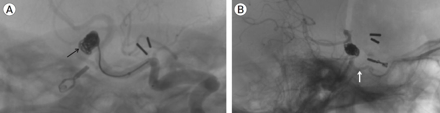
Fig. 4. Cerebral angiography. (A) The introduction of the coils and the occlusion of the aneurysm are shown. (B) Patency of the persistent trigeminal artery is observed after placement of the coils. Black arrow: coils within the aneurysm. White arrow: persistent trigeminal artery.
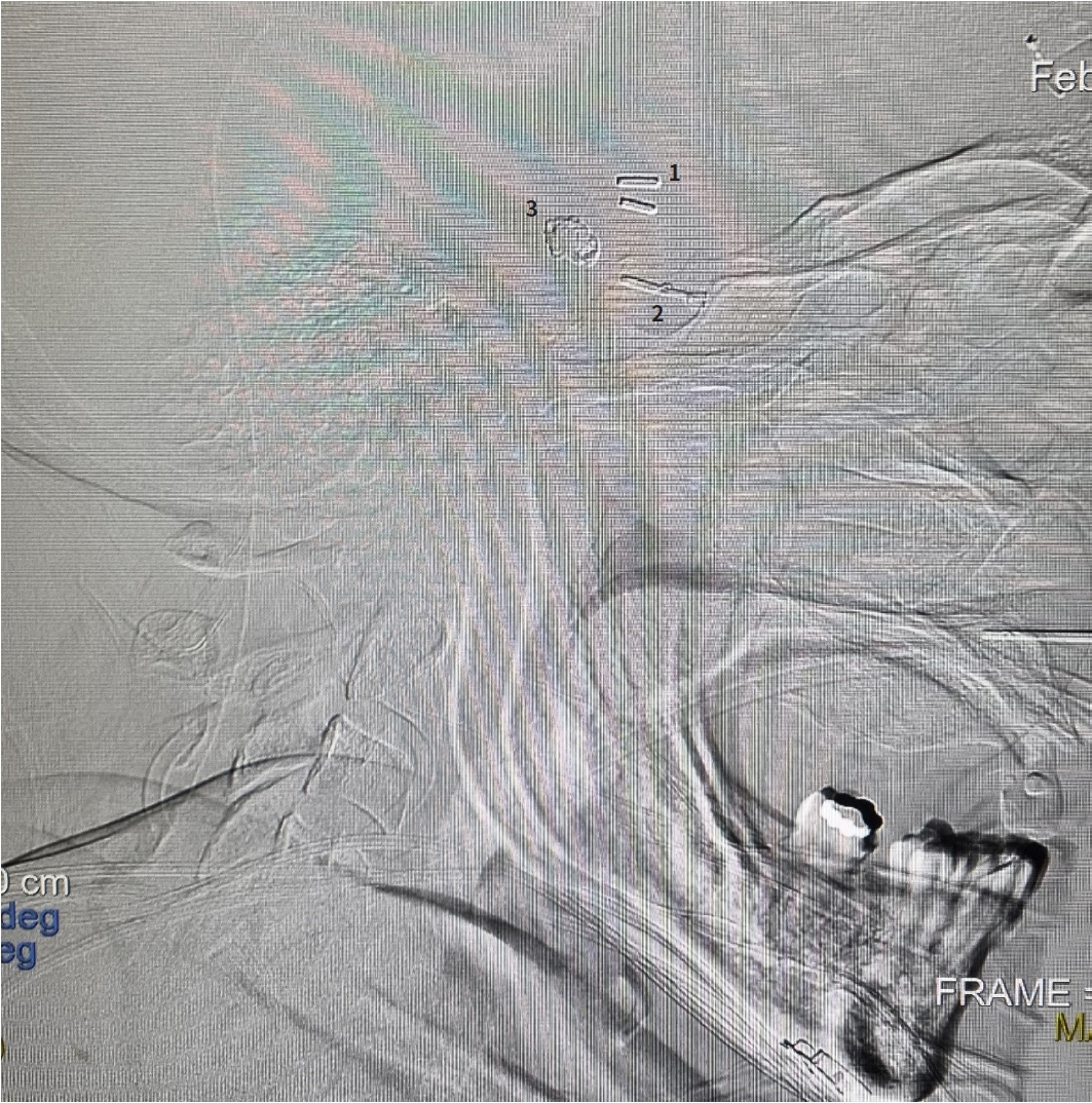
Fig. 5. Cerebral angiography (panoramic lateral view). The three types of materials that have revolutionized the management of cerebral aneurysms throughout history and that were used in this case are shown. 1. Malleable clips, 2. Spring clip, 3. Coils.
Reference
-
1. Bosco D, Consoli D, Lanza PL, Plastino M, Nicoletti F, Ceccotti C. Complete oculomotor palsy caused by persistent trigeminal artery. Neurol Sci. 2010; Oct. 31(5):657–9.
Article2. Burns SK, Brewer KJ, Jenkins C, Miller S. Aneurysmal subarachnoid hemorrhage and vasospasm. AACN Adv Crit Care. 2018; Summer. 29(2):163–74.
Article3. Diana F, Mangiafico S, Valente V, Wlderk A, Grillea G, Colonnese C, et al. Persistent trigeminal artery aneurysms: case report and systematic review. J Neurointerv Surg. 2019; Dec. 11(12):1261–5.
Article4. Fingerlin TJ, Rychen J, Roethlisberger M, Taub E, Mariani L, Guzman R, et al. Long-term aneurysm recurrence and De novo aneurysm formation after surgical treatment of unruptured intracranial aneurysms: a cohort study and systematic review. Neuro Res. 2020; Apr. 42(4):338–45.
Article5. Gokbel A, Secer M, Polat O. Assessment of risk factors in De novo aneurysm development. Brain Circ. 2020; Sep. 6(3):208–10.6. Jersey AM, Foster DM. Cerebral aneurysm, in StatPearls [Internet]. Treasure Island (FL): StatPearls Publishing;2022.7. Komiyama M. Persistent trigeminal artery and its variants. Interv Neuroradiol. 2019; Dec. 25(6):635–37.8. Lai LT, O’Neill AH. History, evolution, and continuing innovations of intracranial aneurysm surgery. World Neurosurg. 2017; Jun. 102:673–81.
Article9. Lawton MT, Vates GE. Subarachnoid hemorrhage. N Engl J Med. 2017; Jul. 377(3):257–66.
Article10. Lee KS, Zhang JJY, Nguyen V, Han J, Johnson JN, Kirollos R, et al. The evolution of intracranial aneurysm treatment techniques and future directions. Neurosurg Rev. 2022; Feb. 45(1):1–25.
Article11. Nasr DM, Fugate J, Brown RD Jr. The genetics of cerebral aneurysms and other vascular malformations. In : Sharma P, Meschia JF, editors. Stroke Genetics. Springer;2017. p. 53–78.12. Petridis AK, Kamp MA, Cornelius JF, Beez T, Beseoglu K, Turowski B, Steiger HJ. Aneurysmal subarachnoid hemorrhage. Dtsch Arztebl Int. 2017; Mar. 114(13):226–36.
Article13. Samuel N, Radovanovic I. Genetic basis of intracranial aneurysm formation and rupture: clinical implications in the postgenomic era. Neurosurg Focus. 2019; Jul. 47(1):E10.
Article14. Sato H, Haraguchi K, Takahashi Y, Ohtaki S, Shimizu T, Matsuura N, et al. Flow-diverter stent for an unruptured aneurysm at the junction of the internal carotid artery and persistent primitive trigeminal artery: case report and literature review. World Neurosurg. 2019; Dec. 132:329–32.
Article15. Shah KA, Katz JM. Ruptured persistent trigeminal artery-basilar artery junction aneurysm: case report and review of literature. World Neurosurg. 2020; Jan. 133:159–62.
Article16. Toth G, Cerejo R. Intracranial aneurysms: review of current science and management. Vasc Med. 2018; Jun. 23(3):276–88.
Article17. Tyagi G, Sadashiva N, Konar S, Aravinda HR, Saini J, Shukla D, et al. Persistent trigeminal artery: neuroanatomic and clinical relevance. World Neurosurg. 2020; Feb. 134:e214–23.
Article18. Xu Z, Rui YN, Hagan JP, Kim DH. Intracranial aneurysms: pathology, genetics, and molecular mechanisms. Neuromolecular Med. 2019; Dec. 21(4):325–43.
Article19. Zenteno M, Lee A, Moscote-Salazar LR. Rupture of persistent primitive trigeminal artery-basilar artery aneurysm managed with stent-assisted coiling. Asian J Neurosurg. 2018; Jul-Sep. 13(3):817–21.
Article
- Full Text Links
-
- Actions
-
Cited
- CITED
-
- Close
- Share
-
- Similar articles
-
- Endovascular Treatment for a Persistent Trigeminal Artery Aneurysm Presenting as Isolated Sixth Nerve Palsy
- Persistent Primitive Trigeminal Artery Aneurysm: A Case Report
- 1 case of relapsed leprosy accompanied by multiple cranial nerve palsies
- Neurovascular Compression Syndrome of the Eighth Cranial Nerve
- Microvascular triple decompression for combined simultaneous trigeminal neuralgia, hemifacial spasm, and pulsatile tinnitus due to separate offending vessels
