Association between the severity of hypodontia and the characteristics of craniofacial morphology in a Chinese population: A cross-sectional study
- Affiliations
-
- 1Department of Orthodontics, State Key Laboratory of Oral Disease, National Clinical Research Center for Oral Disease, West China Hospital of Stomatology, Sichuan University, Chengdu, China
- KMID: 2542810
- DOI: http://doi.org/10.4041/kjod22.073
Abstract
Objective
To investigate craniofacial differences in individuals with hypodontia and explore the relationship between craniofacial features and the number of congenitally missing teeth.
Methods
A cross-sectional study was conducted among 261 Chinese patients (males, 124; females, 137; age, 7–24 years), divided into four groups (without hypodontia: no teeth missing, mild: one or two missing teeth, moderate: three to five missing teeth, severe: six or more missing teeth) according to the number of congenitally missing teeth. Differences in cephalometric measurements among the groups were analyzed. Further, multivariate linear regression and smooth curve fitting were performed to evaluate the relationship between the number of congenitally missing teeth and the cephalometric measurements.
Results
In patients with hypodontia, SNA, NA-AP, FH-NA, ANB, Wits, ANS-Me/N-Me, GoGn-SN, UL-EP, and LL-EP significantly decreased, while Pog-NB, AB-NP, N-ANS, and S-Go/N-Me significantly increased. In multivariate linear regression analysis, SNB, Pog-NB, and S-Go/N-Me were positively related to the number of congenitally missing teeth. In contrast, NA-AP, FH-NA, ANB, Wits, N-Me, ANS-Me, ANS-Me/N-Me, GoGn-SN, SGn-FH (Y-axis), UL-EP, and LL-EP were negatively related, with absolute values of regression coefficients ranging from 0.147 to 0.357. Further, NA-AP, Pog-NB, S-Go/N-Me, and GoGn-SN showed the same tendency in both sexes, whereas UL-EP and LL-EP were different.
Conclusions
Compared with controls, patients with hypodontia tend toward a Class III skeletal relationship, reduced lower anterior face height, flatter mandibular plane, and more retrusive lips. The number of congenitally missing teeth had a greater effect on certain characteristics of craniofacial morphology in males than in females.
Figure
-

Figure 1 Cephalometric landmarks used in the customized analysis. See Table 1 for definitions of each landmark.
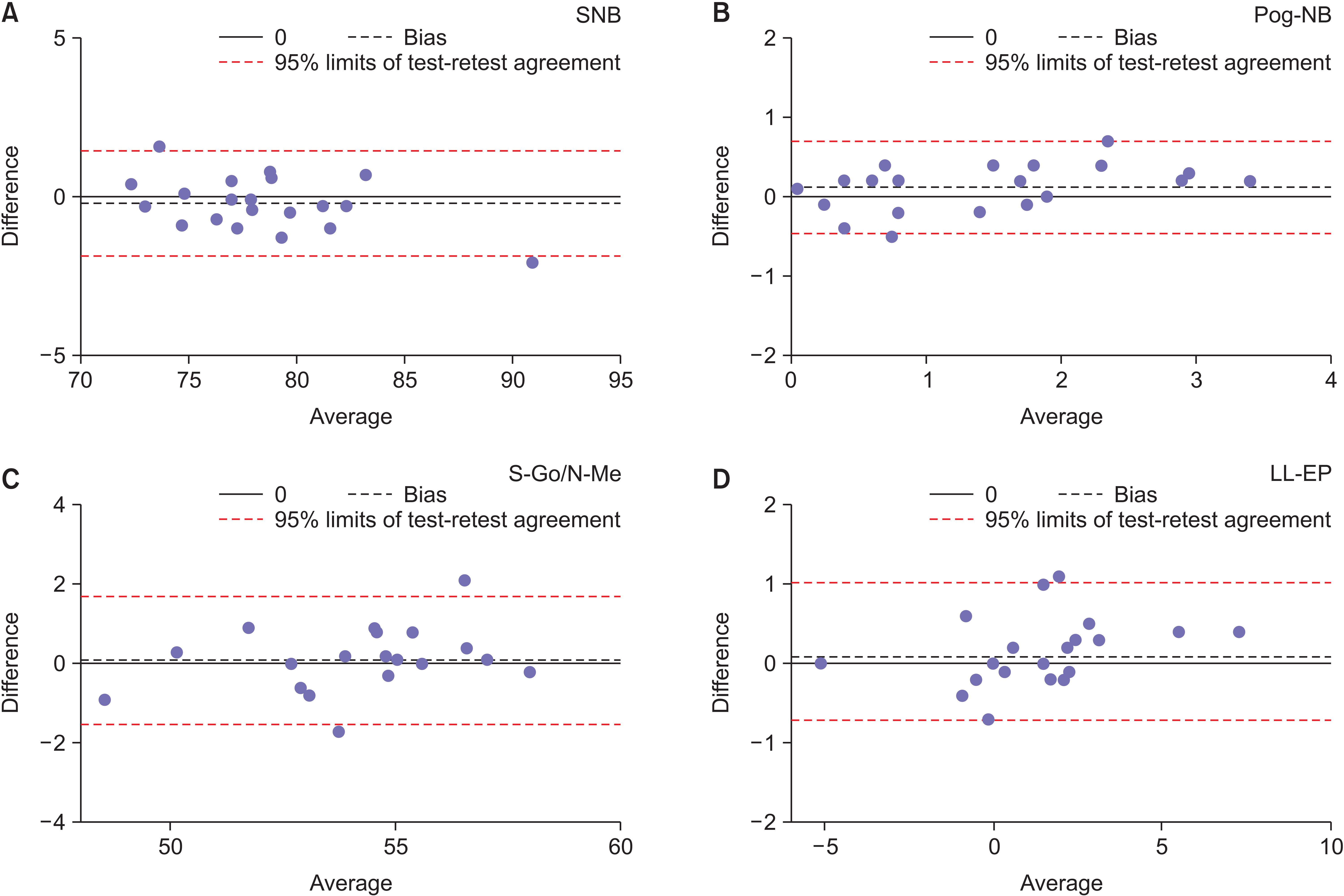
Figure 2 Bland–Altman plots demonstrating the bias for cephalometric measurements. A, SNB. B, Pog-NB. C, S-Go/N-Me. D, LL-EP. Only four measurements are shown in this figure. See Table 1 for definitions of measurement.
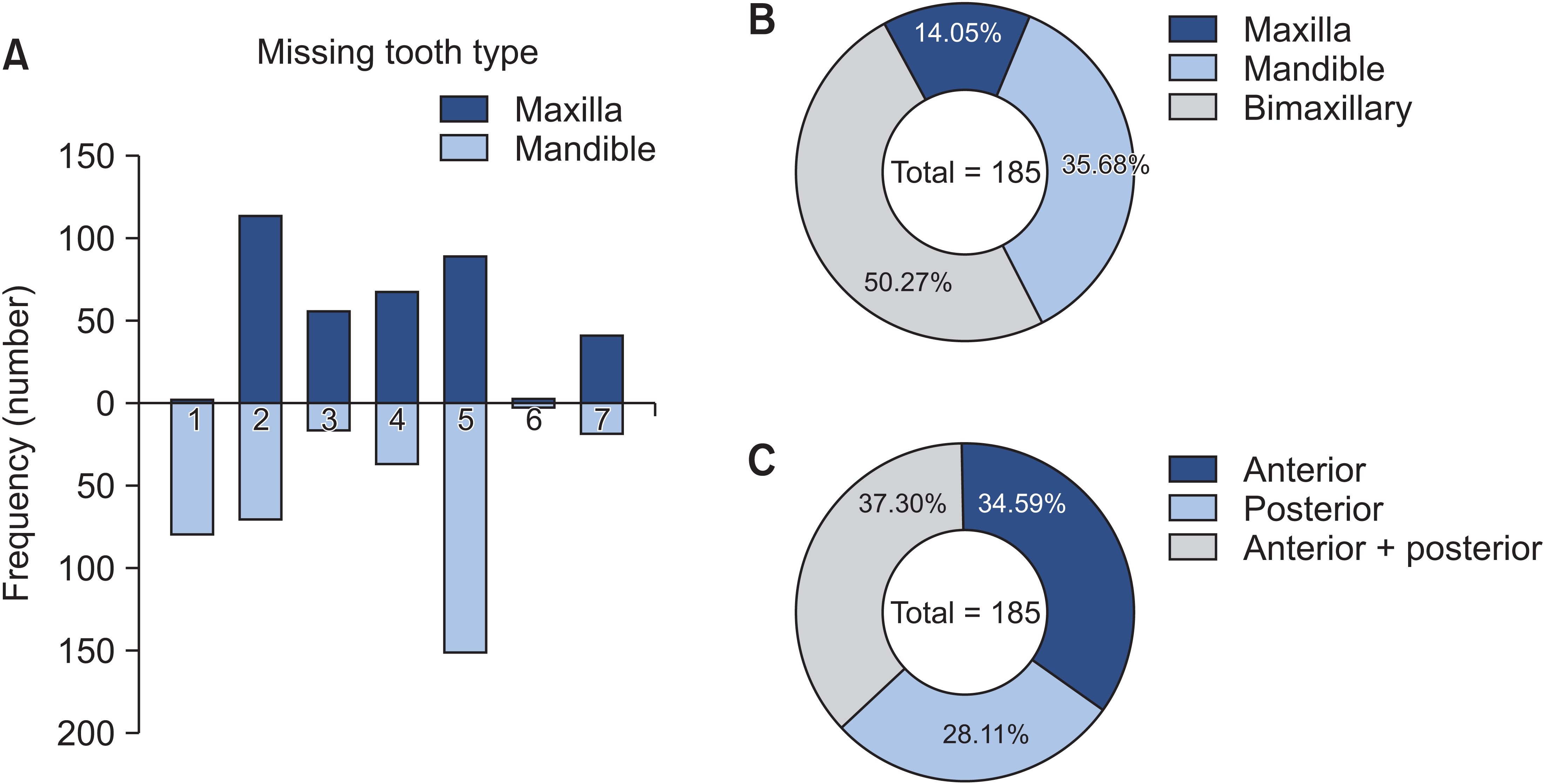
Figure 3 The patterns of permanent tooth missing within the sample. A, The frequency of different types of tooth missing. B, The proportion of different patterns of tooth missing in the maxilla and mandible. C, The proportion of different patterns of tooth missing in the anterior and posterior site of the dental arches.

Figure 4 Smooth curves between the number of congenitally missing teeth and (A) NA-AP, (B) Pog-NB, (C) S-Go/N-Me, (D) GoGn-SN, (E) UL-EP, and (F) LL-EP, stratified by sex. See Table 1 for definitions of measurement.
Reference
-
1. Vucic S, Dhamo B, Kuijpers MA, Jaddoe VW, Hofman A, Wolvius EB, et al. 2016; Craniofacial characteristics of children with mild hypodontia. Am J Orthod Dentofacial Orthop. 150:611–9. https://doi.org/10.1016/j.ajodo.2016.03.021. Erratum in: Am J Orthod Dentofacial Orthop 2017;151:1023. https://doi.org/10.1016/j.ajodo.2017.03.014. DOI: 10.1016/j.ajodo.2017.03.014. PMID: 27692418.
Article2. Bajraktarova Miševska C, Kanurkova L, Bajraktarova Valjakova E, Georgieva S, Bajraktarova B, Georgiev Z, et al. 2016; Craniofacial morphology in individuals with increasing severity of hypodontia. South Eur J Orthod Dentofac Res. 3:12–7. https://doi.org/10.5937/sejodr3-15218. DOI: 10.5937/sejodr3-15218.
Article3. Tunç EŞ, Bayrak S, Koyutürk AE. 2011; Dental development in children with mild-to-moderate hypodontia. Am J Orthod Dentofacial Orthop. 139:334–8. https://doi.org/10.1016/j.ajodo.2009.04.024. DOI: 10.1016/j.ajodo.2009.04.024. PMID: 21392687.
Article4. Rodrigues AS, Antunes LS, Pinheiro LHM, Guimarães LS, Calansans-Maia JA, Küchler EC, et al. 2020; Is dental agenesis associated with craniofacial morphology pattern? A systematic review and meta-analysis. Eur J Orthod. 42:534–43. https://doi.org/10.1093/ejo/cjz087. DOI: 10.1093/ejo/cjz087. PMID: 31783403.
Article5. Polder BJ, Van't Hof MA, Van der Linden FP, Kuijpers-Jagtman AM. 2004; A meta-analysis of the prevalence of dental agenesis of permanent teeth. Community Dent Oral Epidemiol. 32:217–26. https://doi.org/10.1111/j.1600-0528.2004.00158.x. DOI: 10.1111/j.1600-0528.2004.00158.x. PMID: 15151692.
Article6. Al-Ani AH, Antoun JS, Thomson WM, Merriman TR, Farella M. 2017; Hypodontia: an update on its etiology, classification, and clinical management. Biomed Res Int. 2017:9378325. https://doi.org/10.1155/2017/9378325. DOI: 10.1155/2017/9378325. PMID: 28401166. PMCID: PMC5376450.
Article7. Kreczi A, Proff P, Reicheneder C, Faltermeier A. 2011; Effects of hypodontia on craniofacial structures and mandibular growth pattern. Head Face Med. 7:23. https://doi.org/10.1186/1746-160X-7-23. DOI: 10.1186/1746-160X-7-23. PMID: 22142280. PMCID: PMC3248361. PMID: f5d9cea90ca0440791ec8fe5f39e6128.
Article8. Johal A, Huang Y, Toledano S. 2022; Hypodontia and its impact on a young person's quality of life, esthetics, and self-esteem. Am J Orthod Dentofacial Orthop. 161:220–7. https://doi.org/10.1016/j.ajodo.2020.07.039. DOI: 10.1016/j.ajodo.2020.07.039. PMID: 34538709.
Article9. Costa AMG, Trevizan M, Matsumoto MAN, da Silva RAB, da Silva LAB, Horta KC, et al. 2017; Association between tooth agenesis and skeletal malocclusions. J Oral Maxillofac Res. 8:e3. https://doi.org/10.5037/jomr.2017.8203. DOI: 10.5037/jomr.2017.8203. PMID: 28791079. PMCID: PMC5541988. PMID: fa88637d22864192a97c9dd055a63253.
Article10. Acharya PN, Jones SP, Moles D, Gill D, Hunt NP. 2010; A cephalometric study to investigate the skeletal relationships in patients with increasing severity of hypodontia. Angle Orthod. 80:511–8. https://doi.org/10.2319/072309-411.1. DOI: 10.2319/072309-411.1. PMID: 20482356. PMCID: PMC8966445.
Article11. Dhamo B, Vucic S, Kuijpers MA, Jaddoe VW, Hofman A, Wolvius EB, et al. 2016; The association between hypodontia and dental development. Clin Oral Investig. 20:1347–54. https://doi.org/10.1007/s00784-015-1622-1. Erratum in: J Orthod 2017;44:74. https://doi.org/10.1080/14653125.2016.1275610. DOI: 10.1080/14653125.2016.1275610. PMID: 28079474.
Article12. Yu M, Fan Z, Wong SW, Sun K, Zhang L, Liu H, et al. 2021; Lrp6 dynamic expression in tooth development and mutations in oligodontia. J Dent Res. 100:415–22. https://doi.org/10.1177/0022034520970459. DOI: 10.1177/0022034520970459. PMID: 33164649.
Article13. Cunha AS, Dos Santos LV, Marañón-Vásquez GA, Kirschneck C, Gerber JT, Stuani MB, et al. 2021; Genetic variants in tooth agenesis-related genes might be also involved in tooth size variations. Clin Oral Investig. 25:1307–18. https://doi.org/10.1007/s00784-020-03437-8. DOI: 10.1007/s00784-020-03437-8. PMID: 32648061.
Article14. Rodrigues AS, Teixeira EC, Antunes LS, Nelson-Filho P, Cunha AS, Levy SC, et al. 2020; Association between craniofacial morphological patterns and tooth agenesis-related genes. Prog Orthod. 21:9. https://doi.org/10.1186/s40510-020-00309-5. DOI: 10.1186/s40510-020-00309-5. PMID: 32249341. PMCID: PMC7131971. PMID: dd9837aae09041de8e8121bb912d10d1.
Article15. Al-Ani AH, Antoun JS, Thomson WM, Merriman TR, Farella M. 2017; Maternal smoking during pregnancy is associated with offspring hypodontia. J Dent Res. 96:1014–9. https://doi.org/10.1177/0022034517711156. DOI: 10.1177/0022034517711156. PMID: 28535361.
Article16. Taju W, Sherriff M, Bister D, Shah S. 2018; Association between severity of hypodontia and cephalometric skeletal patterns: a retrospective study. Eur J Orthod. 40:200–5. https://doi.org/10.1093/ejo/cjx049. DOI: 10.1093/ejo/cjx049. PMID: 29016739.
Article17. Endo T, Yoshino S, Ozoe R, Kojima K, Shimooka S. 2004; Association of advanced hypodontia and craniofacial morphology in Japanese orthodontic patients. Odontology. 92:48–53. https://doi.org/10.1007/s10266-004-0034-5. DOI: 10.1007/s10266-004-0034-5. PMID: 15490305.
Article18. Chan DW, Samman N, McMillan AS. 2009; Craniofacial profile in Southern Chinese with hypodontia. Eur J Orthod. 31:300–5. https://doi.org/10.1093/ejo/cjn111. DOI: 10.1093/ejo/cjn111. PMID: 19193707.
Article19. Oeschger ES, Kanavakis G, Cocos A, Halazonetis DJ, Gkantidis N. 2022; Number of teeth is related to craniofacial morphology in humans. Biology (Basel). 11:544. https://doi.org/10.3390/biology11040544. DOI: 10.3390/biology11040544. PMID: 35453743. PMCID: PMC9029740. PMID: a0d2f4778979489cb80a89bcc0a41596.
Article20. Gungor AY, Turkkahraman H. 2013; Effects of severity and location of nonsyndromic hypodontia on craniofacial morphology. Angle Orthod. 83:584–90. https://doi.org/10.2319/091012-722.1. DOI: 10.2319/091012-722.1. PMID: 23311600. PMCID: PMC8754045.
Article21. Ben-Bassat Y, Brin I. 2003; Skeletodental patterns in patients with multiple congenitally missing teeth. Am J Orthod Dentofacial Orthop. 124:521–5. https://doi.org/10.1016/s0889-5406(03)00620-6. DOI: 10.1016/S0889-5406(03)00620-6. PMID: 14614419.
Article22. Dermaut LR, Goeffers KR, De Smit AA. 1986; Prevalence of tooth agenesis correlated with jaw relationship and dental crowding. Am J Orthod Dentofacial Orthop. 90:204–10. https://doi.org/10.1016/0889-5406(86)90067-3. DOI: 10.1016/0889-5406(86)90067-3. PMID: 3463196.
Article23. Yelmer ZA, Akbulut S. 2022; Evaluation of the effects of hypodontia on the morphology of craniofacial structures. Orthod Craniofac Res. 25:409–15. https://doi.org/10.1111/ocr.12550. DOI: 10.1111/ocr.12550. PMID: 34837458.
Article24. McNamara JA Jr, Franchi L. 2018; The cervical vertebral maturation method: a user's guide. Angle Orthod. 88:133–43. https://doi.org/10.2319/111517-787.1. DOI: 10.2319/111517-787.1. PMID: 29337631. PMCID: PMC8312535.
Article25. Lu W, Zhang X, Mei L, Wang P, He J, Li Y, et al. 2020; Orthodontic incisor retraction caused changes in the soft tissue chin area: a retrospective study. BMC Oral Health. 20:108. https://doi.org/10.1186/s12903-020-01099-2. DOI: 10.1186/s12903-020-01099-2. PMID: 32295586. PMCID: PMC7160892. PMID: fc4f4769b9134a62b239f59df8d574ff.
Article26. Chung LK, Hobson RS, Nunn JH, Gordon PH, Carter NE. 2000; An analysis of the skeletal relationships in a group of young people with hypodontia. J Orthod. 27:315–8. https://doi.org/10.1093/ortho/27.4.315. DOI: 10.1093/ortho/27.4.315. PMID: 11099569.
Article27. Ogaard B, Krogstad O. 1995; Craniofacial structure and soft tissue profile in patients with severe hypodontia. Am J Orthod Dentofacial Orthop. 108:472–7. https://doi.org/10.1016/s0889-5406(95)70047-1. DOI: 10.1016/S0889-5406(95)70047-1. PMID: 7484966.
Article28. Yüksel S, Uçem T. 1997; The effect of tooth agenesis on dentofacial structures. Eur J Orthod. 19:71–8. https://doi.org/10.1093/ejo/19.1.71. DOI: 10.1093/ejo/19.1.71. PMID: 9071047.
Article29. Higashihori N, Takada JI, Katayanagi M, Takahashi Y, Moriyama K. 2018; Frequency of missing teeth and reduction of mesiodistal tooth width in Japanese patients with tooth agenesis. Prog Orthod. 19:30. https://doi.org/10.1186/s40510-018-0222-4. DOI: 10.1186/s40510-018-0222-4. PMID: 30123921. PMCID: PMC6098995. PMID: e8fc9d6865314dc8a9954c5269008a1e.
Article30. Bassiouny DS, Afify AR, Baeshen HA, Birkhed D, Zawawi KH. 2016; Prevalence of maxillary lateral incisor agenesis and associated skeletal characteristics in an orthodontic patient population. Acta Odontol Scand. 74:456–9. https://doi.org/10.1080/00016357.2016.1193625. DOI: 10.1080/00016357.2016.1193625. PMID: 27306861.
Article31. Lisson JA, Scholtes S. 2005; Investigation of craniofacial morphology in patients with hypo- and oligodontia. J Orofac Orthop. 66:197–207. https://doi.org/10.1007/s00056-005-0437-0. DOI: 10.1007/s00056-005-0437-0. PMID: 15959633.
Article32. Bondarets N, McDonald F. 2000; Analysis of the vertical facial form in patients with severe hypodontia. Am J Phys Anthropol. 111:177–84. https://doi.org/10.1002/(SICI)1096-8644(200002)111:2<177::AID-AJPA4>3.0.CO;2-8. DOI: 10.1002/(SICI)1096-8644(200002)111:2<177::AID-AJPA4>3.0.CO;2-8. PMID: 10640945.
Article33. Bauer N, Heckmann K, Sand A, Lisson JA. 2009; Craniofacial growth patterns in patients with congenitally missing permanent teeth. J Orofac Orthop. 70:139–51. https://doi.org/10.1007/s00056-009-0744-y. DOI: 10.1007/s00056-009-0744-y. PMID: 19322532.
Article34. Takahashi Y, Higashihori N, Yasuda Y, Takada JI, Moriyama K. 2018; Examination of craniofacial morphology in Japanese patients with congenitally missing teeth: a cross-sectional study. Prog Orthod. 19:38. https://doi.org/10.1186/s40510-018-0238-9. DOI: 10.1186/s40510-018-0238-9. PMID: 30270414. PMCID: PMC6165831. PMID: 7244e6206a414cfaa0b9ab723af71417.
Article35. Nodal M, Kjaer I, Solow B. 1994; Craniofacial morphology in patients with multiple congenitally missing permanent teeth. Eur J Orthod. 16:104–9. https://doi.org/10.1093/ejo/16.2.104. DOI: 10.1093/ejo/16.2.104. PMID: 8005197.
Article36. Shiga H, Kobayashi Y, Katsuyama H, Yokoyama M, Arakawa I. 2012; Gender difference in masticatory performance in dentate adults. J Prosthodont Res. 56:166–9. https://doi.org/10.1016/j.jpor.2012.02.001. DOI: 10.1016/j.jpor.2012.02.001. PMID: 22613955.
Article37. Miwa S, Wada M, Murakami S, Suganami T, Ikebe K, Maeda Y. 2019; Gonial angle measured by orthopantomography as a predictor of maximum occlusal force. J Prosthodont. 28:e426–30. https://doi.org/10.1111/jopr.12598. DOI: 10.1111/jopr.12598. PMID: 28913893.
Article38. Koç D, Doğan A, Bek B. 2011; Effect of gender, facial dimensions, body mass index and type of functional occlusion on bite force. J Appl Oral Sci. 19:274–9. https://doi.org/10.1590/s1678-77572011000300017. DOI: 10.1590/S1678-77572011000300017. PMID: 21625746. PMCID: PMC4234342. PMID: 2d4fee5e633e4faa9dfc99d260fa4d78.
Article39. Ikebe K, Matsuda K, Kagawa R, Enoki K, Yoshida M, Maeda Y, et al. 2011; Association of masticatory performance with age, gender, number of teeth, occlusal force and salivary flow in Japanese older adults: is ageing a risk factor for masticatory dysfunction? Arch Oral Biol. 56:991–6. https://doi.org/10.1016/j.archoralbio.2011.03.019. DOI: 10.1016/j.archoralbio.2011.03.019. PMID: 21529776.
Article
- Full Text Links
-
- Actions
-
Cited
- CITED
-
- Close
- Share
-
- Similar articles
-
- Factors Influencing Upper Airway Dimensions in Skeletal Class Ⅱ Children and Adolescents: A CBCT Study
- The relations between craniofacial morphology and dental aesthetic index(DAI)
- Cephalometric study of obstructive sleep apnea patients in the upright and supine positions
- Evolution of craniofacial surgery
- Asian Pacific Craniofacial Association and Archives of Craniofacial Surgery
