Paraneoplastic Pseudoachalasia Complicated with Small Cell Lung Cancer
- Affiliations
-
- 1Department of Internal Medicine, Yeungnam University College of Medicine, Daegu, Korea
- KMID: 2542790
- DOI: http://doi.org/10.4166/kjg.2023.034
Figure
-
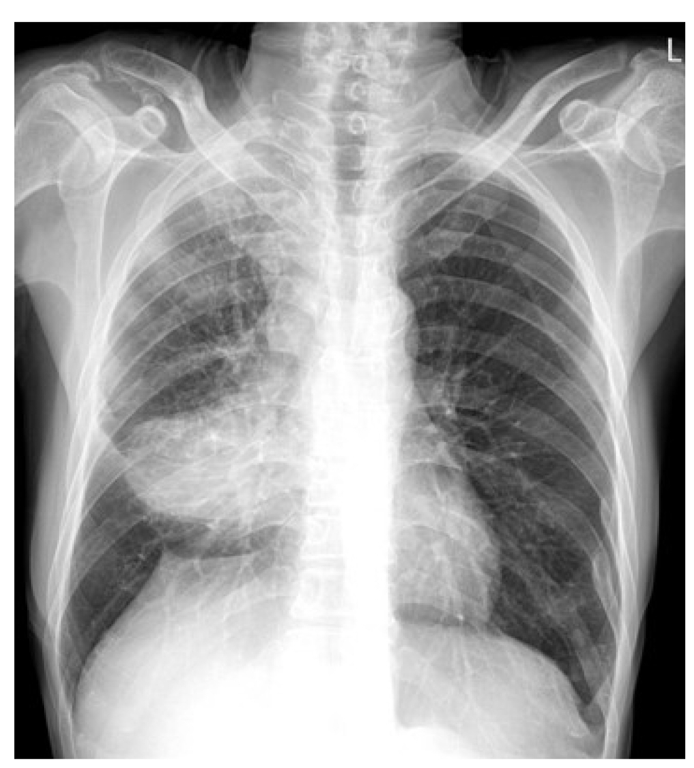
Fig. 1 Chest X-ray image. The image shows tumor shadows at the right middle lung field.

Fig. 2 Thoracic CT images. Initial axial (A, B) and coronal (C, D) CT images obtained at presentation showed a large mass obstructing the right middle lobar bronchus combined with atelectasis and multiple enlarged mediastinal lymph nodes. Although these lesions were close to the esophagus, there was no direct invasion of the esophagus by the lung mass.

Fig. 3 Endoscopy images. Endoscopy images (A, B) show a strongly contracted esophagogastric junction.
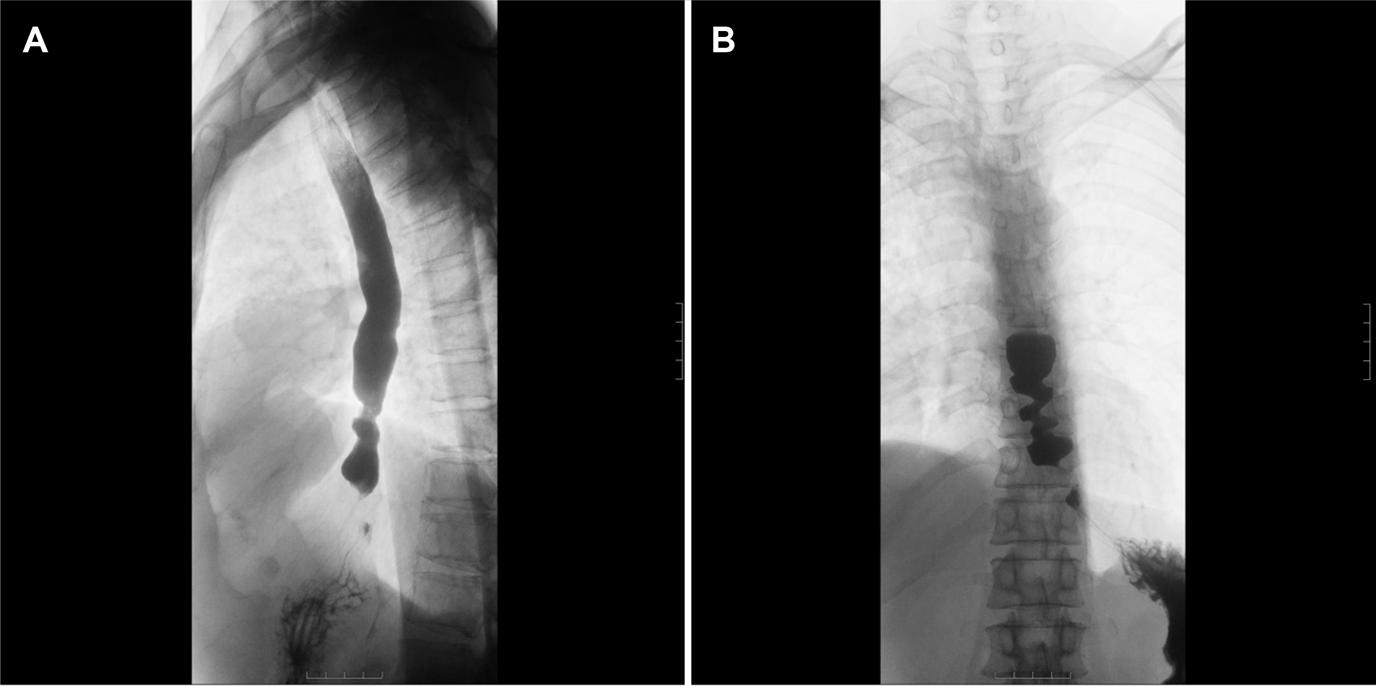
Fig. 4 Barium esophagography image. The images (A, B) show minimal esophageal dilatation with pooling and stasis of contrast and narrowing of the distal esophageal segment.
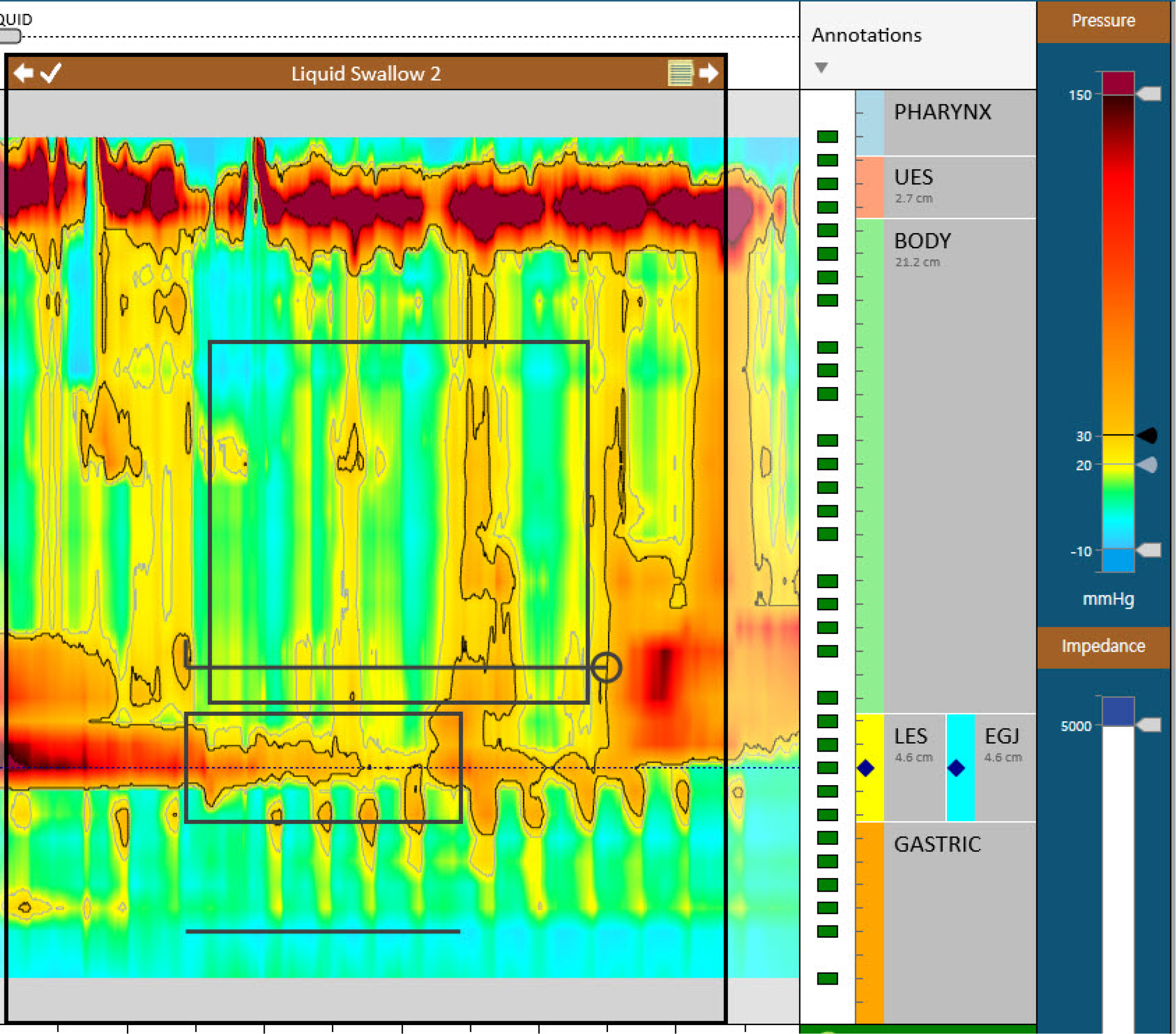
Fig. 5 High-resolution manometry image. The image shows high integrated relaxation pressure (43 mmHg) with failed peristalsis and pan-esophageal pressurization.
Reference
-
1. Savarino E, Bhatia S, Roman S, et al. 2022; Achalasia. Nat Rev Dis Primers. 8:28. DOI: 10.1038/s41572-022-00356-8. PMID: 35513420.
Article2. Campo SM, Zullo A, Scandavini CM, Frezza B, Cerro P, Balducci G. 2013; Pseudoachalasia: A peculiar case report and review of the literature. World J Gastrointest Endosc. 5:450–454. DOI: 10.4253/wjge.v5.i9.450. PMID: 24044045. PMCID: PMC3773858.
Article3. Tucker HJ, Snape WJ Jr, Cohen S. 1978; Achalasia secondary to carcinoma: manometric and clinical features. Ann Intern Med. 89:315–318. DOI: 10.7326/0003-4819-89-3-315. PMID: 686541.
Article4. Gockel I, Eckardt VF, Schmitt T, Junginger T. 2005; Pseudoachalasia: a case series and analysis of the literature. Scand J Gastroenterol. 40:378–385. DOI: 10.1080/00365520510012118. PMID: 16028431.
Article5. Kahrilas PJ, Kishk SM, Helm JF, Dodds WJ, Harig JM, Hogan WJ. 1987; Comparison of pseudoachalasia and achalasia. Am J Med. 82:439–446. DOI: 10.1016/0002-9343(87)90443-8. PMID: 3548347.
Article6. Hejazi RA, Zhang D, McCallum RW. 2009; Gastroparesis, pseudoachalasia and impaired intestinal motility as paraneoplastic manifestations of small cell lung cancer. Am J Med Sci. 338:69–71. DOI: 10.1097/MAJ.0b013e31819b93e5. PMID: 19506460.
Article7. Brown WR, Dee E. 2013; Dysphagia in a patient with recurrent small-cell lung cancer. Gastroenterology. 144:34252–253. DOI: 10.1053/j.gastro.2012.08.007. PMID: 23159301.
Article8. De Giorgio R, Bovara M, Barbara G, et al. 2003; Anti-HuD-induced neuronal apoptosis underlying paraneoplastic gut dysmotility. Gastroenterology. 125:70–79. DOI: 10.1016/S0016-5085(03)00664-4. PMID: 12851872.
Article9. Hirano T, Miyauchi E, Inoue A, et al. 2016; Two cases of pseudo-achalasia with lung cancer: Case report and short literature review. Respir Investig. 54:494–499. DOI: 10.1016/j.resinv.2016.04.006. PMID: 27886865.
Article
- Full Text Links
-
- Actions
-
Cited
- CITED
-
- Close
- Share
-
- Similar articles
-
- A case of Wegener's granulomatosis complicated by non-small cell lung cancer
- A Case of Eosinophilia as a Manifestation of Paraneoplastic Syndrome in Non-Small-Cell Lung Cancer
- A Case of Cancer Associated Retinopathy with Small Cell Lung Carcinoma
- A Case of Intramedullary Myelopathy Associated with Small Cell Lung Cancer
- Paraneoplastic Chorea Associated with Anti-Hu Antibody and Small Cell Lung Carcinoma
