Medial Arterial Calcification and the Risk of Amputation of Diabetic Foot Ulcer in Patients With Diabetic Kidney Disease
- So JM
 1
1 - Park JH1
- Kim JG1
- Park IR2
- Ha EY2
- Chung SM2
- Moon JS2
- Park CH3
- Yun WS4
- Kim TG5
- Kim W6
- Yoon JS2
- Won KC2
- Lee HW2
- Affiliations
-
- 1College of Medicine, Yeungnam University, Daegu, Korea
- 2Division of Endocrinology and Metabolism, Department of Internal Medicine, Yeungnam University College of Medicine, Daegu, Korea
- 3Department of Orthopedic Surgery, Yeungnam University College of Medicine, Daegu, Korea
- 4Department of Surgery, Kyungpook National University School of Medicine, Daegu, Korea
- 5Department of Plastic Surgery, Yeungnam University College of Medicine, Daegu, Korea
- 6Division of Cardiology, Department of Internal Medicine, Yeungnam University College of Medicine, Daegu, Korea
- KMID: 2542597
- DOI: http://doi.org/10.3346/jkms.2023.38.e160
Abstract
- We assessed the risk factors for major amputation of diabetic foot ulcers (DFUs) in patients with diabetic kidney disease (DKD) stages 3b–5. For DFU assessment, in addition to DFU location and presence of infection, ischemia, and neuropathy, vascular calcification was assessed using the medial arterial calcification (MAC) score. Of 210 patients, 26 (12.4%) underwent major amputations. Only the location and extension of DFU, represented by Texas grade differed between the minor and major amputation groups. However, after adjusting for covariates, ulcer location of mid- or hindfoot (vs. forefoot, odds ratio [OR] = 3.27), Texas grades 2 or 3 (vs. grade 0, OR = 5.78), and severe MAC (vs. no MAC, OR = 4.46) was an independent risk factor for major amputation (all P < 0.05). The current use of antiplatelets was a possible protective factor for major amputations (OR = 0.37, P = 0.055). In conclusion, DFU with severe MAC is associated with major amputation in patients with DKD.
Figure
-
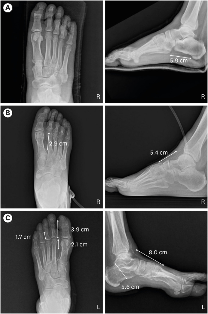
Fig. 1 Assessment of vascular calcification using MAC. MAC was assessed in five arterial sites. A positive score of 1 was given for ≥ 2 cm calcification in the 1) dorsalis pedis, 2) lateral plantar, and 3) first metatarsal artery, or ≥ 1 cm calcification in the 4) first toe and 5) other toe arteries. (A) The anteroposterior (left) and lateral (right) views of “no MAC.” A total of one positive score was assigned to the lateral plantar artery (5.9 cm). (B) The anteroposterior (left) and lateral (right) views of “moderate MAC.” Two positive scores were assigned to the dorsalis pedis artery (5.4 cm) and first metatarsal artery (2.8 cm). (C) The anteroposterior (left) and lateral (right) views of “severe MAC.” Five positive scores were assigned for the dorsalis pedis artery (8.0 cm), lateral plantar artery (5.6 cm), first metatarsal artery (2.1 cm), first toe artery (3.9 cm), and third toe artery (1.7 cm).MAC = medial arterial calcification.
Reference
-
1. Armstrong DG, Swerdlow MA, Armstrong AA, Conte MS, Padula WV, Bus SA. Five year mortality and direct costs of care for people with diabetic foot complications are comparable to cancer. J Foot Ankle Res. 2020; 13(1):16. PMID: 32209136.2. Ibrahim A. IDF Clinical Practice Recommendation on the Diabetic Foot: a guide for healthcare professionals. Diabetes Res Clin Pract. 2017; 127:285–287. PMID: 28495183.3. Papanas N, Liakopoulos V, Maltezos E, Stefanidis I. The diabetic foot in end stage renal disease. Ren Fail. 2007; 29(5):519–528. PMID: 17654312.4. Guzman RJ, Bian A, Shintani A, Stein CM. Association of foot ulcer with tibial artery calcification is independent of peripheral occlusive disease in type 2 diabetes. Diabetes Res Clin Pract. 2013; 99(3):281–286. PMID: 23305901.5. Huang CL, Wu IH, Wu YW, Hwang JJ, Wang SS, Chen WJ, et al. Association of lower extremity arterial calcification with amputation and mortality in patients with symptomatic peripheral artery disease. PLoS One. 2014; 9(2):e90201. PMID: 24587279.6. Clerici G, Faglia E. Saving the limb in diabetic patients with ischemic foot lesions complicated by acute infection. Int J Low Extrem Wounds. 2014; 13(4):273–293. PMID: 25256282.7. Ferraresi R, Ucci A, Pizzuto A, Losurdo F, Caminiti M, Minnella D, et al. A novel scoring system for small artery disease and medial arterial calcification is strongly associated with major adverse limb events in patients with chronic limb-threatening ischemia. J Endovasc Ther. 2021; 28(2):194–207. PMID: 33054496.8. Swartling O, Rydell H, Stendahl M, Segelmark M, Trolle Lagerros Y, Evans M. CKD progression and mortality among men and women: a nationwide study in Sweden. Am J Kidney Dis. 2021; 78(2):190–199.e1. PMID: 33434591.9. Yamazaki T, Mimura I, Tanaka T, Nangaku M. Treatment of diabetic kidney disease: current and future. Diabetes Metab J. 2021; 45(1):11–26. PMID: 33508907.10. Lavery LA, Armstrong DG, Harkless LB. Classification of diabetic foot wounds. J Foot Ankle Surg. 1996; 35(6):528–531. PMID: 8986890.11. Singh N, Armstrong DG, Lipsky BA. Preventing foot ulcers in patients with diabetes. JAMA. 2005; 293(2):217–228. PMID: 15644549.12. Feldman EL, Stevens MJ. Clinical testing in diabetic peripheral neuropathy. Can J Neurol Sci. 1994; 21(4):S3–S7.13. van Netten JJ, Bus SA, Apelqvist J, Lipsky BA, Hinchliffe RJ, Game F, et al. Definitions and criteria for diabetic foot disease. Diabetes Metab Res Rev. 2020; 36(Suppl 1):e3268. PMID: 31943705.14. Weledji EP, Fokam P. Treatment of the diabetic foot - to amputate or not? BMC Surg. 2014; 14(1):83. PMID: 25344293.15. Pena G, Kuang B, Edwards S, Cowled P, Dawson J, Fitridge R. Factors associated with key outcomes in diabetes related foot disease: a prospective observational study. Eur J Vasc Endovasc Surg. 2021; 62(2):233–240. PMID: 34024706.16. Lew E, Nicolosi N, Botek G. Lower extremity amputation risk factors associated with elevated ankle brachial indices and radiographic arterial calcification. J Foot Ankle Surg. 2015; 54(3):473–477. PMID: 25661784.17. Everhart JE, Pettitt DJ, Knowler WC, Rose FA, Bennett PH. Medial arterial calcification and its association with mortality and complications of diabetes. Diabetologia. 1988; 31(1):16–23. PMID: 3350219.18. Lehto S, Niskanen L, Suhonen M, Rönnemaa T, Laakso M. Medial artery calcification. A neglected harbinger of cardiovascular complications in non-insulin-dependent diabetes mellitus. Arterioscler Thromb Vasc Biol. 1996; 16(8):978–983. PMID: 8696962.19. Edmonds ME, Morrison N, Laws JW, Watkins PJ. Medial arterial calcification and diabetic neuropathy. Br Med J (Clin Res Ed). 1982; 284(6320):928–930.20. Jeffcoate WJ, Rasmussen LM, Hofbauer LC, Game FL. Medial arterial calcification in diabetes and its relationship to neuropathy. Diabetologia. 2009; 52(12):2478–2488. PMID: 19756483.21. Temmar M, Liabeuf S, Renard C, Czernichow S, Esper NE, Shahapuni I, et al. Pulse wave velocity and vascular calcification at different stages of chronic kidney disease. J Hypertens. 2010; 28(1):163–169. PMID: 19927012.22. Liu KH, Chu WC, Kong AP, Choi Ko GT, Ma RC, Chan JW, et al. US assessment of medial arterial calcification: a sensitive marker of diabetes-related microvascular and macrovascular complications. Radiology. 2012; 265(1):294–302. PMID: 22843765.23. Tang T, Zhang M, Li W, Hu N, Du X, Ran F, et al. Oral anticoagulant and antiplatelet therapy for peripheral arterial disease: a meta-analysis of randomized controlled trials. Clin Appl Thromb Hemost. 2021; 27:1076029621996810. PMID: 33783251.24. Xiong J, He T, Yu Z, Yang K, Chen F, Cheng J, et al. Antiplatelet therapy for the prevention of atherosclerosis in chronic kidney disease (ALTAS-CKD) patients: study protocol for a randomized clinical trial. Trials. 2021; 22(1):37. PMID: 33413594.
