A modified trans-anconeus approach to facilitate fixation of a posterior radial head fracture: a cadaveric feasibility study
- Affiliations
-
- 1Department of Anatomy, Faculty of Medicine, Ain Shams University, Cairo
- 2Department of Orthopaedic, Faculty of Medicine, Ain Shams University, Cairo, Egypt
- KMID: 2540983
- DOI: http://doi.org/10.5115/acb.22.201
Abstract
- Fixation of radial head fracture with minimally invasive posterior approach remains a significant challenge. The aim of this study was to determine the feasibility of trans-anconeus posterior elbow approach and to observe lateral ulnar collateral ligament (LUCL) in extended elbows. This cadaveric study was performed in twenty upper limbs of fresh fixed adult male cadavers. An oblique incision was made in the middle segment of anconeus until the lateral ligament complex and the joint capsule had been revealed. A deep dissection was explored to observe the anatomical relationship of the LUCL to the anconeus. Measurements of the LUCL were recorded while the elbow was fully extended. The mean distance between the edge of the radial head and the proximal insertion of the LUCL was 13.3 mm (11.5–16.2 mm); the mean distance between the edge of the radial head and the distal insertion of the LUCL was 20.9 mm (19.2–23.4 mm); the distance between the edge of the radial head and the distal edge of the annular ligament was 11.2 mm (8.22–11.7 mm). By estimate correlation of the previous measurements, the direct and accessible way to expose the posterolateral articular capsule of the elbow joint was through a window in medial 2/3 of the middle segment of anconeus muscle. These trans-anconeus approach is useful. It provides good visualization, facilitates applying the implants, and lessens the risk of radial nerve injury. Awareness of the anatomy is mandatory to avoid injury of LUCL.
Keyword
Figure
-
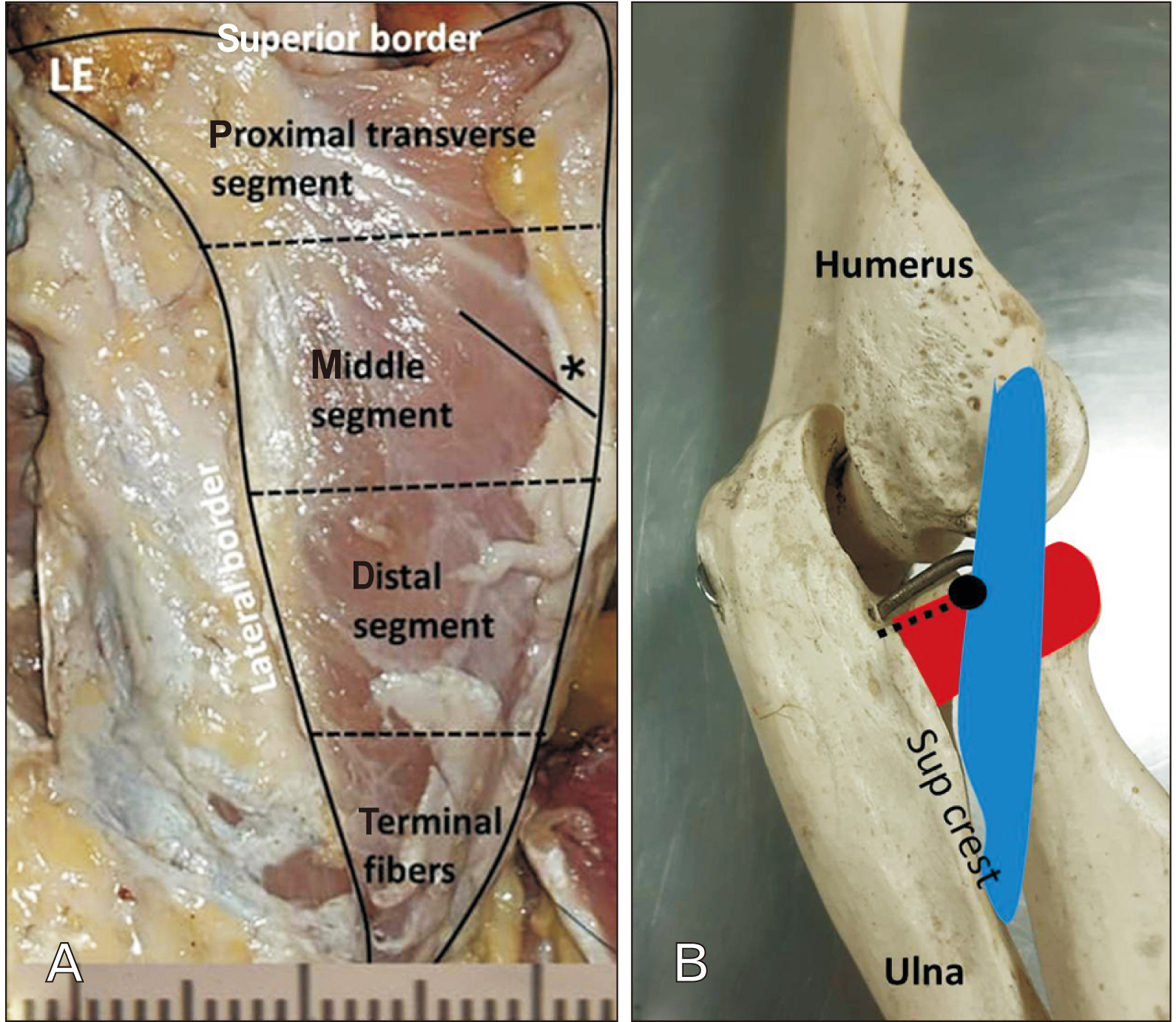
Fig. 1 Posterolateral view of the left proximal forearm in pronation. (A) The anconeus muscle is divided into 4 parts: proximal transverse segment, middle segment, distal segment and terminal fiber. Measurements are generated from the cadaver specimens to determine values for the length of the borders (superior, lateral and base) and anconeus muscle fiber angle (asterisks) of the middle segment with the sagittal plane of ulna. (B) The articulated skeleton showing the LUCL (blue coloured) measurements. The coalescing point of LUCL ligament with the AL (black point) is considered as a reference point to determine the value of the LUCL length to its proximal (LE) and distal (supinator crest) attachments. The dotted line is the distance from the proximal edge of the cartilage of the radial head to the distal edge of the AL (red coloured). LUCL, lateral ulnar collateral ligament; AL, annular ligament; LE, lateral epicondyle.

Fig. 2 Plain x-ray (A) anteroposterior and (B) lateral views of the elbow showing posteromedial radial head fracture.
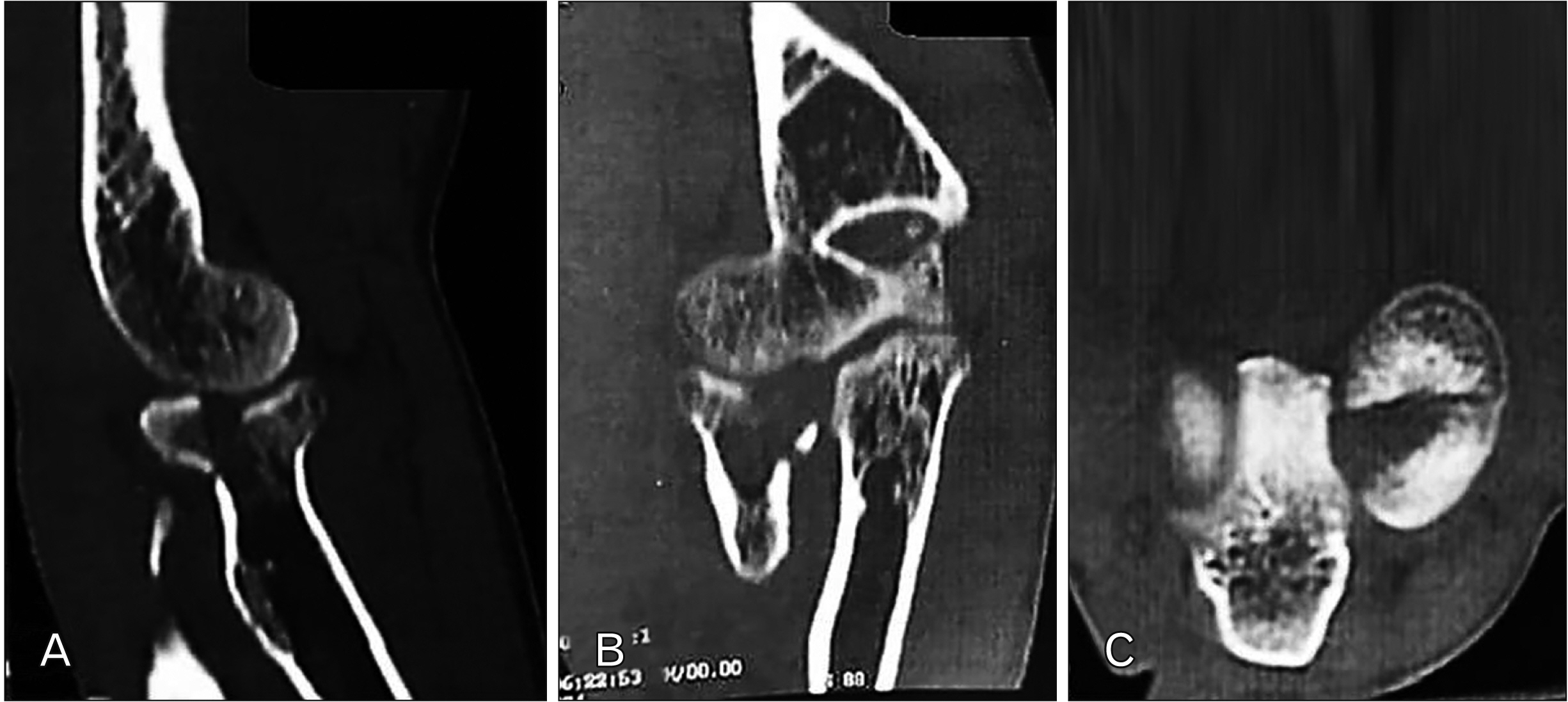
Fig. 3 Computed tomography scan (A)sagittal, (B) coronal and (C) axial cuts respectively showing posteromedial radial head fracture with the axial cut indicating the engagement of the fracture on the posterior edge of the radial notch.

Fig. 4 Posterolateral view of the left proximal forearm in pronation. (A) The anconeus is a right angle triangular muscle attaching proximally to the LE of the humerus with common extensor group. The anconeus muscle is observed to have a sharp, tendinous expansion. (B) The overlying anconeus muscle has been incisied obliquely along the middle segment and extended till the distal segment. (C) The anconeus muscle retracted laterally, the LUCL is identified the window of the anconeus muscle approach exposes the posterior lateral articular capsule of the elbow joint. Cap, capitulum of the humerus; LE, lateral epicondyle; LUCL, lateral ulnar collateral ligament.

Fig. 5 Posterolateral view of elbow joint in pronation. (A) The articulated skeleton showing the LUCL (blue coloured) measurements in relation to anconeus (green coloured) segments. Dotted rectangle is direct way of posterolateral elbow approach (B) lateral view of elbow joint showing direct way of posterolateral elbow approach (metallic probe) passing through the middle segment of anconeus muscle. The superficial extensor muscle was reflected. LE, lateral epicondyle; LUCL, lateral ulnar collateral ligament.
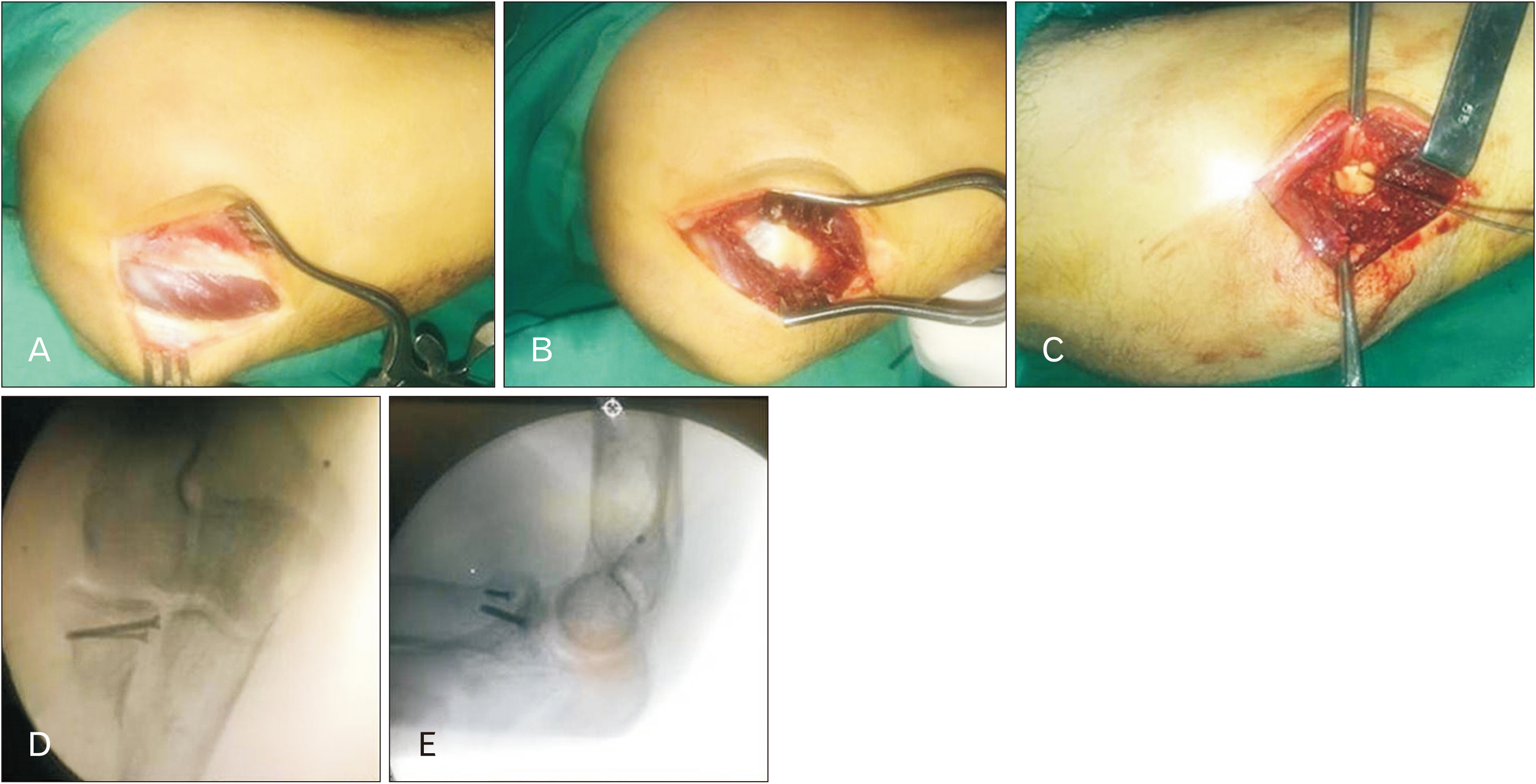
Fig. 6 (A, B) Trans-anconeus approach after incising the overlying fascia. (C) A preliminary anatomical reduction of the fracture by k-wire. (D) Anterior-posterior and lateral. (E) Intraoperative plain x-ray after anatomical reduction .
Reference
-
References
1. Duckworth AD, McQueen MM, Ring D. 2013; Fractures of the radial head. Bone Joint J. 95-B:151–9. DOI: 10.1302/0301-620X.95B2.29877. PMID: 23365021.
Article2. Gawande J, Jain S, Santoshi JA. 2017; Neglected bilateral radial head fracture with a rare presentation: a case report. Chin J Traumatol. 20:246–8. DOI: 10.1016/j.cjtee.2016.07.003. PMID: 28684037. PMCID: PMC5555247.
Article3. Capo JT, Shamian B, Francisco R, Tan V, Preston JS, Uko L, Yoon RS, Liporace FA. 2015; Fracture pattern characteristics and associated injuries of high-energy, large fragment, partial articular radial head fractures: a preliminary imaging analysis. J Orthop Traumatol. 16:125–31. DOI: 10.1007/s10195-014-0331-x. PMID: 25542062. PMCID: PMC4441642.
Article4. Moritomo H, Murase T, Arimitsu S, Oka K, Yoshikawa H, Sugamoto K. 2007; The in vivo isometric point of the lateral ligament of the elbow. J Bone Joint Surg Am. 89:2011–7. DOI: 10.2106/00004623-200709000-00017. PMID: 17768199.5. Stipp WN, Ribeiro FR, Tenor Junior AC, Filardi Filho CS, Molin DCD, Petros RSB, Brasil Filho R. 2013; Anatomical parameters in the lateral ulnar collateral ligament reconstruction: a cadaver study. Rev Bras Ortop. 48:52–6. DOI: 10.1016/j.rbo.2012.05.005. PMID: 31304111. PMCID: PMC6565991.
Article6. Wegmann K, Burkhart KJ, Zimmermann J, Dargel J, Nijs S, Konerding MA, Müller LP. 2014; The interference of distal humeral plating with the medial and lateral collateral ligaments of the elbow. Arch Orthop Trauma Surg. 134:501–7. DOI: 10.1007/s00402-014-1952-5. PMID: 24531976.
Article7. Hackl M, Bercher M, Wegmann K, Müller LP, Dargel J. 2016; Functional anatomy of the lateral collateral ligament of the elbow. Arch Orthop Trauma Surg. 136:1031–7. DOI: 10.1007/s00402-016-2479-8. PMID: 27245451.
Article8. Coriolano M, Lins OG, Amorim MJ, Amorim AA. 2009; Anatomy and functional architecture of the anconeus muscle. Int J Morphol. 27:1009–12. DOI: 10.4067/S0717-95022009000400008.
Article9. Mellema JJ, Eygendaal D, van Dijk CN, Ring D, Doornberg JN. 2016; Fracture mapping of displaced partial articular fractures of the radial head. J Shoulder Elbow Surg. 25:1509–16. DOI: 10.1016/j.jse.2016.01.030. PMID: 27052270.
Article10. Kim DK, Kim MJ, Kim YS, Oh CS, Lee SS, Lim SB, Ki HC, Shin DH. 2013; Long bone fractures identified in the Joseon Dynasty human skeletons of Korea. Anat Cell Biol. 46:203–9. DOI: 10.5115/acb.2013.46.3.203. PMID: 24179696. PMCID: PMC3811853.
Article11. Mason ML. 1954; Some observations on fractures of the head of the radius with a review of one hundred cases. Br J Surg. 42:123–32. DOI: 10.1002/bjs.18004217203. PMID: 13209035.
Article12. Broberg MA, Morrey BF. 1987; Results of treatment of fracture-dislocations of the elbow. Clin Orthop Relat Res. (216):109–19. DOI: 10.1097/00003086-198703000-00017. PMID: 3102139.
Article13. Johnston GW. 1962; A follow-up of one hundred cases of fracture of the head of the radius with a review of the literature. Ulster Med J. 31:51–6. PMID: 14452145. PMCID: PMC2384652.14. Iwasaki N, Kato H, Ishikawa J, Masuko T, Funakoshi T, Minami A. 2010; Autologous osteochondral mosaicplasty for osteochondritis dissecans of the elbow in teenage athletes: surgical technique. J Bone Joint Surg Am. 92(Suppl 1 Pt 2):208–16. DOI: 10.2106/JBJS.J.00214. PMID: 20844176.15. Pereira BP. 2013; Revisiting the anatomy and biomechanics of the anconeus muscle and its role in elbow stability. Ann Anat. 195:365–70. DOI: 10.1016/j.aanat.2012.05.007. PMID: 22874649.
Article16. Jiménez-Díaz V, Aragonés P, García-Lamas L, Barco-Laakso R, Quinones S, Konschake M, Gemmell C, Sanudo JR, Cecilia-López D. 2021; The anconeus muscle revisited: double innervation pattern and its clinical implications. Surg Radiol Anat. 43:1595–601. DOI: 10.1007/s00276-021-02750-5. PMID: 33881559.
Article17. Chaware PN, Santoshi JA, Patel M, Ahmad M, Rathinam BAD. 2018; Surgical implications of innervation pattern of the triceps muscle: a cadaveric study. J Hand Microsurg. 10:139–42. DOI: 10.1055/s-0038-1660771. PMID: 30483020. PMCID: PMC6255738.
Article18. Van Den Broek M, Van Riet R. 2017; Intra-articular capacity of the elbow joint. Clin Anat. 30:795–8. DOI: 10.1002/ca.22915. PMID: 28514501.
Article19. Bartoníček J, Naňka O, Tuček M. 2015; Kocher approach to the elbow and its options. Rozhl Chir. 94:405–14. Czech. DOI: 10.1002/ca.22915. PMID: 26556018.
- Full Text Links
-
- Actions
-
Cited
- CITED
-
- Close
- Share
-
- Similar articles
-
- Treatment of Radial Head Fracture
- Operative Treatement of Comminuted Fracture of the Radial Head
- Comparison of radial head excision and open reduction & internal fixation for comminuted radial head & neck fracture
- Acute Traumatic Irreducible Anterior Dislocation and Fracture of the Radial Head in an Adult
- Swanson Prosthesis Replacement of the Comminuted Radial Head Fracture Associated with Posterior Dislocation of the Elbow: 3 Cases Experienced
