Morphometric evaluation of great vein of Galen and its clinical implications
- Affiliations
-
- 1Department of Anatomy, Jawaharlal Institute of Postgraduate Medical Education and Research, Puducherry
- 2Department of Radiology, Jawaharlal Institute of Postgraduate Medical Education and Research, Puducherry, India
- KMID: 2540982
- DOI: http://doi.org/10.5115/acb.22.051
Abstract
- The Galenic venous system plays a vital role in the drainage of blood from deeper parts of the brain. This venous system is contributed by many major veins. These veins are located closer to the pineal gland making the surgical approach in this region difficult. Any accidental injury or occlusion of the vein of Galen could lead to devasting results. Thus, studying the dimensions of the vein of Galen is more important. Hence, we aimed to evaluate the morphometry and trajectory to the vein of Galen. About 100 computed tomographic venography records were evaluated and the length, diameter of vein of Galen, angle between straight sinus and vein of Galen and distance from internal occipital protuberance and roof of fourth ventricle to vein of Galen were studied. The mean length and diameter of vein of Galen were 9.8±2.7 and 4.08±1.04 respectively. The mean angle between straight sinus and vein of Galen was 64.2°. The mean distance between external occipital protuberance and roof of fourth ventricle to vein of Galen were 52±6.9 and 33.3±4.5 respectively. No significant morphometric differences were observed between the age groups as well as between the sexs. The results obtained from this study may be helpful for the neurosurgeons in better understanding of the anatomy of the Galenic venous system and to adopt a safe surgical approach to improve the efficacy of the surgeries of the pineal gland and also in the region of vein of Galen.
Figure
-
Fig. 1 Measurement of length and diameter of vein of Galen. Red line, length of vein of Galen; yellow line, diameter of vein of Galen.
Fig. 2 Measurement of angle between vein of Galen and straight sinus. Red line, vein of Galen and straight sinus; yellow line, angle between the vein of Galen and straight sinus.
Fig. 3 Measurement of distance between EOP and roof of fourth ventricle to vein of Galen. Red line, distance between EOP and vein of Galen; yellow line, distance between roof of fourth ventricle to vein of Galen; EOP, external occipital protuberance.
Fig. 4 Comparison of distance from EOP and roof of fourth ventricle to vein of Galen between sexs. EOP, external occipital protuberance.

Fig. 5 Comparison of distance from EOP and roof of fourth ventricle to vein of Galen between age groups. EOP, external occipital protuberance.
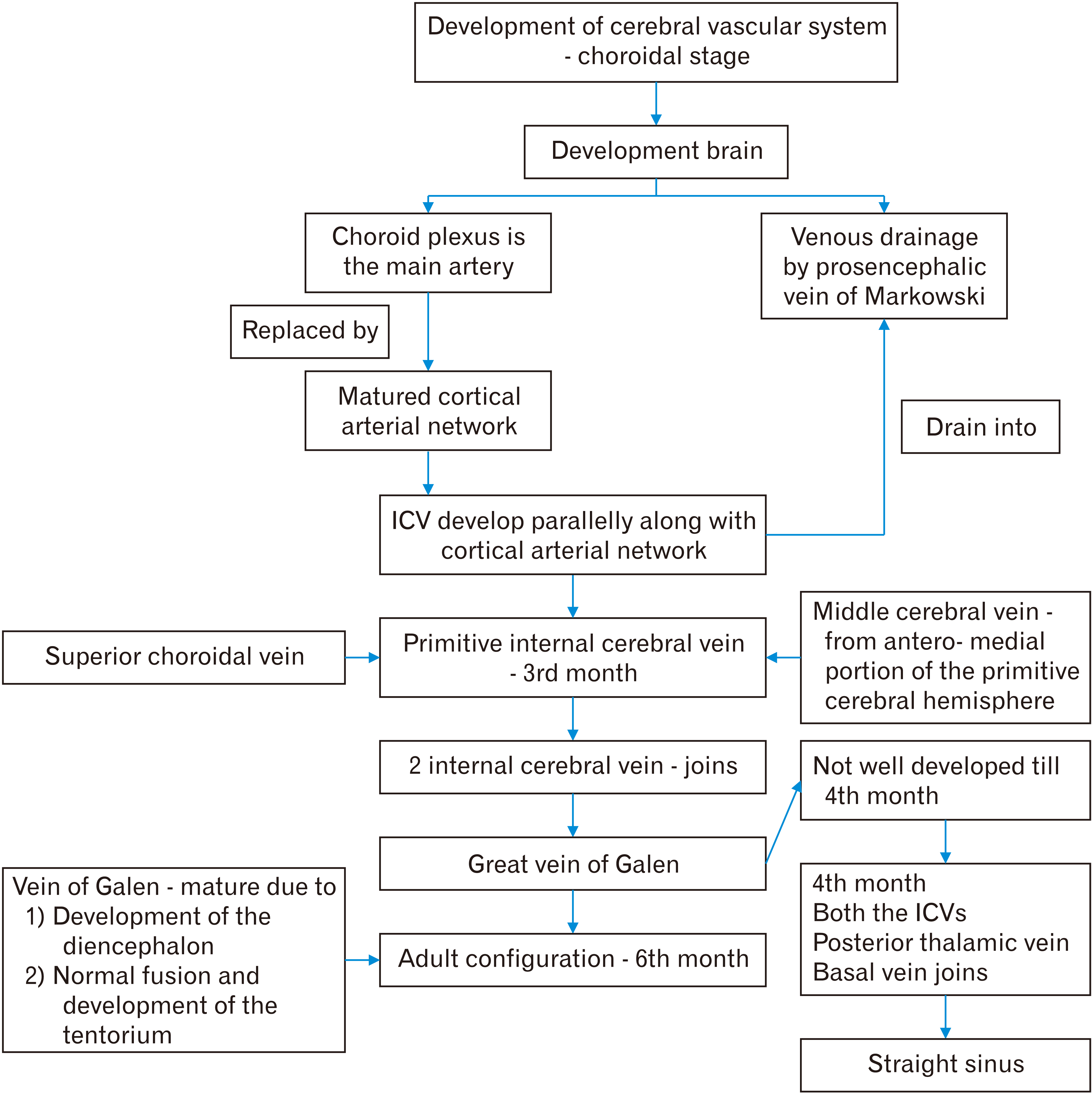
Fig. 6 Embryology of vein of Galen.
Reference
-
References
1. Ono M, Rhoton AL Jr, Peace D, Rodriguez RJ. 1984; Microsurgical anatomy of the deep venous system of the brain. Neurosurgery. 15:621–57. DOI: 10.1227/00006123-198411000-00002. PMID: 6504279.
Article2. Standring S. 2016. Gray's anatomy: the anatomical basis of clinical practice. 41st ed. Elsevier;Philadelphia (PA): p. 289. DOI: 10.1097/00006123-198411000-00002.3. Youssef AS, Downes AE, Agazzi S, Van Loveren HR. 2011; Life without the vein of Galen: clinical and radiographic sequelae. Clin Anat. 24:776–85. DOI: 10.1002/ca.21176. PMID: 21438020.
Article4. Chaynes P. 2003; Microsurgical anatomy of the great cerebral vein of Galen and its tributaries. J Neurosurg. 99:1028–38. DOI: 10.3171/jns.2003.99.6.1028. PMID: 14705731.
Article5. Berenstein A, Paramasivam S, Sorscher M, Molofsky W, Meila D, Ghatan S. 2019; Vein of Galen aneurysmal malformation: advances in management and endovascular treatment. Neurosurgery. 84:469–78. Erratum in: Neurosurgery 2018;83:593. DOI: 10.1093/neuros/nyy100. PMID: 29860355.6. D'Amico A, Tinari S, D'Antonio F, Rizzo G, Liberati M, Vasciaveo L, Buca D. 2021; Jan. 28. Outcome of fetal vein Galen aneurysmal malformations: a systematic review and meta-analysis. J Matern Fetal Neonatal Med. [Epub]. https://doi.org/10.1080/14767058.2021.1878494. DOI: 10.1080/14767058.2021.1878494. PMID: 33508985.7. Browder J, Kaplan HA, Krieger AJ. 1976; Anatomical features of the straight sinus and its tributaries. J Neurosurg. 44:55–61. DOI: 10.3171/jns.1976.44.1.0055. PMID: 1244434.
Article8. Yamamoto I, Kageyama N. 1980; Microsurgical anatomy of the pineal region. J Neurosurg. 53:205–21. DOI: 10.3171/jns.1980.53.2.0205. PMID: 7431059.
Article9. Deniz C, Gokce E, Acu B, Kuyucu YE. 2019; Comparative evaluation of dural venous sinuses and cerebral veins using contrast-enhanced spoiled gradient recalled echo and time-of-flight magnetic resonance venography. J Contemp Med. 9:214–21. DOI: 10.16899/jcm.556044.
Article10. Ghali WM, Rafla MF, Ekladious EY, Ibrahim KA. 1989; A study of the junction between the straight sinus and the great cerebral vein. J Anat. 164:49–54. PMID: 2606794. PMCID: PMC1256597.11. Hernesniemi J, Romani R, Albayrak BS, Lehto H, Dashti R, Ramsey C 3rd, Karatas A, Cardia A, Navratil O, Piippo A, Fujiki M, Toninelli S, Niemelä M. 2008; Microsurgical management of pineal region lesions: personal experience with 119 patients. Surg Neurol. 70:576–83. DOI: 10.1016/j.surneu.2008.07.019. PMID: 19055952.
Article12. Gutierrez S, Iwanaga J, Dumont AS, Tubbs RS. 2020; Direct drainage of the basal vein of Rosenthal into the superior petrosal sinus: a literature review. Anat Cell Biol. 53:379–84. DOI: 10.5115/acb.20.199. PMID: 33148874. PMCID: PMC7769095.
Article13. Widjaja E, Griffiths PD. 2004; Intracranial MR venography in children: normal anatomy and variations. AJNR Am J Neuroradiol. 25:1557–62. PMID: 15502138. PMCID: PMC7976417.14. Hoang S, Choudhri O, Edwards M, Guzman R. 2009; Vein of Galen malformation. Neurosurg Focus. 27:E8. DOI: 10.3171/2009.8.FOCUS09168. PMID: 19877798.
Article15. Sasidharan CK, Anoop P, Vijayakumar M, Jayakrishnan MP, Reetha G, Sindhu TG. 2004; Spectrum of clinical presentations of vein of Galen aneurysm. Indian J Pediatr. 71:459–63. DOI: 10.1007/BF02725643. PMID: 15163883.
Article
