A Case of Forehead Flap Nasal Reconstruction for Dog Bite Nasal Injury
- Affiliations
-
- 1Department of Otorhinolaryngology-Head and Neck Surgery, Asan Medical Center, University of Ulsan College of Medicine, Seoul, Republic of Korea
- KMID: 2540856
- DOI: http://doi.org/10.18787/jr.2022.00426
Abstract
- A dog bite is the most common trauma among animal bites, and it has varying severity, from simple skin lacerations to defects in whole tissue layers. Considering the aesthetic and functional importance of the nose, an appropriate reconstruction should be conducted for large and full-thickness tissue defects. Although this is quite common, literature detailing surgical reconstruction is lacking, especially in domestic journals. A 45-year-old male patient visited an outpatient clinic due to nasal trauma caused by a dog bite. The patient’s nose showed whole-layer tissue defects, with necrotic tissues at the periphery. Nasal reconstruction surgery was conducted using a forehead flap and ear cartilage composite graft. Pedicle division was conducted three weeks after primary surgery. The functional and aesthetic outcomes of the surgery were satisfactory. Our experience demonstrates that the forehead flap is a useful option for reconstructing a nose badly injured by a dog bite.
Figure
-
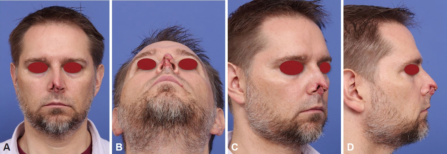
Fig. 1. Preoperative clinical photography. A: Frontal view. B: Basal view. C: Oblique view. D: Profile view. Upper 1/2 of columella of patient was destructed. Skin and cartilage portion of right lower lateral cartilage defects were identified. Left medial crus of lower lateral cartilage has necrotic tissue with atrophy.
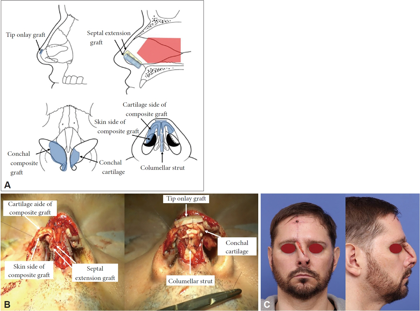
Fig. 2. Image of reconstruction procedure, surgical field view, and postoperative clinical photography after forehead flap. A: Image of nasal reconstruction procedure. B: Surgical view of nasal reconstruction. C: Postoperative clinical photography after forehead flap nasal reconstruction. After reconstruction of bony, cartilage and right inner skin defect sites, soft tissue defect was reconstructed by forehead flap.
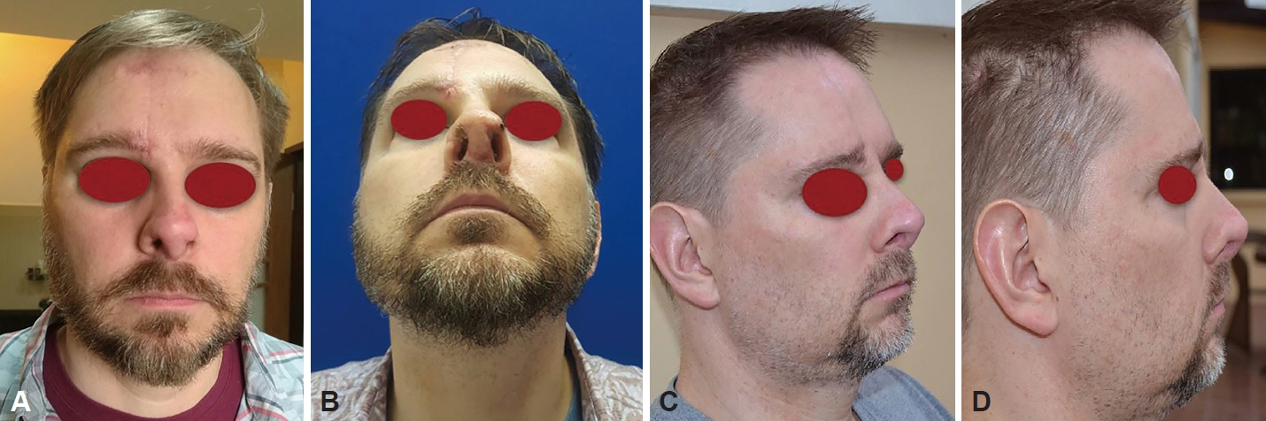
Fig. 3. Postoperative patient’s clinical photography. A: Frontal view—postoperative 6 months. B: Basal view—postoperative 1.5 months. C: Oblique view—postoperative 6 months. D: Profile view—postoperative 6 months. After reconstruction, patient showed successful nasal shape and function. Because the patient lived in Philippines, he sent photos 6 months after surgery and we can compare postoperative results with preoperative photos.
Reference
-
References
1. Mendoza JM, Chi JJ. Reconstruction of animal bite injuries to the head and neck. Curr Opin Otolaryngol Head Neck Surg. 2019; 27(5):407–12.
Article2. Rothe K, Tsokos M, Handrick W. Animal and human bite wounds. Dtsch Arztebl Int. 2015; 112(25):433–42. quiz 443.
Article3. Senturk E, Dagistanli N, Calim OF, Ozturan O. Nasal reconstruction following a dog bite. J Craniofac Surg. 2019; 30(7):2233–5.
Article4. Kalmar CL, Nguyen PD, Taylor JA. Subtotal nasal reconstruction after traumatic avulsion. Plast Reconstr Surg Glob Open. 2020; 8(11):e3239.
Article5. Cavadas PC, Torres A. Total nasal reconstruction with prefabricated and prelaminated free flap. Ann Plast Surg. 2019; 83(6):e35–8.
Article6. Shipkov H, Traikova N, Stefanova P, Pazardjikliev D, Simov R. The forehead flap for immediate reconstruction of the nose after bite injuries: indications, advantages, and disadvantages. Ann Plast Surg. 2014; 73(3):358.
Article7. Piccart F, Dormaar JT, Coropciuc R, Schoenaers J, Bila M, Politis C. Dog bite injuries in the head and neck region: a 20-year review. Craniomaxillofac Trauma Reconstr. 2019; 12(3):199–204.
Article8. Huang AH, Wong MS. Acute nasal reconstruction with forehead flap after dog bite. Ann Plast Surg. 2013; 70(4):401–5.
Article9. Menick FJ. Nasal reconstruction with a forehead flap. Clin Plast Surg. 2009; 36(3):443–59.
Article10. Shaye DA, Tollefson TT. What is the optimal timing for dividing a forehead flap? Laryngoscope. 2020; 130(10):2303–4.
Article11. Yim S, Eun SC. Chondrocutaneous preauricular free flap for reconstruction of nasal defects aided by interposition vascular graft. J Craniofac Surg. 2017; 28(7):1842–6.
Article12. Boettcher-Haberzeth S, Schiestl C. Management of avulsion injuries. Eur J Pediatr Surg. 2013; 23(5):359–64.
Article13. Ferreira S, Ayres Quaresma LE, Timóteo CA, da Silva Fabris AL, Faverani LP, Francisconi GB, et al. The primary closure approach of dog bite injuries of the nose. J Craniofac Surg. 2014; 25(3):e216–8.
Article
- Full Text Links
-
- Actions
-
Cited
- CITED
-
- Close
- Share
-
- Similar articles
-
- Reconstruction of Postburn Nasal Alar Defect by Paramedian Forehead Flap
- Reconstruction of a large nasal defect using a folded forehead flap: a case report
- Simultaneous Upper and Lower Eyelid Reconstruction for Eyelid Defects Following a Dog Bite
- Forehead Island Flap For Nasal Reconstruction
- A case report of the external nose reconstruction using forehead flap and auricular composite grafts
