Negative-pressure helmet restores cerebral hemodynamic parameters in sinking skin flap syndrome: a case report
- Affiliations
-
- 1Department of Intensive Care, Geneva University Hospitals, Université Catholique de Louvain, Geneva, Switzerland
- 2Department of Intensive Care, Geneva University Hospitals, Faculty of Medicine Geneva, Geneva, Switzerland
- 3Division of Neurosurgery, Department of Clinical Neurosciences, Geneva University Hospitals, Faculty of Medicine Geneva, Geneva, Switzerland
- KMID: 2538770
- DOI: http://doi.org/10.18700/jnc.220072
Abstract
- Background
Sinking skin flap syndrome (SSFS) is a rare complication of decompressive craniectomy (DC) and causes a wide range of neurological deficits. Its pathophysiology remains debatable, however cranioplasty may decrease the symptoms of SSFS by reducing the direct effect of atmospheric pressure on the brain and allowing the correction of cerebral blood flow (CBF) and cerebral metabolism.
Case Report
A 36-year-old woman underwent DC for a right frontal hematoma and signs of increased intracranial pressure following the resection of a right frontal arteriovenous malformation. Subsequently, she developed SSFS, altered neurological status with a Glasgow Coma Scale (GCS) score of 8/15, and bacterial-resistant meningitis. We applied a custom-shaped negative pressure helmet on the bone defect and used invasive and noninvasive techniques to measure the changes in intracranial pressure, CBF and cerebral oxygen saturation. Despite improvements in the cerebral physiological parameters, the neurological status did not improve (GCS score, 8/15).
Conclusions
To our knowledge, this is the first reported case of SSFS treated with a negative-pressure helmet and subsequent changes in cerebral parameters that were monitored invasively and noninvasively.
Figure
-
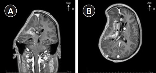
Fig. 1. Magnetic resonance imaging T1-weighted images showing the bone defect area of the skull of the patient. (A) Coronal T1-weighted view shows a left-sided deviation of the midline and a filling of the posterior fossa with amygdala herniation (asterisk). (B) Axial T1 view shows left-sided deviation (arrows) and shrinking of lateral ventricles (white cross). Sup, superior; R, right; Ant, anterior.

Fig. 2. The Lenoir Orthopédie negative-pressure helmet with a manual vacuum pump. Images obtained from Lenoir Orthopédie user manual (Lenoir Orthopédie, Daniel Robert Orthopédie, Geneva, Switzerland).
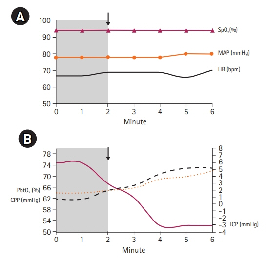
Fig. 3. Hemodynamic and cerebral monitoring before and after the application of negative pressure using a Lenoir Orthopédie helmet. Grey zone, the time periods before the application of the negative pressure; arrow, start application of negative pressure. The procedure was hemodynamically supported and revealed reduction of intracranial pressure (ICP) to normal values and improvements in cerebral perfusion pressure (CPP) and cerebral oxygen saturation. (A) Hemodynamic parameters before and after applying negative pressure. (B) Cerebral hemodynamic parameters before and after applying negative pressure. SpO2, pulse oximeter; MAP, mean arterial pressure; HR, heart rate; PbtO2, brain tissue oxygen tension.

Fig. 4. Increase in cerebral blood flow (CBF) after applying negative pressure. The CBF increased from 50 mL/g to 65 mL/g.
Reference
-
1. Yamaura A, Makino H. Neurological deficits in the presence of the sinking skin flap following decompressive craniectomy. Neurol Med Chir (Tokyo). 1977; 17(1 Pt 1):43–53.
Article2. Annan M, De Toffol B, Hommet C, Mondon K. Sinking skin flap syndrome (or syndrome of the trephined): a review. Br J Neurosurg. 2015; 29:314–8.
Article3. Di Rienzo A, Colasanti R, Gladi M, Pompucci A, Della Costanza M, Paracino R, et al. Sinking flap syndrome revisited: the who, when and why. Neurosurg Rev. 2020; 43:323–35.
Article4. Khan NA, Ullah S, Alkilani W, Zeb H, Tahir H, Suri J. Sinking skin flap syndrome: phenomenon of neurological deterioration after decompressive craniectomy. Case Rep Med. 2018; 2018:9805395.
Article5. Cholet C, André A, Law-Ye B. Sinking skin flap syndrome following decompressive craniectomy. Br J Neurosurg. 2018; 32:73–4.
Article6. Ashayeri K, M Jackson E, Huang J, Brem H, Gordon CR. Syndrome of the trephined: a systematic review. Neurosurgery. 2016; 79:525–34.7. Goedemans T, Verbaan D, van der Veer O, Bot M, Post R, Hoogmoed J, et al. Complications in cranioplasty after decompressive craniectomy: timing of the intervention. J Neurol. 2020; 267:1312–20.
Article8. Yoshida K, Furuse M, Izawa A, Iizima N, Kuchiwaki H, Inao S. Dynamics of cerebral blood flow and metabolism in patients with cranioplasty as evaluated by 133Xe CT and 31P magnetic resonance spectroscopy. J Neurol Neurosurg Psychiatry. 1996; 61:166–71.9. Winkler PA, Stummer W, Linke R, Krishnan KG, Tatsch K. The influence of cranioplasty on postural blood flow regulation, cerebrovascular reserve capacity, and cerebral glucose metabolism. Neurosurg Focus. 2000; 8:e9.10. Bijlenga P, Zumofen D, Yilmaz H, Creisson E, de Tribolet N. Orthostatic mesodiencephalic dysfunction after decompressive craniectomy. J Neurol Neurosurg Psychiatry. 2007; 78:430–3.
Article11. Keller E, Wolf M, Martin M, Yonekawa Y. Estimation of cerebral oxygenation and hemodynamics in cerebral vasospasm using indocyaningreen dye dilution and near infrared spectroscopy: a case report. J Neurosurg Anesthesiol. 2001; 13:43–8.12. Vos JJ, Ellermann SF, Scheeren TW. Journal of Clinical Monitoring and Computing 2017/2018 end of year summary: monitoring-and provocation-of the microcirculation and tissue oxygenation. J Clin Monit Comput. 2019; 33:201–9.
Article13. Ku T, Choi C. Noninvasive optical measurement of cerebral blood flow in mice using molecular dynamics analysis of indocyanine green. PLoS One. 2012; 7:e48383.14. Caballero-Lozada AF, Nanwani KL, Pavón F, Zorrilla-Vaca A, Zorrilla-Vaca C. Clinical applications of ultrasonography in neurocritically ill patients. J Intensive Care Med. 2021; 36:627–34.
Article15. Bonow RH, Young CC, Bass DI, Moore A, Levitt MR. Transcranial Doppler ultrasonography in neurological surgery and neurocritical care. Neurosurg Focus. 2019; 47:E2.
- Full Text Links
-
- Actions
-
Cited
- CITED
-
- Close
- Share
-
- Similar articles
-
- Reperfusion Injury after Autologous Cranioplasty in a Patient with Sinking Skin Flap Syndrome
- Sinking Skin Flap Syndrome after Decompressive Craniotomy
- "Syndrome of the Sinking Skin-Flap" Secondary to the Ventriculoperitoneal Shunt after Craniectomy
- Intracerebral Hemorrhagic Infarction after Cranioplasty in a Patient with Sinking Skin Flap Syndrome
- Sinking Skin Flap Syndrome after Craniectomy in a Patient Who Previously Underwent Ventriculoperitoneal Shunt
