Reappraisal of arterial anatomy of thumb
- Affiliations
-
- 1Department of Anatomy, Indira Gandhi Medical College and Research Institute, Puducherry, India
- 2Department of Anatomy, Jawaharlal Institute of Postgraduate Medical Education and Research, Puducherry, India
- KMID: 2537460
- DOI: http://doi.org/10.5115/acb.22.052
Abstract
- The nomenclature of arteries supplying the thumb and its sources arteries differs between the studies. This makes difficulty in understanding the irrigation pattern to the thumb. The main purpose of this study was to identify the proper digital arteries supplying the thumb on its radial and ulnar side from both the palmar and dorsal aspect. Also this study aimed to trace its source and classify with proper definition with the review of blood supply to thumb. Dissection was carried out in 55 hands form 28 freshly embalmed adult human cadavers of both genders. The proper digital arteries to the thumb and its source arteries were carefully traced and defined. Thumb receives its dominant blood supply mostly from its palmar side. The ulnar palmar digital artery was seen in all the dissected hand (100.0%) whereas; the radial palmar digital artery was present in 53 hands (96.4%). The radial dorsal digital artery and ulnar dorsal digital artery were observed in only 10.0% and 7.3%. The most common source of both the palmar digital arteries to thumb was from first palmar metacarpal artery (FPMA). In majority of the hands, in addition to the radial or ulnar palmar digital arteries from the FPMA, there were also additional palmar digital arteries arising from the superficial palmar arterial system. The universal naming of the proper digital arteries to the thumb as well as its source arteries is mandatory for the proper understanding of normal as well as variant arterial anatomy of thumb.
Keyword
Figure
-
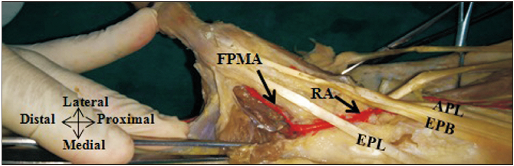
Fig. 1 FPMA arises from the deep branch of radial artery after it pierces the first dorsal interosseous muscle to continue as deep palmar arch. In this figure the first dorsal interosseous muscle is incised to show the course of FPMA running between the first dorsal interosseous and adductor pollicis muscles. APL, abductor pollicis longus; EPB, extensor pollicis brevis; EPL, extensor pollicis longus; FPMA, first palmar metacarpal artery; RA, radial artery.
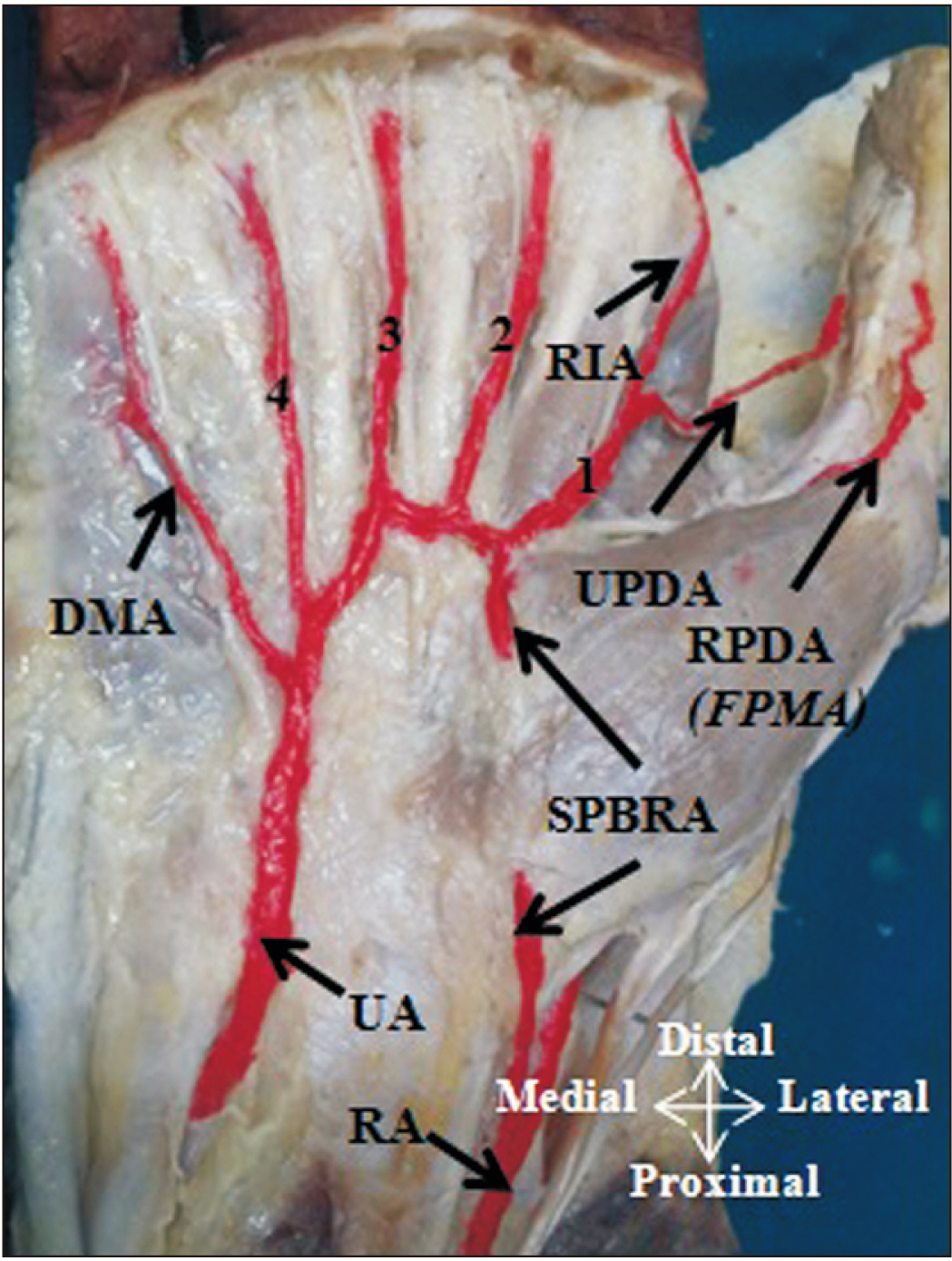
Fig. 2 RPDA from the FPMA and UPDA from the first CPDA from typical SPA. CPDA, common palmar digital artery; 1, 2, 3, 4, CPDA; DMA, digiti minimi artery; FPMA, first palmar metacarpal artery; RA, radial artery; RIA, radialis indicis artery; RPDA, radial palmar digital artery; SPA, superficial palmar arch; SPBRA, superficial palmar branch of radial artery; UA, ulnar artery; UPDA, ulnar palmar digital artery.
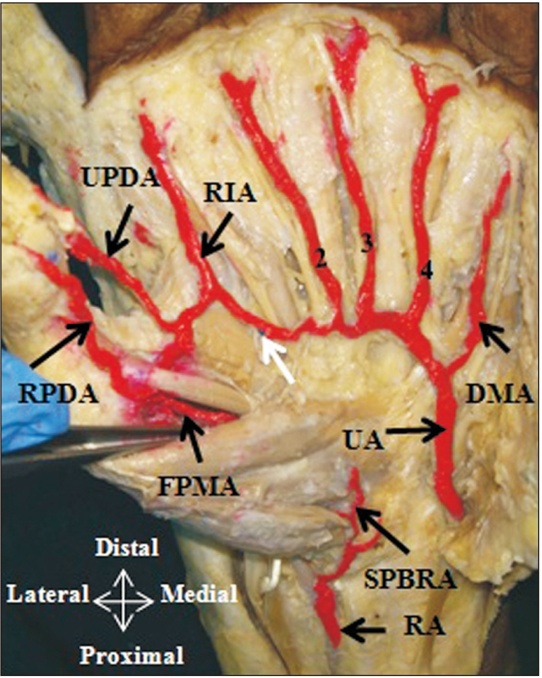
Fig. 3 RPDA and UPDA from the FPMA. CPDA, common palmar digital artery; 1, 2, 3, 4, CPDA; DMA, digiti minimi artery; FPMA, first palmar metacarpal artery; RA, radial artery; RIA, radialis indicis artery; RPDA, radial palmar digital artery; SPBRA, superficial palmar branch of radial artery; UA, ulnar artery; UPDA, ulnar palmar digital artery; white arrow, anastomosis of UA with RIA.
Fig. 4 RPDA arise directly from the UA and UPDA from the UA through first CPDA. CPDA, common palmar digital artery; 1, 2, 3, 4, CPDA; DMA, digiti minimi artery; RA, radial artery; RIA, radialis indicis artery; RPDA, radial palmar digital artery; UA, ulnar artery; UPDA, ulnar palmar digital artery.

Fig. 5 RPDA arises directly from the SPBRA whereas; the UPDA and radialis indicis artery arise from the SPBRA through the first CPDA. CPDA, common palmar digital artery; 1, 2, 3, 4, CPDA; DMA, digiti minimi artery; RA, radial artery; RIA, radialis indicis artery; RPDA, radial palmar digital artery; SPBRA, superficial palmar branch of radial artery; UA, ulnar artery; UPDA, ulnar palmar digital artery.

Fig. 6 UPDA and RPDA from the MA through first CPDA. CPDA, common palmar digital artery; 1, 2, 3, 4, CPDA; DMA, digiti minimi artery; MA, median artery; RA, radial artery; RIA, radialis indicis artery; RPDA, radial palmar digital artery; SPBRA, superficial palmar branch of radial artery; UA, ulnar artery; UPDA, ulnar palmar digital artery.
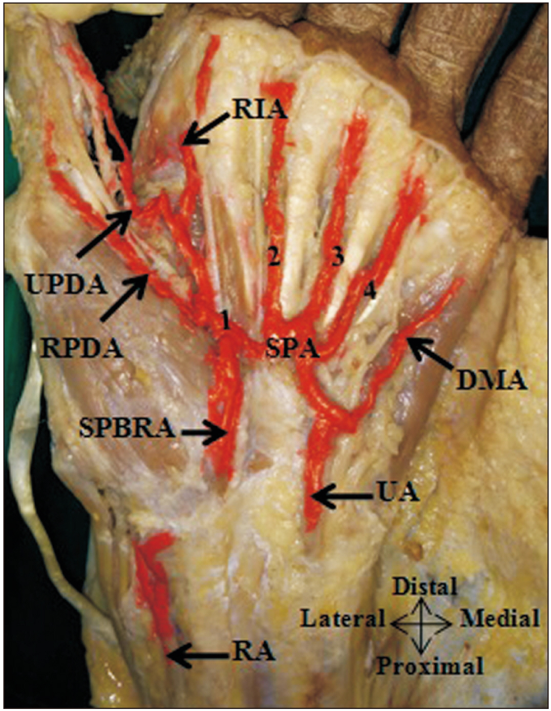
Fig. 7 RPDA, UPDA from the typical SPA through the first CPDA. CPDA, common palmar digital artery; 1, 2, 3, 4, CPDA; DMA, digiti minimi artery; RA, radial artery; RIA, radialis indicis artery; RPDA, radial palmar digital artery; SPA, superficial palmar arch; SPBRA, superficial palmar branch of radial artery; UA, ulnar artery; UPDA, ulnar palmar digital artery.
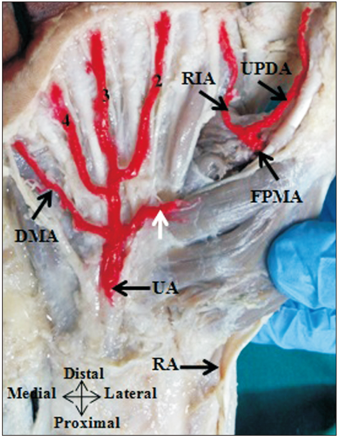
Fig. 8 UPDA and radialis indicis artery arising from the FPMA. CPDA, common palmar digital artery; 2, 3, 4, CPDA; DMA, digiti minimi artery; FPMA, first palmar metacarpal artery; RA, radial artery; RIA, radialis indicis artery; UPDA, ulnar palmar digital artery; UA, ulnar artery; white arrow muscular branch to thenar muscles from UA, according to Gnanasekaran and Veeramani [18]. Adapted from Gnanasekaran and Veeramani. Surg Radiol Anat 2019;41:791-9 [18].
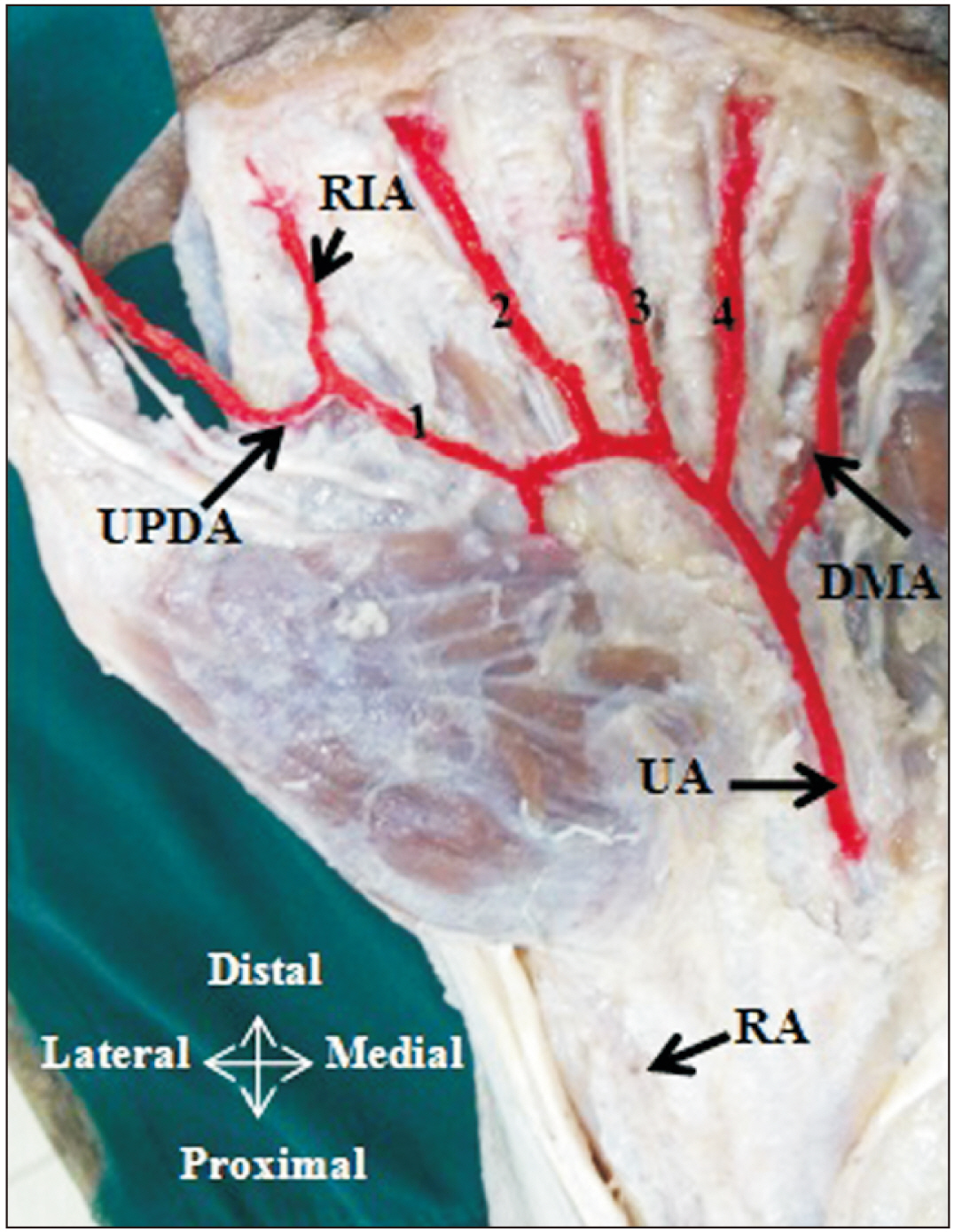
Fig. 9 UPDA arise from the UA through the first common palmar digital. CPDA, common palmar digital artery; 1, 2, 3, 4, CPDA; DMA, digiti minimi artery; RA, radial artery; RIA, radialis indicis artery; UA, ulnar artery; UPDA, ulnar palmar digital artery, according to Gnanasekaran and Veeramani [18]. Adapted from Gnanasekaran and Veeramani. Surg Radiol Anat 2019;41:791-9 [18].
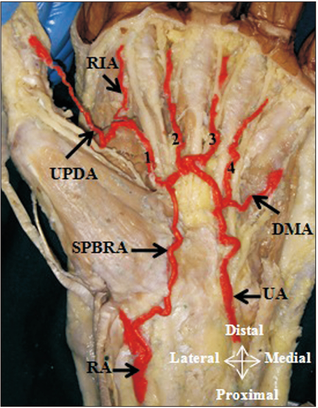
Fig. 10 UPDA arise from typical SPA through the first CPDA. CPDA, common palmar digital artery; 1, 2, 3, 4, CPDA; DMA, digiti minimi artery; RA, radial artery; RIA, radialis indicis artery; SPA, superficial palmar arch; SPBRA, superficial palmar branch of radial artery; UA, ulnar artery; UPDA, ulnar palmar digital artery, according to Gnanasekaran and Veeramani [18]. Adapted from Gnanasekaran and Veeramani. Surg Radiol Anat 2019;41:791-9 [18].

Fig. 11 UPDA and radialis indicis artery arise from the FDMA, a direct branch of radial artery. FDMA, first dorsal metacarpal artery; RA, radial artery; RIA, radialis indicis artery; UPDA, ulnar palmar digital artery, according to Gnanasekaran and Veeramani [18]. Adapted from Gnanasekaran and Veeramani. Surg Radiol Anat 2019;41:791-9 [18].
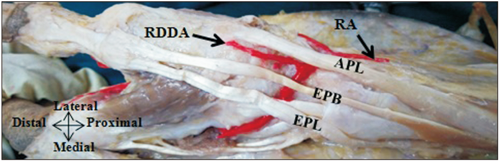
Fig. 12 RDDA arise as a direct branch from the radial artery. APL, abductor pollicis longus; EPB, extensor pollicis brevis; EPL, extensor pollicis longus; RA, radial artery; RDDA, radial dorsal digital artery.
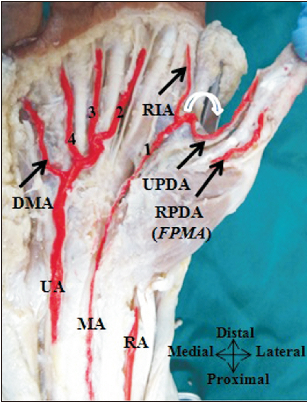
Fig. 13 FDMA anastomosed with the UPDA provided by the MA around the first web space. CPDA, common palmar digital artery; 1, 2, 3, 4, CPDA; DMA, digiti minimi artery; FDMA, first dorsal metacarpal artery; FPMA, first palmar metacarpal artery; MA, median artery; RA, radial artery; RPDA, radial palmar digital artery; RIA, radialis indicis artery; UA, ulnar artery; UPDA, ulnar palmar digital artery, according to Gnanasekaran and Veeramani [18]. Adapted from Gnanasekaran and Veeramani. Surg Radiol Anat 2019;41:791-9 [18].
Reference
-
References
1. Cho SH, Bahar-Moni AS, Park HC. 2016; Thumb replantation using the superficial palmar branch of the radial artery. J Hand Microsurg. 8:106–8. DOI: 10.1055/s-0036-1585058. PMID: 27625540. PMCID: PMC5018973.2. Parks BJ, Arbelaez J, Horner RL. 1978; Medical and surgical importance of the arterial blood supply of the thumb. J Hand Surg Am. 3:383–5. DOI: 10.1016/S0363-5023(78)80044-6. PMID: 681725.3. Hierner R, Putz R, Bishop AT, Shen ZL, Wilhelm K, Brugger U, Kellner L, Rintelen H. 2013. Flaps in hand and upper limb reconstruction: surgical anatomy, operative techniques and differential therapy. Urban & Fischer;Munchen: DOI: 10.1016/s0363-5023(78)80044-6.4. Schlenker JD, Kleinert HE, Tsai TM. 1980; Methods and results of replantation following traumatic amputation of the thumb in sixty-four patients. J Hand Surg Am. 5:63–70. DOI: 10.1016/S0363-5023(80)80046-3. PMID: 7365219.5. Ames EL, Bissonnette M, Acland R, Lister G, Firrell J. 1993; Arterial anatomy of the thumb. J Hand Surg Br. 18:427–36. DOI: 10.1016/0266-7681(93)90141-2. PMID: 8409651.6. Biant LC. Standring S, editor. 2016. Elbow and forearm. Gray's Anatomy: The Anatomical Basis of Clinical Practice. 41st ed. Elsevier Limited;Philadelphia: p. 837–61. DOI: 10.1201/b13469-22.7. Earley MJ. 1986; The arterial supply of the thumb, first web and index finger and its surgical application. J Hand Surg Br. 11:163–74. DOI: 10.1016/0266-7681_86_90253-6. PMID: 3734551.8. Al-Turk M, Metcalf WK. 1984; A study of the superficial palmar arteries using the Doppler ultrasonic flowmeter. J Anat. 138(Pt 1):27–32. PMID: 6706837. PMCID: PMC1164307.9. Bilge O, Pinar Y, Ozer MA, Gövsa F. 2006; A morphometric study on the superficial palmar arch of the hand. Surg Radiol Anat. 28:343–50. DOI: 10.1007/s00276-006-0109-9. PMID: 16642281.10. Coleman SS, Anson BJ. 1961; Arterial patterns in the hand based upon a study of 650 specimens. Surg Gynecol Obstet. 113:409–24. DOI: 10.1097/00006534-196201000-00028. PMID: 13694610.11. Fazan VP, Borges CT, Da Silva JH, Caetano AG, Filho OA. 2004; Superficial palmar arch: an arterial diameter study. J Anat. 204:307–11. DOI: 10.1111/j.0021-8782.2004.00281.x. PMID: 15061757. PMCID: PMC1571293.12. Ikeda A, Ugawa A, Kazihara Y, Hamada N. 1988; Arterial patterns in the hand based on a three-dimensional analysis of 220 cadaver hands. J Hand Surg Am. 13:501–9. DOI: 10.1016/S0363-5023(88)80085-6. PMID: 3418051.13. Murakami T, Takaya K, Outi H. 1969; The origin, course and distribution of arteries to the thumb, with special reference to the so-called A. princeps pollicis. Okajimas Folia Anat Jpn. 46:123–37. DOI: 10.2535/ofaj1936.46.2-3_123. PMID: 5820050.14. Nunley JA, Goldner RD, Urbaniak JR. 1985; Thumb reconstruction by the wrap-around method. Clin Orthop Relat Res. 195:97–103. DOI: 10.1097/00003086-198505000-00009. PMID: 3884213.15. Ramírez AR, Gonzalez SM. 2012; Arteries of the thumb: description of anatomical variations and review of the literature. Plast Reconstr Surg. 129:468e–76e. DOI: 10.1097/PRS.0b013e3182402d43. PMID: 22373995.16. Miletin J, Sukop A, Baca V, Kachlik D. 2017; Arterial supply of the thumb: systemic review. Clin Anat. 30:963–73. DOI: 10.1002/ca.22973. PMID: 28791730.17. Cunningham DJ. 2005. Cunningham's manual of practical anatomy. 15th ed. Vol 1:Oxford University Press;Oxford: DOI: 10.1002/ca.22973.18. Gnanasekaran D, Veeramani R. 2019; Newer insights in the anatomy of superficial palmar arch. Surg Radiol Anat. 41:791–9. DOI: 10.1007/s00276-019-02223-w. PMID: 30923841.19. Tan RES, Lahiri A. 2020; Vascular anatomy of the hand in relation to flaps. Hand Clin. 36:1–8. DOI: 10.1016/j.hcl.2019.08.001. PMID: 31757342.20. Omokawa S, Tanaka Y, Ryu J, Kish VL. 2005; The anatomical basis for reverse first to fifth dorsal metacarpal arterial flaps. J Hand Surg Br. 30:40–4. DOI: 10.1016/J.JHSB.2004.09.006. PMID: 15620490.
- Full Text Links
-
- Actions
-
Cited
- CITED
-
- Close
- Share
-
- Similar articles
-
- Morphometric study of pulleys of the thumb
- Clinical experiences in Thumb reconstruction
- Thumb Reconstruction with a Osteocutaneous Free Flap Transfer with Partial 1 st Matatarsus (Case Report )
- A Case of Bilateral Congenital Clasped Thumb
- Functional reconstruction of the thumb by heterotopic thumb-to-thumb replantation and secondary opponensplasty in bilateral amputation in the upper extremities: a case report
