Variant pectoralis minor muscle: a case study with clinical relevance
- Affiliations
-
- 1Department of Anatomy, AIIMS Bibinagar, Hyderabad, India
- KMID: 2537459
- DOI: http://doi.org/10.5115/acb.22.076
Abstract
- The pectoralis minor (PMn) muscle originates from the third, fourth, and fifth ribs near the costochondral junctionusually and gets inserted on the medial margin and upper surface of the coracoid process of the scapula. To look at the morphological insertion patterns and sites of attachment of the PMn muscle in the donated cadavers. Over all 19 limbs were included in the study (9 right and 10 left). Out of 19 limbs, 10 belonged to female and 9 belonged to male cadavers. The cadavers were meticulously dissected to determine the morphological insertion types and location of the attachment of the muscle. Unusual pattern of insertion was observed in 6 limbs (31.6%) out of total 19 limbs included in the study. The variations we observed does not fall completely in the classification by Le Double, hence variations we observed can be considered as new and rare variant which to our knowledge is not reported in literature. We propose this new variant to be type 4 of Le Double classification. The potential of ectopic PMn tendon should be taken into consideration and tested out, especially in patients with shoulder discomfort and stiffening who have ruled out the more frequent diseases. For proper surgical planning, a preoperative magnetic resonance imaging or USG examination of the shoulder joint is required considering the prevalence of variation in the insertion pattern of PMn muscle. Preoperative identification of any abnormal PMn insertion can help to reduce the risk of iatrogenic tendon injury and post-operative problems.
Keyword
Figure
-
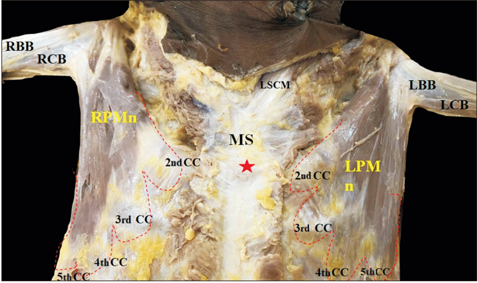
Fig. 1 Showing dissected pectoral region, with the variation in the origin of pectoralis minor (PMn) muscle bilaterally. Right pectoralis minor (RPMn) and left pectoralis minor (LPMn) muscles taking origin from second to fifth ribs near their costochondral junction. MS, manubrium sternum (red asterisk, sternal angle); LBB, left biceps brachi; LCB, left coracobrachialis; RBB, right biceps brachi; RCB, right coracobrachialis; CC, costal cartilage; LSCM, left sternocleidomastoid muscle.
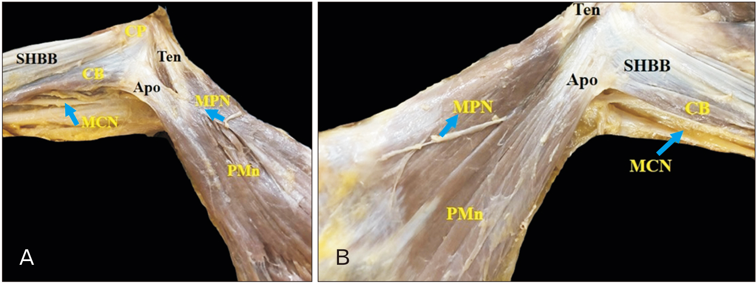
Fig. 2 Showing variant form of insertion of pectoralis minor (PMn) muscle in cadaver-1. Dual insertion of PMn in right side (A) and in left side (B), in the form of medial tendon (Ten) and lateral aponeurosis (Apo). MCN, musculocutaneous nerve; MPN, medial pectoral nerve; SHBB, short head of biceps brachii; CB, coracobrachialis; CP, coracoid process.
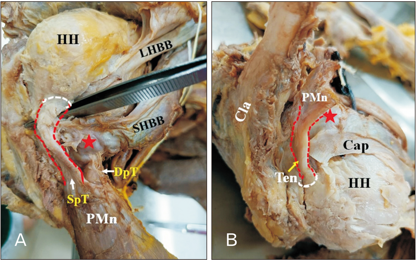
Fig. 3 Showing variant form of insertion of pectoralis minor (PMn) muscle in cadaver-2. (A) Right PMn is with two tendons, superficial tendon (SpT) and deep tendon (DpT). DpT is inserted on the coracoid process (red asterisk) along with coracobrachialis (CB) and short head of biceps brachii (SHBB). SpT got merged with the capsule of the shoulder joint and inserted at upper part of greater tubercle along with supraspinatus muscle. (B) Left PMn muscle (cut) inserted as a single tendon gets merged with the capsule of the shoulder joint and was inserted at upper part of greater tubercle. Cap, capsule of shoulder joint; Cla, clavicle; HH, humeral head; LHBB, long head of biceps brachii.
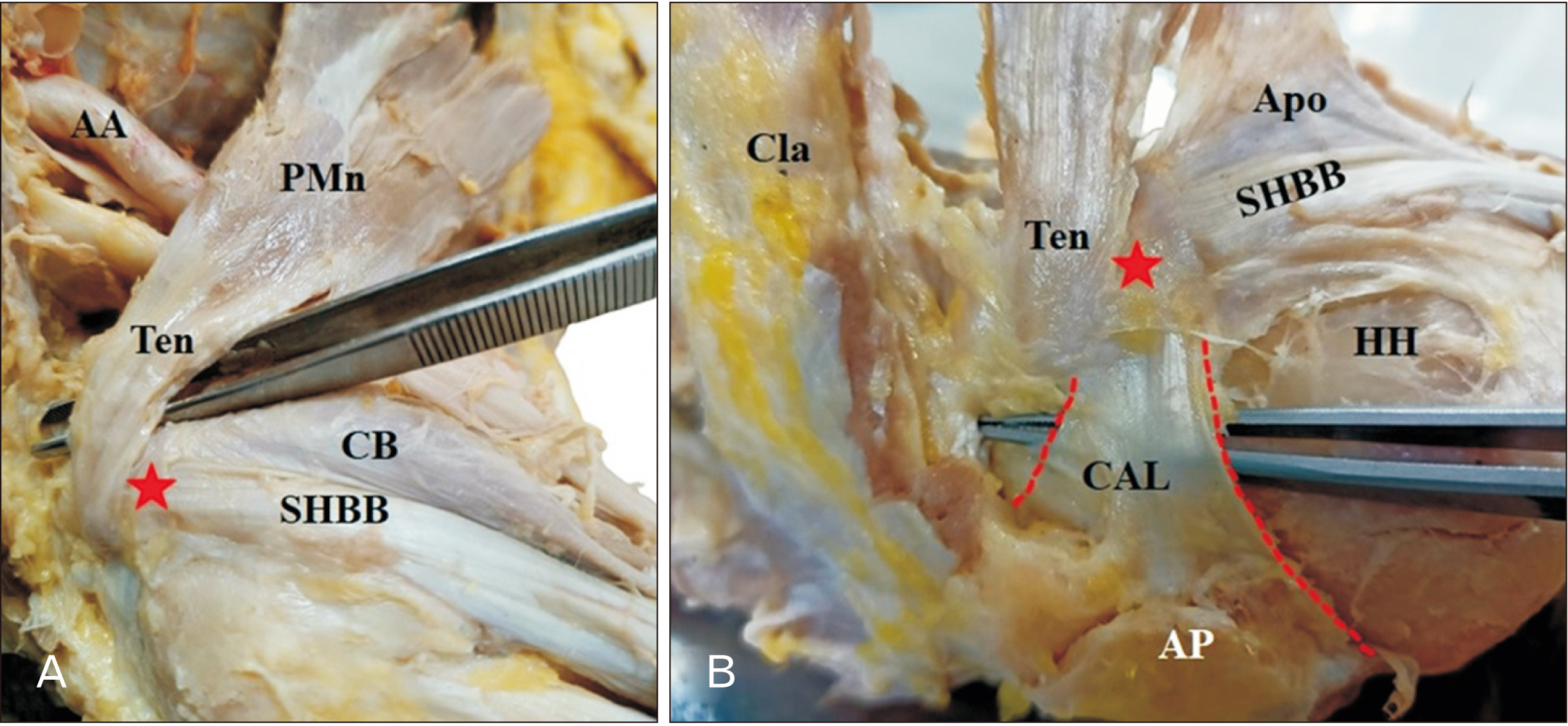
Fig. 4 Showing variant form of insertion of pectoralis minor (PMn) muscle in cadaver-1. (A) Right PMn where tendon (Ten) part is crossing the coracoid process (red asterisk). (B) Left side PMn Ten crossing the coracoid process (red asterisk) and entering deep to coracoacromial ligament (CAL). SHBB, short head of biceps brachii; CB, coracobrachialis; AA, axillary artery; Cla, clavicle; HH, humeral head; Apo, aponeurosis; AP, acromion process.
Reference
-
References
1. 2020. Gray's anatomy 42nd edition [Internet]. Medicon;Edinburg: Available from: https://topmedicon.com/grays-anatomy-42nd-edition-pdf-free-download/. cited 2022 Apr 4.2. 2008. Gray's anatomy the anatomical basis of clinical practice, 40th Ed [Internet]. Yumpu;Edinburg: Available from: https://www.yumpu.com/xx/document/view/55652992/grays-anatomy-the-anatomical-basis-of-clinical-practice-40th-ed-pdftahir99-vrg. cited 2022 Apr 4.3. Chen S, Yang D, Sun Q, Guan Z, Tan P, Zhang K, Mao X. 2022; Effect of pectoralis minor relaxation on the prognosis of rotator cuff injury under arthroscopy. Ann Palliat Med. 11:77–84. DOI: 10.21037/apm-21-3959. PMID: 35144400.4. Lambert AE. 1925; A rare variation in the pectoralis minor muscle. Anat Rec. 31:193–200. DOI: 10.1002/ar.1090310303.5. Schwarz GM, Hirtler L. 2019; Ectopic tendons of the pectoralis minor muscle as cause for shoulder pain and motion inhibition-explaining clinically important variabilities through phylogenesis. PLoS One. 14:e0218715. DOI: 10.1371/journal.pone.0218715. PMID: 31226146. PMCID: PMC6588231. PMID: b1657ad813e84b8c9f66b54c093ecc70.6. Lee SJ, Ha DH, Lee SM. 2010; Unusual variation of the rotator interval: insertional abnormality of the pectoralis minor tendon and absence of the coracohumeral ligament. Skeletal Radiol. 39:1205–9. DOI: 10.1007/s00256-010-0926-0. PMID: 20401480.7. Lim TK, Koh KH, Yoon YC, Park JH, Yoo JC. 2015; Pectoralis minor tendon in the rotator interval: arthroscopic, magnetic resonance imaging findings, and clinical significance. J Shoulder Elbow Surg. 24:848–53. DOI: 10.1016/j.jse.2015.03.003. PMID: 25979554.8. Low SC, Tan SC. 2010; Ectopic insertion of the pectoralis minor muscle with tendinosis as a cause of shoulder pain and clicking. Clin Radiol. 65:254–6. DOI: 10.1016/j.crad.2009.11.004. PMID: 20152284.9. Moinfar AR, Murthi AM. 2007; Anatomy of the pectoralis minor tendon and its use in acromioclavicular joint reconstruction. J Shoulder Elbow Surg. 16:339–46. DOI: 10.1016/j.jse.2006.09.007. PMID: 17408976.10. Shibata T, Izaki T, Miyake S, Doi N, Arashiro Y, Shibata Y, Irie Y, Tachibana K, Yamamoto T. 2019; Predictors of safety margin for coracoid transfer: a cadaveric morphometric analysis. J Orthop Surg Res. 14:174. DOI: 10.1186/s13018-019-1212-z. PMID: 31182130. PMCID: PMC6558900. PMID: 61da6dc7e0f94f03ba1642c619365b23.11. Le Double AF. 1897. Treats variations of the muscular system of man and their significance from the point of view of anthropology, zoology [Internet]. Biodiversity Heritage Library;Paris: Available from: https://www.biodiversitylibrary.org/bibliography/44649. cited 2022 Apr 4.12. Moineau G, Cikes A, Trojani C, Boileau P. 2008; Ectopic insertion of the pectoralis minor: implication in the arthroscopic treatment of shoulder stiffness. Knee Surg Sports Traumatol Arthrosc. 16:869–71. DOI: 10.1007/s00167-008-0535-9. PMID: 18641969.13. Asghar A, Narayan RK, Satyam A, Naaz S. 2021; Prevalence of anomalous or ectopic insertion of pectoralis minor: a systematic review and meta-analysis of 4146 shoulders. Surg Radiol Anat. 43:631–43. DOI: 10.1007/s00276-020-02610-8. PMID: 33165647.14. Krause W. 1880. Handbuch der menschlichen Anatomy [Internet]. Hahn'sche Hofbuchhandlung;Halle: Available from: http://archive.org/details/handbuchdermens09kraugoog. cited 2022 Mar 15.15. Le Double AF. 1897. Prevalence and Le Double classification relative to previous studies. N = number of included shoulders. [Internet]. ResearchGate;Berlin: Available from: https://www.researchgate.net/figure/Prevalence-and-Le-Double-classification-relative-to-previous-studies-N-number-of_tbl1_333937589. cited 2022 Mar 15.16. Tubbs RS, Oakes WJ, Salter EG. 2005; Unusual attachment of the pectoralis minor muscle. Clin Anat. 18:302–4. DOI: 10.1002/ca.20113. PMID: 15832348.17. Turgut HB, Anil A, Peker T, Barut C. 2000; Insertion abnormality of bilateral pectoralis minimus. Surg Radiol Anat. 22:55–7. DOI: 10.1007/s00276-000-0055-x. PMID: 10863749.18. Uzel AP, Bertino R, Caix P, Boileau P. 2008; Bilateral variation of the pectoralis minor muscle discovered during practical dissection. Surg Radiol Anat. 30:679–82. DOI: 10.1007/s00276-008-0382-x. PMID: 18612582.19. Lee KW, Choi YJ, Lee HJ, Gil YC, Kim HJ, Tansatit T, Hu KS. 2018; Classification of unusual insertion of the pectoralis minor muscle. Surg Radiol Anat. 40:1357–61. DOI: 10.1007/s00276-018-2107-0. PMID: 30306210.20. Soni S, Rath G, Suri R, Kumar H. 2008; Anomalous pectoral musculature. Anat Sci Int. 83:310–3. DOI: 10.1111/j.1447-073X.2008.00234.x. PMID: 19159367.21. Musso F, Azeredo R, Tose D, Marchiori JGT. 2004; Pectoralis minor muscle. An unusual insertion. Braz J Morphol Sci. 21:139–40.22. Taylor AE. 1898; Case of clavicular insertion of the pectoralis minor. J Anat Physiol. 32(Pt 2):218. PMID: 17232299. PMCID: PMC1327880.23. Shane Tubbs R, Shoja MM, Loukas M. 2016. Bergman's comprehensive encyclopedia of human anatomic variation. John Wiley & Sons;Hoboken: DOI: 10.1002/9781118430309.24. Turan-Ozdemir S, Cankur NS. 2004; Unusual variation of the inferior attachment of the pectoralis minor muscle. Clin Anat. 17:416–7. DOI: 10.1002/ca.20010. PMID: 15176040.25. Bland-Sutton J. 1897. Ligaments their nature and morphology [Internet]. AbeBooks;München, London: Available from: https://www.abebooks.co.uk/book-search/title/ligaments-their-nature-and-morphology/author/j-bland-sutton/. cited 2022 Mar 28.26. Simovitch RW, Bal GK, Basamania CJ. 2006; Thoracic outlet syndrome in a competitive baseball player secondary to the anomalous insertion of an atrophic pectoralis minor muscle: a case report. Am J Sports Med. 34:1016–9. DOI: 10.1177/0363546505284386. PMID: 16476910.27. Zvijac JE, Zikria B, Botto-van Bemden A. 2009; Isolated tears of pectoralis minor muscle in professional football players: a case series. Am J Orthop (Belle Mead NJ). 38:145–7. PMID: 19377649.28. Dumontier C, Sautet A, Gagey O, Apoil A. 1999; Rotator interval lesions and their relation to coracoid impingement syndrome. J Shoulder Elbow Surg. 8:130–5. DOI: 10.1016/S1058-2746(99)90005-8. PMID: 10226964.29. Sanders RJ, Annest SJ. 2017; Pectoralis minor syndrome: subclavicular brachial plexus compression. Diagnostics (Basel). 7:46. DOI: 10.3390/diagnostics7030046. PMID: 28788065. PMCID: PMC5617946.
- Full Text Links
-
- Actions
-
Cited
- CITED
-
- Close
- Share
-
- Similar articles
-
- Accessory Tendon of Biceps Brachii Originated from Pectoralis Major
- The Third Head of the Biceps Brachii Muscle Originated from the Pectoralis Major Muscle
- Untrapped: bilateral hypoplasia of the trapezius muscle
- Pectoralis Major Muscle as an Alternate for Reconstruction of Shoulder Fourth-Degree Burn
- Rupture of the Pectoralis Major Muscle during Exercise
