Korean Circ J.
2022 Dec;52(12):903-905. 10.4070/kcj.2022.0192.
A Case of a Long-term Survivor of Myocardial Infarction With Extensive Dystrophic Myocardial Calcification
- Affiliations
-
- 1Department of Internal Medicine, Catholic Medical Center, The Catholic University of Korea, Seoul, Korea
- 2Division of Cardiology, Department of Internal Medicine, Daejeon St. Mary’s Hospital, The Catholic University of Korea, Seoul, Korea
- 3Catholic Research Institute for Intractable Cardiovascular Disease (CRID), College of Medicine, The Catholic University of Korea, Seoul, Korea
- KMID: 2536794
- DOI: http://doi.org/10.4070/kcj.2022.0192
Figure
-

Figure 1 Radiographic images. (A) Chest X-ray showed a large, well-demarcated, spherical calcified mass (red arrows) at LV. (B) Chest computed tomography demonstrated an extensive myocardial calcification (red arrow) along with the aneurysm of LV.LV = left ventricle.
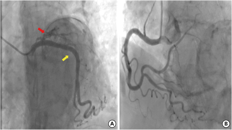
Figure 2 Coronary angiography images. (A) Coronary angiography showed the chronic total occlusion at the ostium of the left anterior descending artery (red arrow). The left circumflex coronary artery (yellow arrow) was normal. (B) Right coronary artery showed no stenosis.
Reference
-
1. Freundlich IM, Lind TA. Calcification of the heart and great vessels. CRC Crit Rev Clin Radiol Nucl Med. 1975; 6:171–216. PMID: 238789.2. Nance JW Jr, Crane GM, Halushka MK, Fishman EK, Zimmerman SL. Myocardial calcifications: pathophysiology, etiologies, differential diagnoses, and imaging findings. J Cardiovasc Comput Tomogr. 2015; 9:58–67. PMID: 25456525.
- Full Text Links
-
- Actions
-
Cited
- CITED
-
- Close
- Share
-
- Similar articles
-
- Dystrophic Endocardial Calcification Associated with Prior Myocardial Infarction
- Long-term outcome in young adults with myocardial infarction
- Invasive Treatment of Acute Myocardial Infarction: What is the Optimal Therapy for Acute Myocardial Infarction?
- Myocardial Infarction in a Patient with Myocardial Bridge and Pheochromocytoma: A case report
- A Case of Q Wave Acute Myocardial Infarction in Patients with Myocardial Bridging Caused by Fibrous Band
