Reconstruction of a high-energy penetrating injury from the abdomen to the sacral area using a latissimus dorsi free flap with monofilament polypropylene mesh and pedicled flap rotation: a case report
- Affiliations
-
- 1Department of Plastic and Reconstructive Surgery, Ajou University School of Medicine, Suwon, Korea
- KMID: 2536225
- DOI: http://doi.org/10.12790/ahm.22.0063
Abstract
- A 50-year-old man was transferred to a level I trauma center for penetrating injury. Industrial metal had penetrated his trunk, and he was injured in internal organs. The injured internal organs were treated by the trauma surgery team. The peritoneum was reconstructed with artificial dermal matrix graft. The wound was managed with negative-pressure wound therapy, and several debridement procedures were performed. The full-thickness abdominal defect was covered with monofilament polypropylene mesh(Parietene mesh, 30×30 cm). A latissimus dorsi flap was elevated with a musculocutaneous flap measured 50×30 cm, and 6-cm thoracodorsal artery pedicle. Microvascular anastomosis was performed using the thoracodorsal and left femoral arteries. Two weeks later, we performed local flap rotation based on gluteal artery perforator in the sacral area. Polypropylene mesh was successfully inserted without complications. Combining a latissimus dorsi free flap on a polypropylene mesh can be an effective method for reconstructing large penetrating wounds on the trunk.
Keyword
Figure
-

Fig. 1. Initial status of the patient upon arrival at the trauma bay after a penetrating injury by a compressing machine.
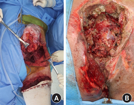
Fig. 2. Wounds after 2 weeks of multiple wound perfusion and debridement. (A) Abdominal side. (B) Sacral side.
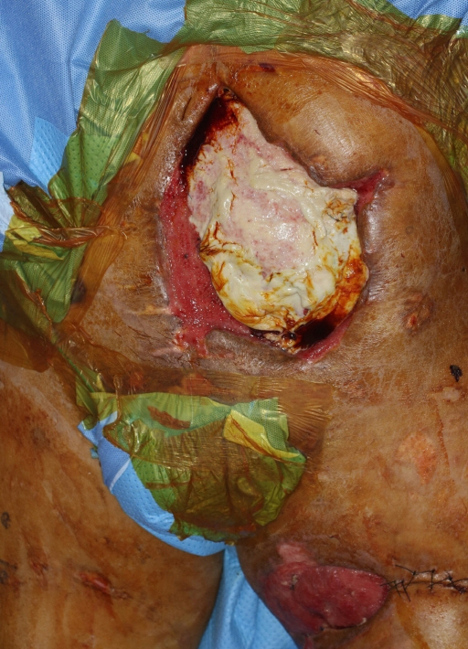
Fig. 3. Abdominal wound applied with acellular dermal matrix for maintenance of the intestinal injury recovery and temporary intestinal coverage.
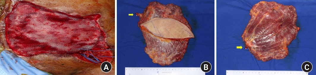
Fig. 4. Abdominal wound repaired with mesh and latissimus dorsi musculocutaneous flap with monofilament polypropylene mesh coverage. (A) Wounds with monofilament polypropylene mesh applied. (B, C) Elevated latissimus dorsi musculocutaneous flap. Yellow arrows indicate the thoracodorsal artery.
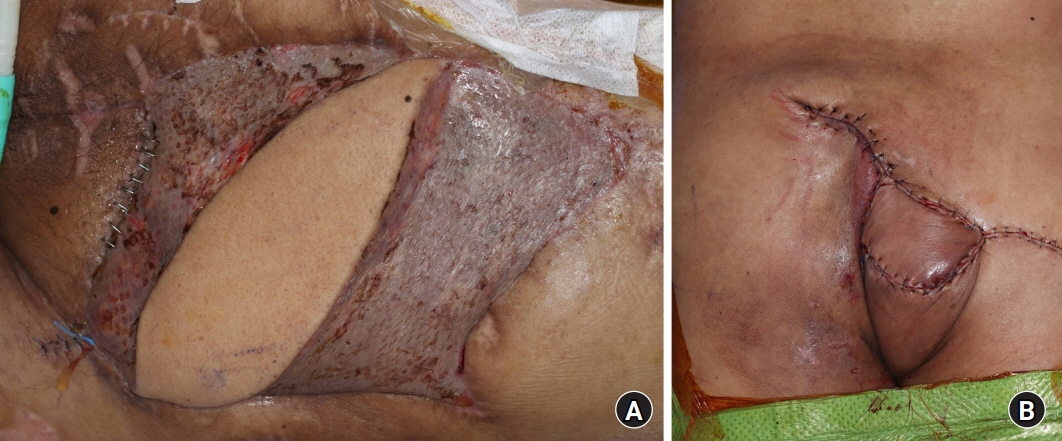
Fig. 5. Image at 2 months postoperatively. (A) Abdominal side. (B) Sacral side.
Cited by 1 articles
-
Reconstruction of an abdominal wall defect using a latissimus dorsi musculocutaneous free flap after high-intensity focused ultrasound: a case report
Dae Kwan Kim, Youn Hwan Kim
Arch Hand Microsurg. 2023;28(2):110-113. doi: 10.12790/ahm.23.0008.
Reference
-
References
1. Gad MA, Saber A, Farrag S, Shams ME, Ellabban GM. Incidence, patterns, and factors predicting mortality of abdominal injuries in trauma patients. N Am J Med Sci. 2012; 4:129–34.
Article2. Rohrich RJ, Lowe JB, Hackney FL, Bowman JL, Hobar PC. An algorithm for abdominal wall reconstruction. Plast Reconstr Surg. 2000; 105:202–16.
Article3. Wong CH, Lin CH, Fu B, Fang JF. Reconstruction of complex abdominal wall defects with free flaps: indica-tions and clinical outcome. Plast Reconstr Surg. 2009; 124:500–9.
Article4. Arnaud E, Couturaud B, Revol M, Servant JM, Banzet P. [Surgical repair of abdominal wall]. Ann Chir Plast Esthet. 1999; 44:357–72. French.5. Servant JM, Arnault E, Revol M, Danino A. Reconstruction of large thoracoabdominal defects using two-stage free tissue transfers and prosthetic materials. J Plast Reconstr Aesthet Surg. 2006; 59:360–5.
Article6. Carlson GL, Patrick H, Amin AI, et al. Management of the open abdomen: a national study of clinical outcome and safety of negative pressure wound therapy. Ann Surg. 2013; 257:1154–9.7. Köckerling F, Alam NN, Antoniou SA, et al. What is the evidence for the use of biologic or biosynthetic meshes in abdominal wall reconstruction? Hernia. 2018; 22:249–69.
Article
- Full Text Links
-
- Actions
-
Cited
- CITED
-
- Close
- Share
-
- Similar articles
-
- Six Cases of Reconstruction with Latissimus Dorsi Pedicled Flap for Head and Neck Defects in the Era of Free Flap Reconstruction
- Axillary Reconstruction Using a Pedicled Thoracodorsal Artery Perforator Flap Including Latissimus Dorsi Muscle Strip
- A Combined Scapular Flap and Latissimus Dorsi Flap
- Reconstruction of an abdominal wall defect using a latissimus dorsi musculocutaneous free flap after high-intensity focused ultrasound: a case report
- Scalp Reconstruction and Cranioplasty using the Latissimus Dorsi Musculocutaneous Flap in a Patient with Recurrent Wound Dehiscence Accompanied by MRSA Infection
