Clin Endosc.
2022 Nov;55(6):824-825. 10.5946/ce.2022.151.
All that elongates is not a polyp
- Affiliations
-
- 1Department of Endoscopy, Graduate School of Medicine, University of the Ryukyus, Okinawa, Japan
- KMID: 2536085
- DOI: http://doi.org/10.5946/ce.2022.151
Figure
-
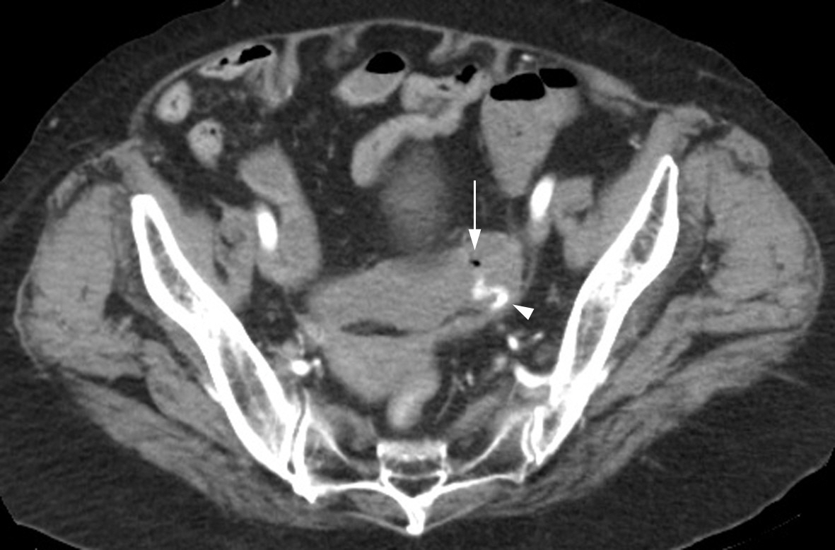
Fig. 1. The abdominal computed tomography image showing an air bubble (arrow) and an extravasation of contrast (arrowhead) in the sigmoid colon.

Fig. 2. Colonoscopy revealing a polypoid lesion with an exposed vessel.
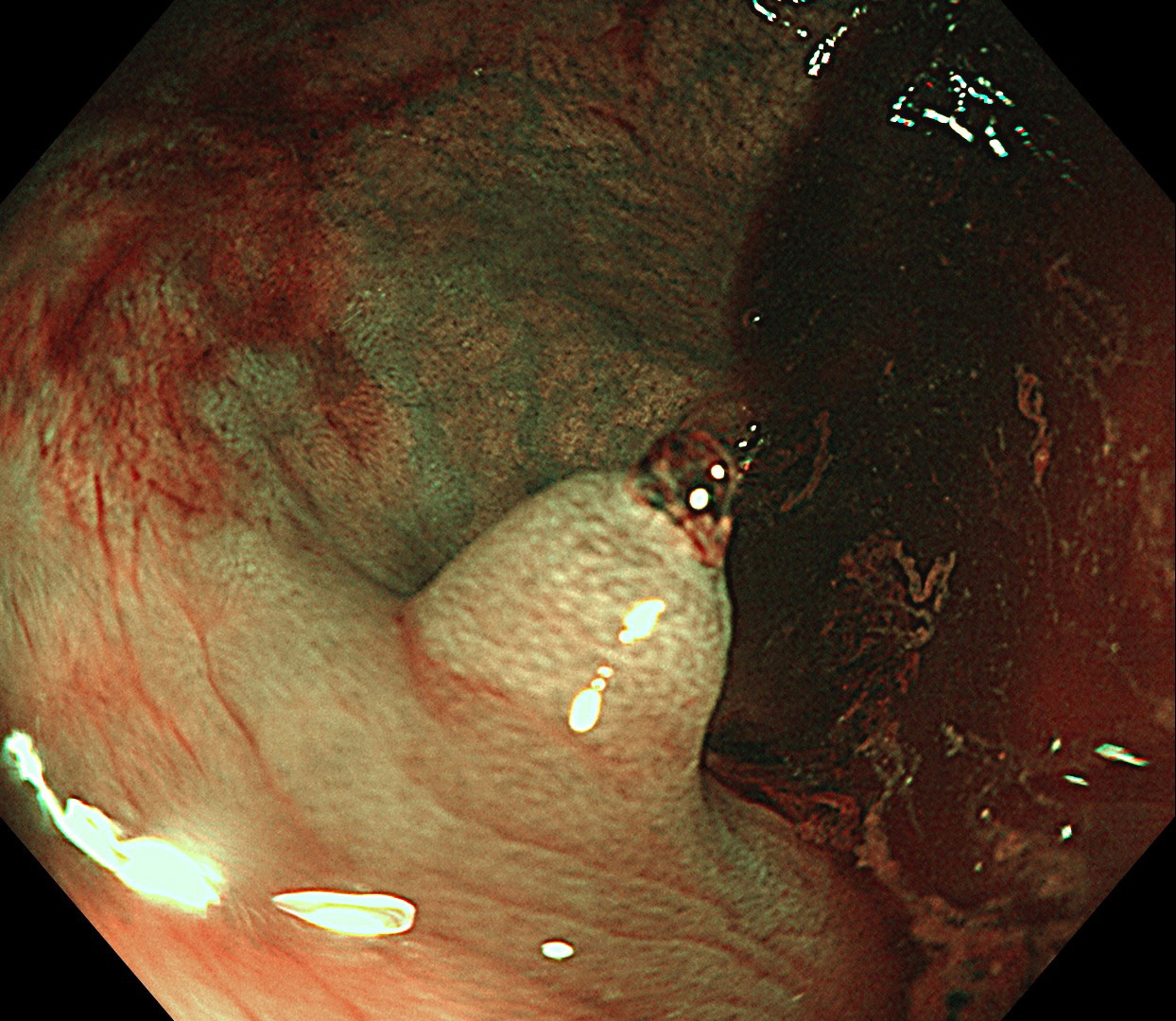
Fig. 3. Enhancement of the normal mucosal pattern and the concentric rings surrounding the polypoid lesion in narrow-band imaging.
Reference
-
1. Triadafilopoulos G. Inverted colonic diverticulum. N Engl J Med. 1999; 341:1508.
Article2. Share MD, Avila A, Dry SM, et al. Aurora rings: a novel endoscopic finding to distinguish inverted colonic diverticula from colon polyps. Gastrointest Endosc. 2013; 77:308–312.
Article3. Adioui T, Seddik H. Inverted colonic diverticulum. Ann Gastroenterol. 2014; 27:411.
- Full Text Links
-
- Actions
-
Cited
- CITED
-
- Close
- Share
-
- Similar articles
-
- Choanal Polyps Originating from the Ethmoid Sinus: Ethmochoanal Polyps?
- Solitary Juvenile Polyp Manifesting as Spontaneous Resection with Rectal Bleeding in a Child
- A Case of Grannlomatous Polyp in Larynx Following Endotracheal Intubation
- Local Production of IgE in Nasal Polyp
- Lymphoid Polyp in the Rectum
