Sinus mucosal healing pattern according to pterygomaxillary disjunction type after Le Fort I osteotomy
- Affiliations
-
- 1Department of Oral and Maxillofacial Surgery, Kyung Hee University Dental Hospital, Seoul, Korea
- 2Department of Oral and Maxillofacial Surgery, School of Dentistry, Kyung Hee University, Seoul, Korea
- KMID: 2534809
- DOI: http://doi.org/10.5125/jkaoms.2022.48.5.292
Abstract
Objectives
During Le Fort I osteotomy, the separation of the pterygomaxillary junction (PMJ) is a difficult procedure for most surgeons because it is invisible. In this process, damage to the posterior structures constituting the sinus or those adjacent to it, including the maxillary sinus posterior wall and pterygoid plate, may occur. We would like to investigate the effects of this on the inside of the maxillary sinus after surgery and whether there are complications.
Materials and Methods
One-hundred patients who underwent Le Fort I osteotomy from 2013 to 2020 using cone-beam computed tomography images were classified into two groups (clean-cut type and fractured type) according to the PMJ cutting pattern. In addition, the mucosal thickness in the maxillary sinus was divided into preoperative, postoperative three months, one year, and the change over the course of surgery was evaluated retrospectively.
Results
Of the total 100 cases, the clean-cut type numbered 28 cases and the fractured type totaled 72 cases. Among the fracture types, part of the sinus wall and the pterygoid plate were broken in 69 cases, and the maxillary sinus posterior wall was detached in three cases. There was no statistically significant difference in sinus mucosal thickening between the clean-cut type and fractured type of the PMJ, three months and one year after surgery between the two groups. However, there was a significant difference in sinus mucosal thickness at postoperative one year in the case where a partial detachment of the maxillary sinus posterior wall occurred compared to not.
Conclusion
Even if there is some damage to the structures behind the PMJ, it may not be reasonable to spend some time on the PMJ separation process considering the overall postoperative complications, if there is no significant difference inside the sinus, or increased probability of postoperative complications.
Figure
-
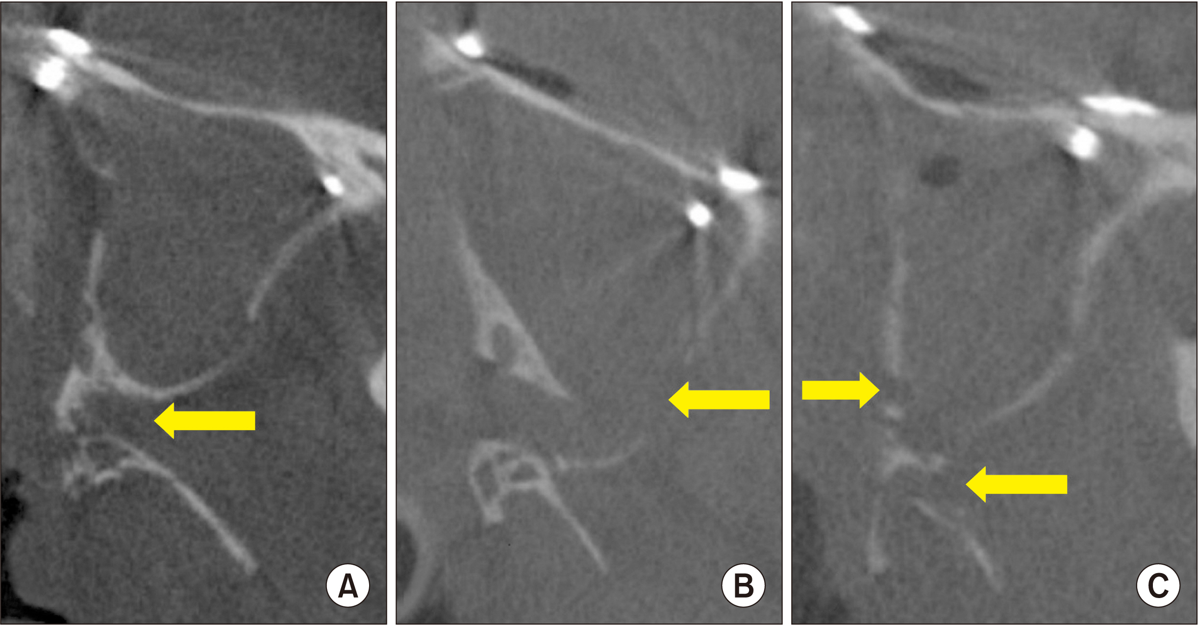
Fig. 1 Pterygomaxillary junction (PMJ) cutting type in postoperative cone-beam computed tomography. A. Type I: the cutting line within the PMJ (arrow). B. Type II-1: the posterior wall of maxillary sinus detached (arrow). C. Type II-2: a part of posterior wall of the maxillary sinus (left arrow) and pterygoid (right arrow) fracture occurred after PMJ separation.
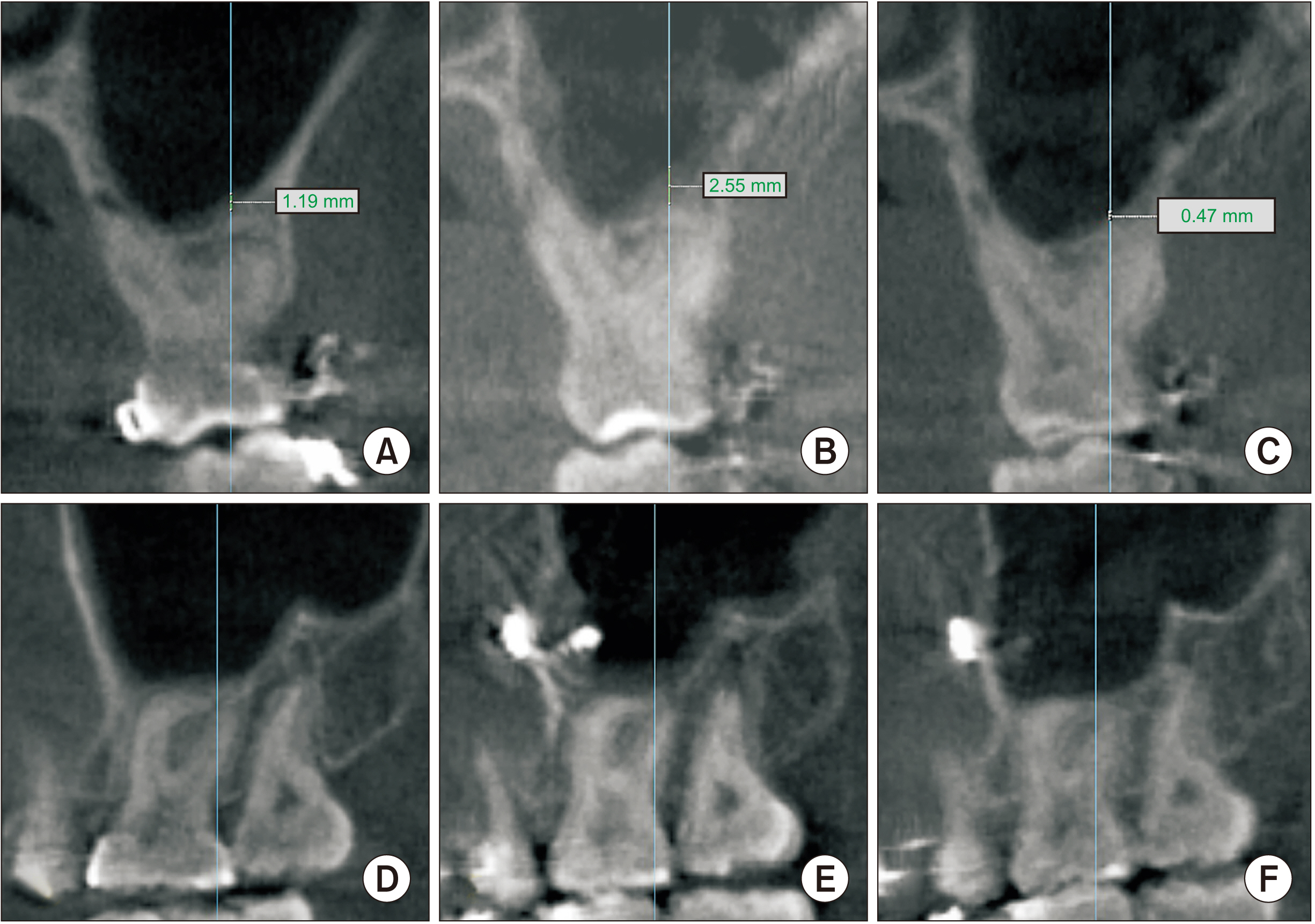
Fig. 2 Measurement of maxillary sinus mucosal thickness over time. A, D. Preoperative. B, E. Postoperative 3 months. C, F. Postoperative 1 year.

Fig. 3 Sinus mucosal thickness by two groups (Type I and Type II).

Fig. 4 Type II was classified into subgroups (Type II-1 and Type II-2) and mucosal thickness was compared. There was a significant difference in the mean mucosal thickness of Type II-1 at 1 year after surgery.
Reference
-
References
1. Iwamoto M, Watanabe M, Yamamoto M, Narita M, Kamio T, Takaki T, et al. 2019; Prognostic factors for maxillary sinus mucosal thickening following Le Fort I osteotomy: a retrospective analysis. Maxillofac Plast Reconstr Surg. 41:12. https://doi.org/10.1186/s40902-019-0195-3. DOI: 10.1186/s40902-019-0195-3. PMID: 30915318. PMCID: PMC6409288.
Article2. Joh Y, Park HS, Yang HJ, Hwang SJ. 2019; Horizontal change of philtrum after orthognathic surgery in patients with facial asymmetry. Maxillofac Plast Reconstr Surg. 41:48. https://doi.org/10.1186/s40902-019-0232-2. DOI: 10.1186/s40902-019-0232-2. PMID: 31799219. PMCID: PMC6851271.
Article3. Chin YP, Leno MB, Dumrongwongsiri S, Chung KH, Lin HH, Lo LJ. 2017; The pterygomaxillary junction: an imaging study for surgical information of LeFort I osteotomy. Sci Rep. 7:9953. https://doi.org/10.1038/s41598-017-10592-8. DOI: 10.1038/s41598-017-10592-8. PMID: 28855714. PMCID: PMC5577125.
Article4. Dadwal H, Shanmugasundaram S, Krishnakumar Raja VB. 2015; Preoperative and postoperative CT scan assessment of pterygomaxillary junction in patients undergoing Le Fort I osteotomy: comparison of pterygomaxillary dysjunction technique and trimble technique-a pilot study. J Maxillofac Oral Surg. 14:713–9. https://doi.org/10.1007/s12663-014-0720-y. DOI: 10.1007/s12663-014-0720-y. PMID: 26225067. PMCID: PMC4511914.
Article5. Bin LR, Filho LI, Yamashita AL, de Souza Pinto GN, Mendes RA, Ramos AL, et al. 2020; How does bimaxillary orthognathic surgery change dimensions of maxillary sinuses and pharyngeal airway space? Angle Orthod. 90:715–22. https://doi.org/10.2319/120919-782.1. DOI: 10.2319/120919-782.1. PMID: 33378484. PMCID: PMC8032267.
Article6. Panou E, Motro M, Ateş M, Acar A, Erverdi N. 2013; Dimensional changes of maxillary sinuses and pharyngeal airway in class III patients undergoing bimaxillary orthognathic surgery. Angle Orthod. 83:824–31. https://doi.org/10.2319/100212-777.1. DOI: 10.2319/100212-777.1. PMID: 23438197. PMCID: PMC8744528.
Article7. Shanbhag S, Karnik P, Shirke P, Shanbhag V. 2013; Association between periapical lesions and maxillary sinus mucosal thickening: a retrospective cone-beam computed tomographic study. J Endod. 39:853–7. https://doi.org/10.1016/j.joen.2013.04.010. DOI: 10.1016/j.joen.2013.04.010. PMID: 23791251.
Article
- Full Text Links
-
- Actions
-
Cited
- CITED
-
- Close
- Share
-
- Similar articles
-
- Le Fort I osteotomy as treatment for traumatic class III malocclusion caused by Le Fort III fracture: A case report
- Prognostic factors for maxillary sinus mucosal thickening following Le Fort I osteotomy: a retrospective analysis
- A case report of surgical correction of mandibular prognathism with midfacial deficiency using Le Fort III osteotomy
- Repetitive Postoperative Infection after Le Fort I Osteotomy in a Patient with a History of Non-allergic Rhinitis
- Management of Le Fort I fracture
