Heterotopic mesenteric ossification: a report of two cases
- Affiliations
-
- 11Arkadi M. Rywlin M.D. Department of Pathology and Laboratory Medicine, Mount Sinai Medical Center, Miami Beach, FL, USA
- 2Herbert Wertheim College of Medicine, Florida International University, Miami, FL, USA
- 3Department of General Surgery, Mount Sinai Medical Center, Miami Beach, FL, USA
- KMID: 2533706
- DOI: http://doi.org/10.4132/jptm.2022.07.23
Abstract
- Heterotopic mesenteric ossification (HMO) is abnormal bone formation in tissues which usually do not undergo ossification. There are approximately 75 cases reported worldwide. We present two cases of HMO. The first case is that of a 39-year-old man who presented with abdominal pain and a computerized tomography scan of the abdomen and pelvis revealed an apple core lesion resulting in small bowel obstruction. The second case is that of a 36-year-old woman who presented 2 months after undergoing robotic gastric sleeve resection complaining of weakness and emesis. An esophagogram revealed kinking at the distal esophagus. Surgical resection was performed in both, yielding the diagnosis of HMO. There are various theories as to the pathophysiology of HMO, but no clearly defined mechanism has been established. Management should be conservative whenever possible to prevent further ossification with subsequent surgical intervention.
Keyword
Figure
-

Fig. 1 Computerized tomography scan of the abdomen and pelvis with intravenous contrast. Results showed segmental concentric thickening of the jejunum in the right upper quadrant of the abdomen (with apple core configuration, white arrowhead) resulting in small bowel obstruction and stranding of the surrounding mesentery.

Fig. 2 Gross examination of the resected segment of small intestine. (A) An area of luminal constriction is seen. (B) Induration in the surrounding mesenteric fat showing chalky white cut surface.

Fig. 3 Microscopic examination showing heterotopic ossification. Histopathologic examination of the resected segment of small intestine demonstrated focal mucosal erosion (A) with acute and chronic inflammation in the surrounding peri-intestinal soft and adipose tissue, granulation tissue, fat necrosis (B), fibrosis, hemosiderin deposition, focal foreign body giant cell reaction (C), and new bone formation (D).

Fig. 4 Exploratory laparoscopy showing significant abdominal scar tissue and previous sleeve gastrectomy.
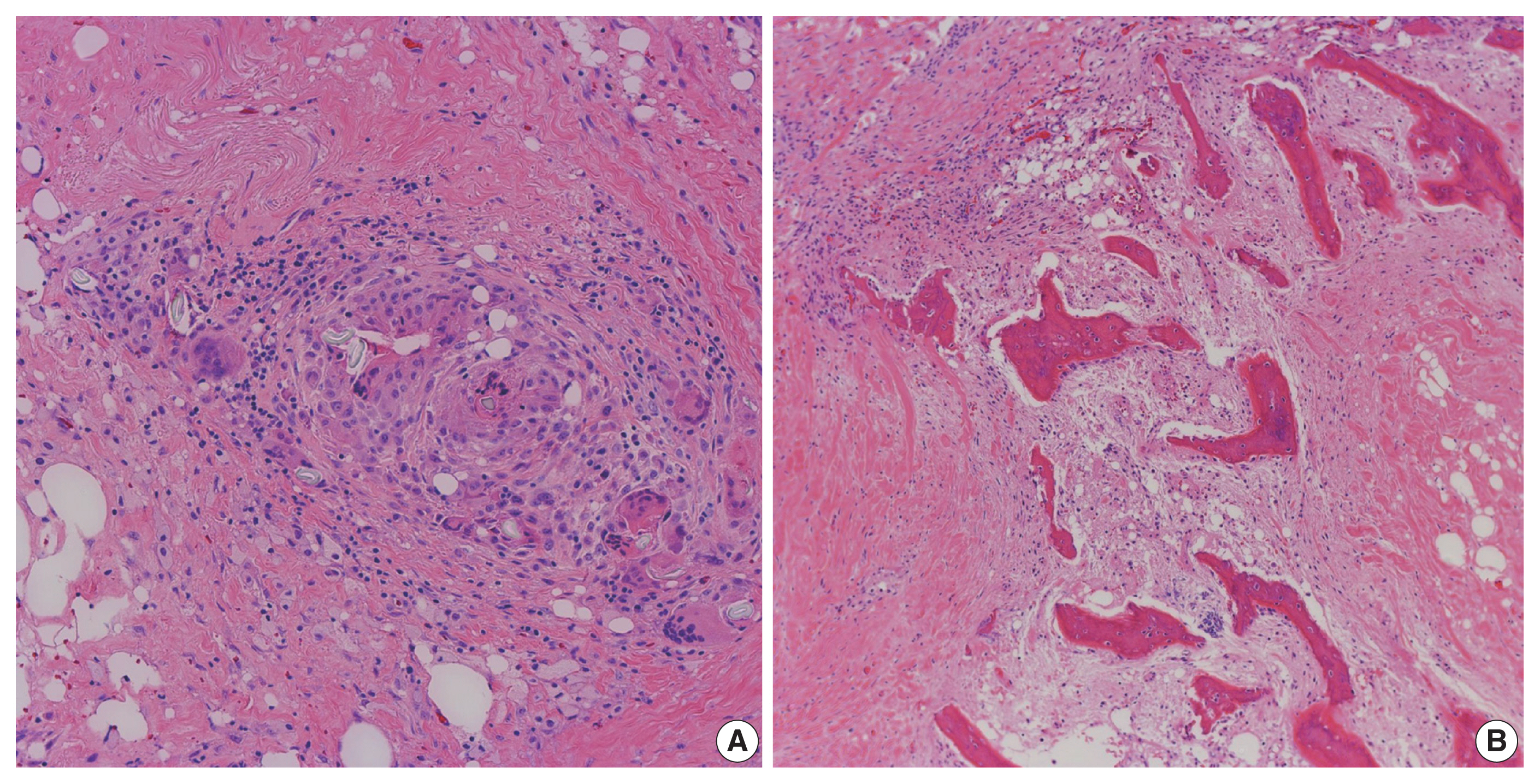
Fig. 5 Microscopic examination showing heterotopic ossification. Histopathologic examination of the resected segments of stomach and small intestine demonstrated chronic inflammation, suture granulomas, foreign body giant cell reaction (A), and new bone formation (B).

Fig. 6 Schematic diagram illustrating the proposed hypotheses for the formation of heterotopic mesenteric ossification. ECM, extracellular matrix; ALP, alkaline phosphatase; BMP, bone morphogenetic protein; TGF-β, transforming growth factor-β; PTH, parathyroid hormone; IGH-1, insulin-like growth factor 1; FGF, fibroblast growth factor.
Reference
-
References
1. Balboni TA, Gobezie R, Mamon HJ. Heterotopic ossification: pathophysiology, clinical features, and the role of radiotherapy for prophylaxis. Int J Radiat Oncol Biol Phys. 2006; 65:1289–99.
Article2. Naraghi FF, DeCoster TA, Moneim MS, Miller RA, Rivero D. Heterotopic ossification. Orthopedics. 1996; 19:145–51.
Article3. Lemeshev Y, Lahr CJ, Denton J, Kent SP, Diethelm AG. Heterotopic bone formation associated with intestinal obstruction and small bowel resection. Ala J Med Sci. 1983; 20:314–7.4. Hansen O, Sim F, Marton PF, Gruner OP. Heterotopic ossification of the intestinal mesentery: report of a case following intraabdominal surgery. Pathol Res Pract. 1983; 176:125–30.5. Wilson JD, Montague CJ, Salcuni P, Bordi C, Rosai J. Heterotopic mesenteric ossification (‘intraabdominal myositis ossificans’): report of five cases. Am J Surg Pathol. 1999; 23:1464–70.6. Ferreira C, Gomes C, Melo A, et al. Heterotopic mesenteric and abdominal wall ossification: two case reports in one institution. Int J Surg Case Rep. 2017; 37:22–5.7. Althaqafi RM, Assiri SA, Aloufi RA, Althobaiti F, Althobaiti B, Al Adwani M. A case report and literature review of heterotopic mesenteric ossification. Int J Surg Case Rep. 2021; 82:105905.
Article8. Sarraf K, Newman O, Mirkazemi M, Serena T. Laparoscopic enterolysis of congenital band precipitating pathogenic heterotopic mesenteric ossification requiring hemicolectomy: a case report. Am J Case Rep. 2022; 23:e934910.
Article9. McCarthy EF, Sundaram M. Heterotopic ossification: a review. Skeletal Radiol. 2005; 34:609–19.
Article10. Binesh F, Akhavan A, Navabii H, Ostadi M. Heterotopic mesenteric ossification: report of a case and review of the literature. BMJ Case Rep. 2012; 2012:bcr0220125793.
Article11. Gaffey MJ, Winston DC. Heterotopic ossification of soft tissue: a review with emphasis on ossification within abdominal surgical scars. Anat Pathol. 1998; 3:195–208.12. Xu Y, Huang M, He W, et al. Heterotopic ossification: clinical features, basic researches, and mechanical stimulations. Front Cell Dev Biol. 2022; 10:770931.
Article13. Myers MA, Minton JP. Heterotopic ossification within the small-bowel mesentery. Arch Surg. 1989; 124:982–3.
Article14. Hicks CW, Velopulos CG, Sacks JM. Mesenteric calcification following abdominal stab wound. Int J Surg Case Rep. 2014; 5:476–9.
Article15. Leblanc E, Trensz F, Haroun S, et al. BMP-9-induced muscle heterotopic ossification requires changes to the skeletal muscle microenvironment. J Bone Miner Res. 2011; 26:1166–77.
Article16. Guo X, Wang XF. Signaling cross-talk between TGF-beta/BMP and other pathways. Cell Res. 2009; 19:71–88.
Article17. Koolen PG, Schreinemacher MH, Peppelenbosch AG. Heterotopic ossifications in midline abdominal scars: a critical review of the literature. Eur J Vasc Endovasc Surg. 2010; 40:155–9.
Article18. Kan L, Lounev VY, Pignolo RJ, et al. Substance P signaling mediates BMP-dependent heterotopic ossification. J Cell Biochem. 2011; 112:2759–72.
Article
- Full Text Links
-
- Actions
-
Cited
- CITED
-
- Close
- Share
-
- Similar articles
-
- Heterotopic Mesenteric Ossification: A Case Report
- Heterotopic Ossification of the Elbow after Medial Epicondylectomy
- Osteomyelitis in Heterotopic Ossification after Trochanteric Pressure Sore Reconstruction: A Case Report
- Heterotopic Ossification of a Partially Ruptured Achilles Tendon (A Case Report)
- Heterotopic Mesenteric Ossification Following Intraabdominal Surgery
