A case report of an accessory flexor digitorum profundus indicis contributing the first lumbrical
- Affiliations
-
- 1Department of Anatomy, Lake Erie College of Osteopathic Medicine, Erie, PA, USA
- KMID: 2533368
- DOI: http://doi.org/10.5115/acb.22.014
Abstract
- Variations of the musculature within the upper extremity have been widely documented, with clinical implications ranging from motor dysfunction to compressive neuropathies. Herein, we described an aberrant muscle that originated from the anterior proximal forearm, formed a tendon that coursed through the carpal tunnel, and converged with the flexor digitorum profundus muscle to contribute to the first lumbrical. Additionally, the second lumbrical consisted of two heads, originating from the index and middle finger tendons of flexor digitorum profundus. Documentation and recognition of such anatomic variants is important, as this anatomic pattern may contribute to anterior interosseous or median nerve compression, incoordination, complications during surgery, and other clinical manifestations.
Keyword
Figure
-
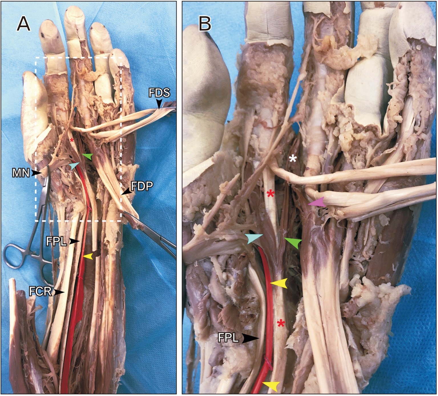
Fig. 1 Shown in (A) is the distal forearm and hand from the dissected specimen. The median nerve (MN), flexor digitorum superficialis (FDS), and flexor digitorum profundus (FDP) are displaced. The aberrant muscle is indicated by a yellow arrowhead and is colored red. It courses between flexor pollicis longus (FPL) and FDP. The slip of FDP to the index finger is indicated by a red asterisk. The aberrant muscle contributed to the first lumbrical (cyan arrowhead). Shown in (B) is an enlarged view of the region indicated by the white box in (A). Again, the muscle is colored red and indicated by yellow arrowheads. The image in (B) clearly indicated the proximal attachments of the lumbricals. The union of the two slips of the second lumbrical is indicated by the white asterisk. FCR, flexor carpi radialis.

Fig. 2 Shown in (A) is the distal forearm and hand from the dissected specimen. The region indicated by the white dashed lines is shown enlarged in (B). In (A), the accessory muscle (yellow arrowheads; colored red) is seen to have proximal attachment to the interosseous membrane (green arrowhead) and distally from the anterior surface of the radius (cyan arrowheads). The support of the accessory muscle is shown in (B). The proximal muscle belly is colored red and indicated by a yellow arrowhead. The muscle received innervation from the anterior interosseous nerve (AIN) and arterial supply from the anterior interosseous artery (red arrowhead). The ulnar artery is indicated by the magenta arrowheads. MN, median nerve; FPL, flexor pollicis longus.
Cited by 1 articles
-
Degenerated lumbricals in the feet of adult human cadavers: case series
Hare Krishna, Rati Tandon, Tony George Jacob
Anat Cell Biol. 2023;56(2):288-292. doi: 10.5115/acb.22.225.
Reference
-
References
1. al-Qattan MM. 1996; Gantzer's muscle. An anatomical study of the accessory head of the flexor pollicis longus muscle. J Hand Surg Br. 21:269–70. DOI: 10.1016/S0266-7681(96)80114-8. PMID: 8732417.2. Hemmady MV, Subramanya AV, Mehta IM. 1993; Occasional head of flexor pollicis longus muscle: a study of its morphology and clinical significance. J Postgrad Med. 39:14–6. PMID: 8295137.3. Linburg RM, Comstock BE. 1979; Anomalous tendon slips from the flexor pollicis longus to the flexor digitorum profundus. J Hand Surg Am. 4:79–83. DOI: 10.1016/S0363-5023(79)80110-0. PMID: 759509.
Article4. Basu SS, Hazary S. 1960; Variations of the lumbrical muscles of the hand. Anat Rec. 136:501–4. DOI: 10.1002/ar.1091360409. PMID: 13797470.
Article5. Mehta HJ, Gardner WU. 1961; A study of lumbrical muscles in the human hand. Am J Anat. 109:227–38. DOI: 10.1002/aja.1001090302. PMID: 14472150.
Article6. Zhang G, Fenderson BA. 2015; Bilateral accessory flexor muscle of the forearm giving rise to a variant head of the first lumbrical. Int J Anat Var. 8:4–6.7. Butler B Jr, Bigley EC Jr. 1971; Aberrant index (first) lumbrical tendinous origin associated with carpal-tunnel syndrome. A case report. J Bone Joint Surg Am. 53:160–2. DOI: 10.2106/00004623-197153010-00018. PMID: 5540152.8. Degreef I, De Smet L. 2004; Anterior interosseous nerve paralysis due to Gantzer's muscle. Acta Orthop Belg. 70:482–4. PMID: 15587039.9. Eriksen J. 1973; A case of carpal tunnel syndrome on the basis of an abnormally long lumbrical muscle. Acta Orthop Scand. 44:275–7. DOI: 10.3109/17453677308988692. PMID: 4768793.
Article10. Javed S, Woodruff M. 2014; Carpal tunnel syndrome secondary to an accessory flexor digitorum superficialis muscle belly: case report and review of the literature. Hand (N Y). 9:554–5. DOI: 10.1007/s11552-014-9622-1. PMID: 25414622. PMCID: PMC4235923.
Article11. Sbai M, Arab R, Essid L, Gallas A, Khelil K, Boussen M, Maalla R. 2019; Carpal tunnel syndrome caused by anatomic anomalies muscles: a three cases report. Asian J Res Surg. 2:1–6.12. Jones M, Abrahams PH, Sañudo JR, Campillo M. 1997; Incidence and morphology of accessory heads of flexor pollicis longus and flexor digitorum profundus (Gantzer's muscles). J Anat. 191(Pt 3):451–5. DOI: 10.1046/j.1469-7580.1997.19130451.x. PMID: 9419002. PMCID: PMC1467702.
Article13. Buford WL Jr, Koh S, Andersen CR, Viegas SF. 2005; Analysis of intrinsic-extrinsic muscle function through interactive 3-dimensional kinematic simulation and cadaver studies. J Hand Surg Am. 30:1267–75. DOI: 10.1016/j.jhsa.2005.06.019. PMID: 16344187.
Article14. Winckler G, Foroglou C. 1965; Comparative study on the neuromuscular spindles of the lumbrical muscles in certain mammals and in man. Arch Anat Histol Embryol. 48:1–17. French.15. Marzke MW. 1997; Precision grips, hand morphology, and tools. Am J Phys Anthropol. 102:91–110. DOI: 10.1002/(SICI)1096-8644(199701)102:1<91::AID-AJPA8>3.0.CO;2-G. PMID: 9034041.
Article
- Full Text Links
-
- Actions
-
Cited
- CITED
-
- Close
- Share
-
- Similar articles
-
- An accessory muscle of flexor digitorum profundus with bipennate first lumbrical: a unique variation of clinical significance
- Avulsion Injury of the Flexor Digitorum Profundus Tendon: A Case Report
- Double Gantzer's Muscles by Four Muscle Bellies and Its Clinical Significance: A Case Report
- The prevalence and distribution of the variants of Gantzer’s muscle: a meta-analysis of cadaveric studies
- Congenital Absence of the Flexor Digitorum Profundus: A Case Report
