Korean J Gastroenterol.
2022 Sep;80(3):135-141. 10.4166/kjg.2022.042.
Prediction of Helicobacter pylori Infection by Endoscopic Severity of Erythematous/exudative Gastritis in Asymptomatic Adults
- Affiliations
-
- 1Department of Internal Medicine, Uijeongbu Eulji Medical Center, Eulji University School of Medicine, Uijeongbu, Korea.
- 2Department of Internal Medicine, Hansol Hospital, Seoul, Korea.
- 3Department of Internal Medicine, Hanyang University Guri Hospital, Hanyang University College of Medicine, Guri, Korea.
- KMID: 2533316
- DOI: http://doi.org/10.4166/kjg.2022.042
Abstract
- Background/Aims
Helicobacter pylori (H. pylori) infection highly correlates with erythematous/exudative gastritis, which is one of the endoscopic findings of the Sydney classification system. The present study aimed to evaluate the association between endoscopic severity of erythematous/exudative gastritis and H. pylori infection.
Methods
We prospectively enrolled asymptomatic adults who were diagnosed with erythematous/exudative gastritis during screening esophagogastroduodenoscopy. A rapid urease test was performed in all participants to diagnose H. pylori infection. The severity of erythematous/exudative gastritis was determined based on the Sydney classification system. Two investigators independently evaluated the endoscopic findings. The primary endpoint was H. pylori infection rate according to the severity of erythematous/exudative gastritis (mild vs. moderate-to-severe).
Results
A total of 177 patients with erythematous/exudative gastritis were included. The rate of H. pyloriinfection was 86.4% in all patients. Of 177 included patients, 78 were at mild degree, 48 were at moderate degree, and 51 were at severe degree. The inter-observer variation was 4.6% and kappa value was 0.593. H. pylori infection rate was similar between patients with mild erythematous/exudative gastritis and those with moderate-to-severe erythematous/exudative gastritis (91.0% vs. 82.8%, p=0.115). Even after adjusting potential confounding variables, the severity of erythematous/exudative gastritis was not associated with H. pylori infection rate.
Conclusions
H. pylori infection is commonly observed in patients with erythematous/exudative gastritis. However, the severity of erythematous/exudative gastritis is not associated with H. pylori infection rate.
Figure
-
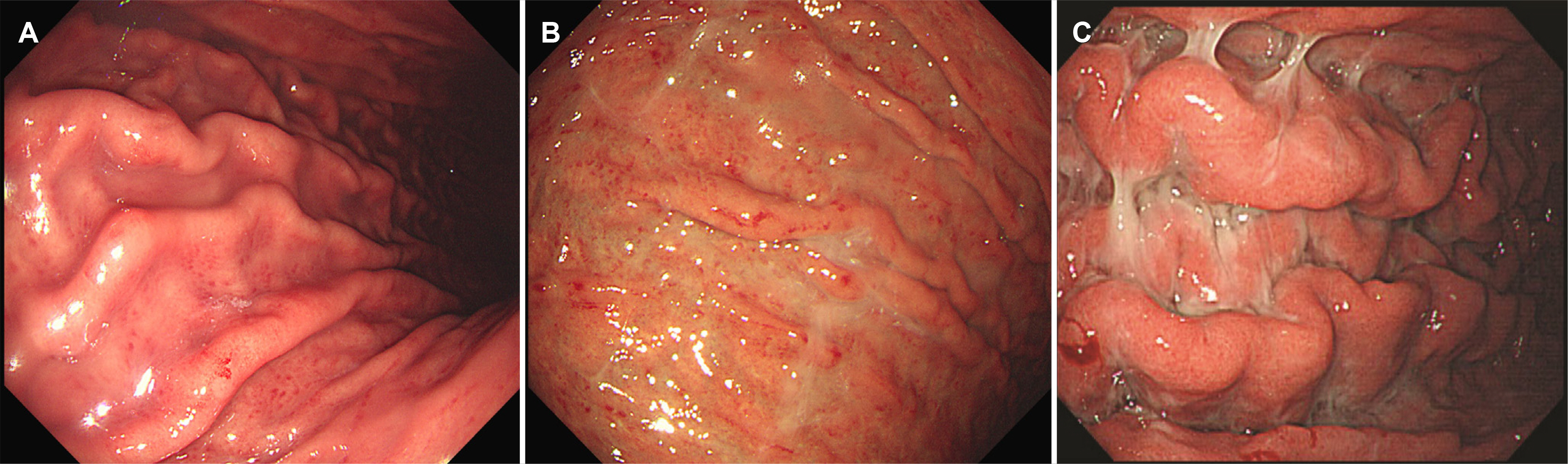
Fig. 1 Gastroscopy findings of erythematous/exudative gastritis. (A) Mild erythematous/exudative gastritis is called when a few punctate exudates are visible. (B) Moderate erythematous/exudative gastritis is called when focally patches or plaques of exudate are seen. (C) Severe erythematous/exudative gastritis is called when extensive areas are covered by exudate.
Reference
-
1. Kim BJ, Kim JG. 2015; Epidemiology and pathophysiology of Helicobacter pylori infections in Korea. Korean J Med. 89:133–141. DOI: 10.3904/kjm.2015.89.2.133.2. Leodolter A, Kulig M, Brasch H, Meyer-Sabellek W, Willich SN, Malfertheiner P. 2001; A meta-analysis comparing eradication, healing and relapse rates in patients with Helicobacter pylori-associated gastric or duodenal ulcer. Aliment Pharmacol Ther. 15:1949–1958. DOI: 10.1046/j.1365-2036.2001.01109.x. PMID: 11736726.3. Ford AC, Yuan Y, Forman D, Hunt R, Moayyedi P. 2020; Helicobacter pylori eradication for the prevention of gastric neoplasia. Cochrane Database Syst Rev. 7:CD005583. DOI: 10.1002/14651858.CD005583.pub3. PMID: 32628791. PMCID: PMC7389270.4. Kim JH, Kim HY, Kim NY, et al. 2001; Seroepidemiological study of Helicobacter pylori infection in asymptomatic people in South Korea. J Gastroenterol Hepatol. 16:969–975. DOI: 10.1046/j.1440-1746.2001.02568.x. PMID: 11595059.5. Yim JY, Kim N, Choi SH, et al. 2007; Seroprevalence of Helicobacter pylori in South Korea. Helicobacter. 12:333–340. DOI: 10.1111/j.1523-5378.2007.00504.x. PMID: 17669107.6. Lim SH, Kwon JW, Kim N, et al. 2013; Prevalence and risk factors of Helicobacter pylori infection in Korea: nationwide multicenter study over 13 years. BMC Gastroenterol. 13:104. DOI: 10.1186/1471-230X-13-104. PMID: 23800201. PMCID: PMC3702482.7. Lee HJ, Chung JM, Seo EH, Jeon SW. 2008; Clinicopathologic characteristics of gastric cancer diagnosed at health screening. Korean J Med. 75:665–672.8. Cho SJ. 2010; Screening of gastric cancer. Korean J Med. 79:219–223. DOI: 10.4174/jkss.2010.79.3.223.9. Kamada T, Haruma K, Inoue K, Shiotani A. 2015; Helicobacter pylori infection and endoscopic gastritis -Kyoto classification of gastritis. Nihon Shokakibyo Gakkai Zasshi. 112:982–993.10. Lee SY. 2019; Helicobacter pylori Infection and the Kyoto classification of gastritis. Korean J Helicobacter Up Gastrointest Res. 19:81–87. DOI: 10.7704/kjhugr.2019.19.2.81.11. Tytgat GN. 1991; The Sydney System: endoscopic division. Endoscopic appearances in gastritis/duodenitis. J Gastroenterol Hepatol. 6:223–234. DOI: 10.1111/j.1440-1746.1991.tb01469.x. PMID: 1912432.12. Ohkusa T, Okayasu I, Yamada M, et al. 1991; A high frequency of detection of Helicobacter pylori in whitish exudate of gastric ulcer. J Clin Gastroenterol. 13:649–655. DOI: 10.1097/00004836-199112000-00008. PMID: 1722231.13. Lim SH, Kim N, Kwon JW, et al. 2018; Trends in the seroprevalence of Helicobacter pylori infection and its putative eradication rate over 18 years in Korea: a cross-sectional nationwide multicenter study. PLoS One. 13:e0204762. DOI: 10.1371/journal.pone.0204762. PMID: 30332428. PMCID: PMC6192591.14. Lim E, Jo IH, Kim YJ, Chung WC. 2021; In situ diagnosis of Helicobacter pylori infection using the endoscopic Kyoto scoring system. Korean J Helicobacter Up Gastrointest Res. 21:322–332. DOI: 10.7704/kjhugr.2021.0046.15. Kato T, Yagi N, Kamada T, et al. 2013; Diagnosis of Helicobacter pylori infection in gastric mucosa by endoscopic features: a multicenter prospective study. Dig Endosc. 25:508–518. DOI: 10.1111/den.12031. PMID: 23369058.
- Full Text Links
-
- Actions
-
Cited
- CITED
-
- Close
- Share
-
- Similar articles
-
- Prediction of Helicobacter pylori Infection by Endoscopic Severity of Erythematous Gastritis in Asymptomatic Adults
- Endoscopic diagnosis of Helicobacter pylori infection
- The value of Helicobacter pylori IgG antibody in estimating the severity of gastritis in children
- Immune Response to Helicobacter pylori Infection
- Nodular Gastritis and Pathologic Findings in Children and Young Adults with Helicobacter pylori Infection
