Ewha Med J.
2022 Jul;45(3):e7. 10.12771/emj.2022.e7.
Minute Colon Perforation by a Nonabsorbable Suture Knot after Uterine Myomectomy
- Affiliations
-
- 1Department of Surgery, Ewha Womans University College of Medicine, Seoul, Korea
- 2Division of Colon and Rectal Surgery, Department of Surgery, Ewha Womans University Mokdong Hospital, Seoul, Korea
- KMID: 2532213
- DOI: http://doi.org/10.12771/emj.2022.e7
Abstract
- We report a rare case of suture material-related colon perforation. A 60-year-old woman visited clinics because of the nonspecific abdominal discomfort for several months. There were no specific medical history except previous laparoscopic myomectomy 15 years ago. Colonoscopy and abdomen-pelvis computed tomography revealed an unknown foreign body penetrating the sigmoid colon wall adjacent to the uterus. We performed laparoscopic exploration with foreign body removal and primary colon wall repair. The foreign body was identified as a non-absorbable suture material suggestive of used in previous myomectomy. With recent trends for minimally invasive procedures in the field of pelvic organ surgery, surgeons, especially those without sufficient training have to pay attention to selecting the proper surgical suture materials. (Ewha Med J 2022;45(3):e7)
Keyword
Figure
-
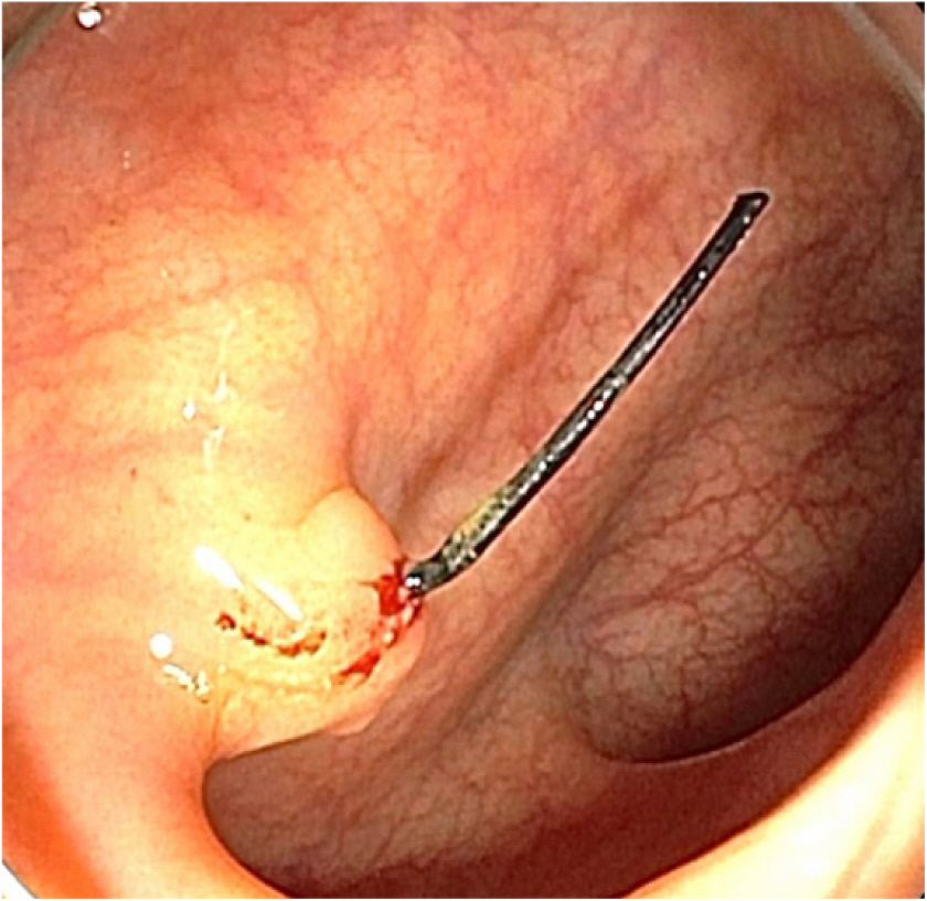
Fig. 1. Colonoscopic findings. A polypoid lesion with an unknown foreign body was found in sigmoid colon.
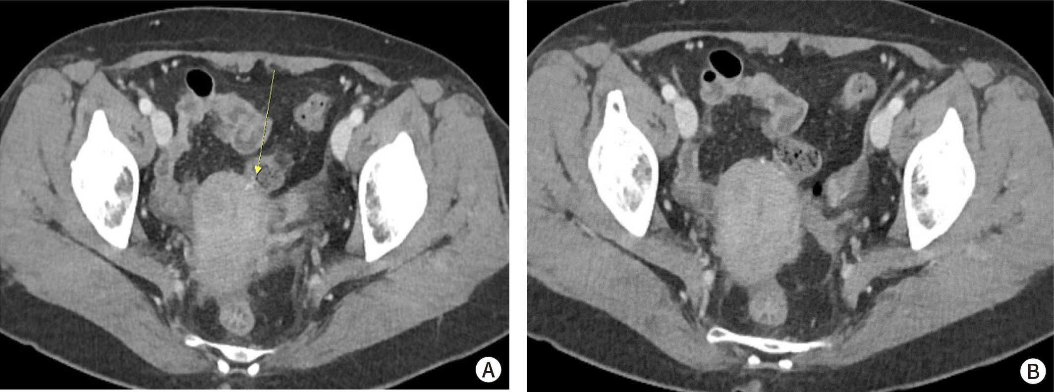
Fig. 2. Pelvic CT findings. (A) A radiopaque foreign body was probably penetrating the uterus and abutting the sigmoid colon (yellow arrow). (B) There was no free air and fluid collection around the penetrated site.
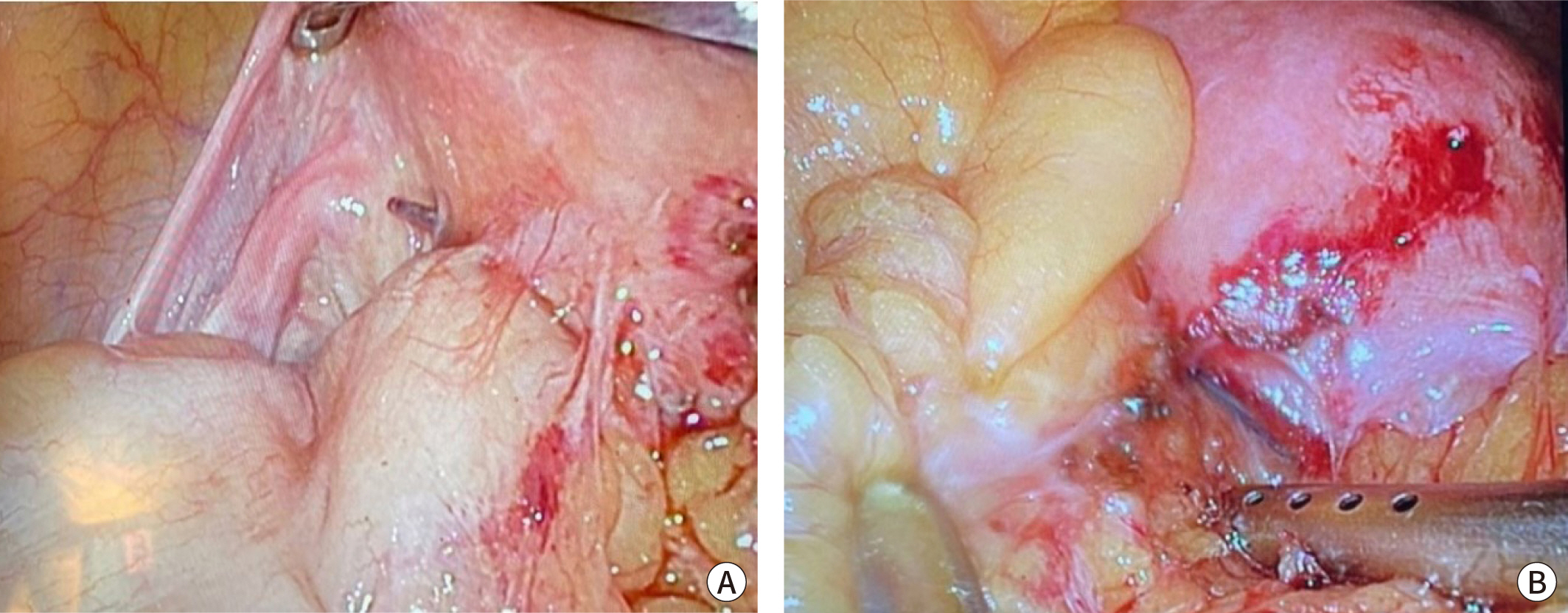
Fig. 3. Operative findings. (A) A suture knot at the left-upper uterus wall formed an adhesion with the sigmoid colon that was predicted to penetrate the abutting colon wall. (B) A suture knot at the mid-upper uterus wall formed adhesion with nearby omentum.
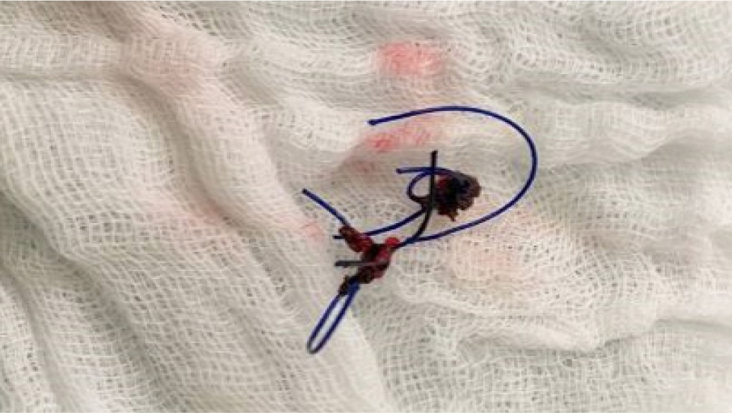
Fig. 4. The removed suture knot that was penentrating the colon wall, and was predicted to be a non-absorbable monofilament suture.
Reference
-
References
1. Talpur AA, Awan MS, Surhio AR. Closure of elective abdominal incisions with monofilament, non-absorbable suture material versus polyfilament absorbable suture material. J Ayub Med Coll Abbottabad. 2011; 23:51–54.2. Pal KMI. Urinary bladder wall repair: what suture to use? Br J Urol. 1998; 82:196–198. DOI: 10.1046/j.1464-410X.1998.00722.x. PMID: 9722753.
Article3. Parell GJ, Becker GD. Comparison of absorbable with nonabsorbable sutures in closure of facial skin wounds. Arch Facial Plast Surg. 2003; 5:488–490. DOI: 10.1001/archfaci.5.6.488. PMID: 14623686.
Article4. Khanal N, Thapa A, Raut A, Ghimire B, Pradhan S, Shrestha S. Intra-abdominal propylene suture fragment leading to complete small bowel obstruction: a case report. Int J Surg Case Rep. 2021; 85:106174. DOI: 10.1016/j.ijscr.2021.106174. PMID: 34274756. PMCID: PMC8319363.
Article5. Wang L, Maejima T, Fukahori S, Nishihara S, Yoshikawa D, Kono T. Bowel obstruction and perforation secondary to barbed suture after minimally invasive inguinal hernia repair: report of two cases and literature review. Surg Case Rep. 2021; 7:161. DOI: 10.1186/s40792-021-01249-w. PMID: 34255201. PMCID: PMC8276904.
Article6. Nakano T, Endo S, Negishi H, Yamamoto S. Penetration of bronchial wall by knot of absorbable monofilament suture. Interact Cardiovasc Thorac Surg. 2017; 24:468–470. DOI: 10.1093/icvts/ivw402. PMID: 28040761.
Article
- Full Text Links
-
- Actions
-
Cited
- CITED
-
- Close
- Share
-
- Similar articles
-
- Simplified suturing method using Hem-o-lock in two port laparoscopic myomectomy
- Obstetric outcomes after uterine myomectomy: Laparoscopic versus laparotomic approach
- Repetitive Spontaneous Uterine Rupture in the First Trimester after Laparoscopic Myomectomy: A Case Report and Review of Literature
- Can It be Said that this Case Confirmed the Phenomenon of Subacromial Suture Knot Impingement after Arthroscopic Rotator Cuff Repair?: A Case Report
- A New Slip Knot with Locking Loop
