Rhino-Orbito-Cerebral Mucormycosis in an Immunocompromised Patient
- Affiliations
-
- 1Department of Otolaryngology-Head & Neck Surgery, Gachon University Gil Medical Center, Gachon University College of Medicine, Incheon, Republic of Korea
- KMID: 2532016
- DOI: http://doi.org/10.18787/jr.2022.00410
Abstract
- Rhino-orbito-cerebral mucormycosis (ROCM) is an invasive fungal infection that usually occurs in immunocompromised patients. It is aggressive and has a high risk of mortality. With unclear guidelines, ROCM is treated in various ways. We present a patient who underwent kidney transplant and who treated for ROCM without major complications.
Keyword
Figure
-
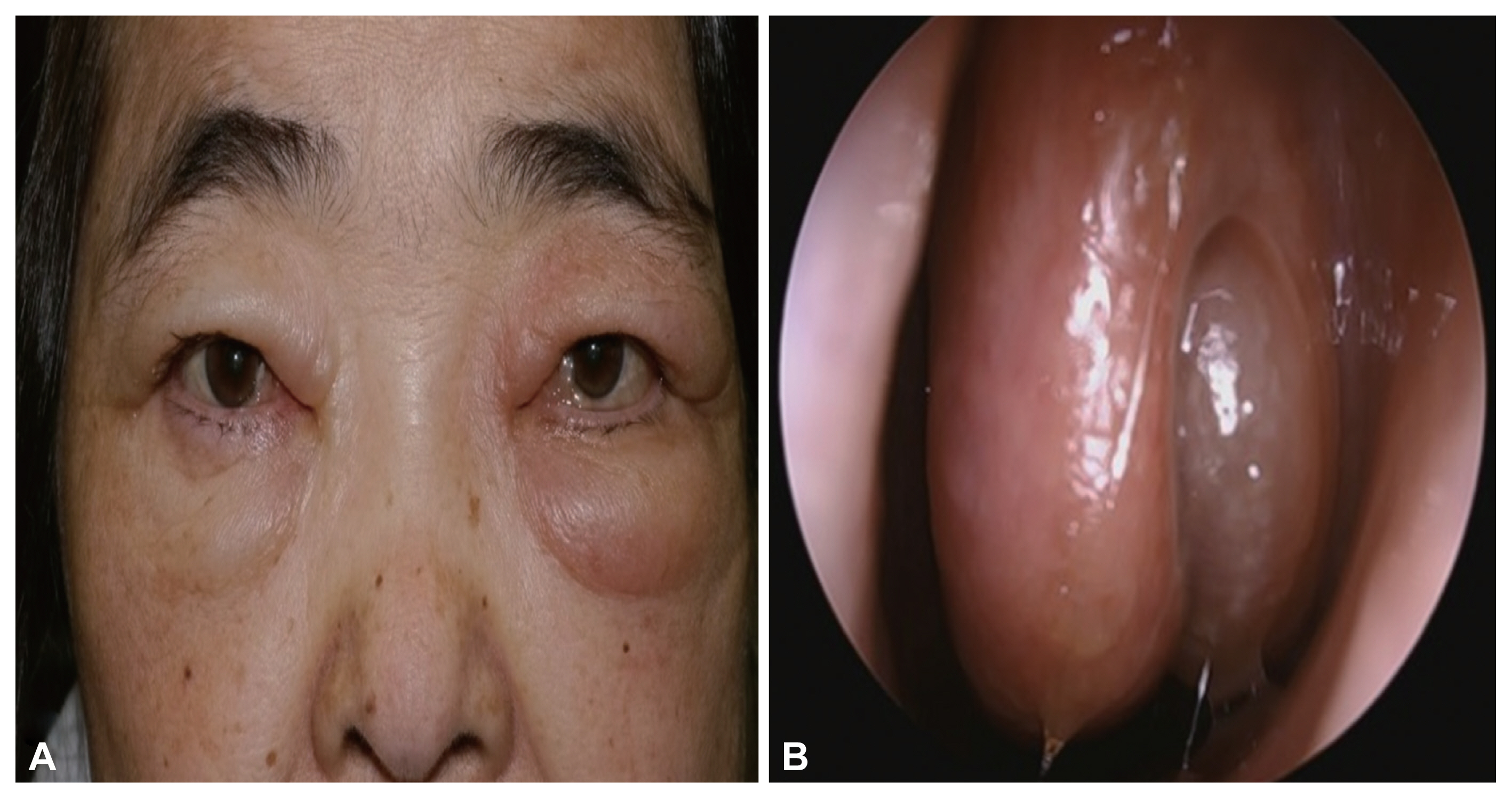
Fig. 1 Preoperative patient’s face pictures. Left periorbital swelling was observed. A: Patient’s left nasal cavity. B: A bulging mass filling the left middle meatus.
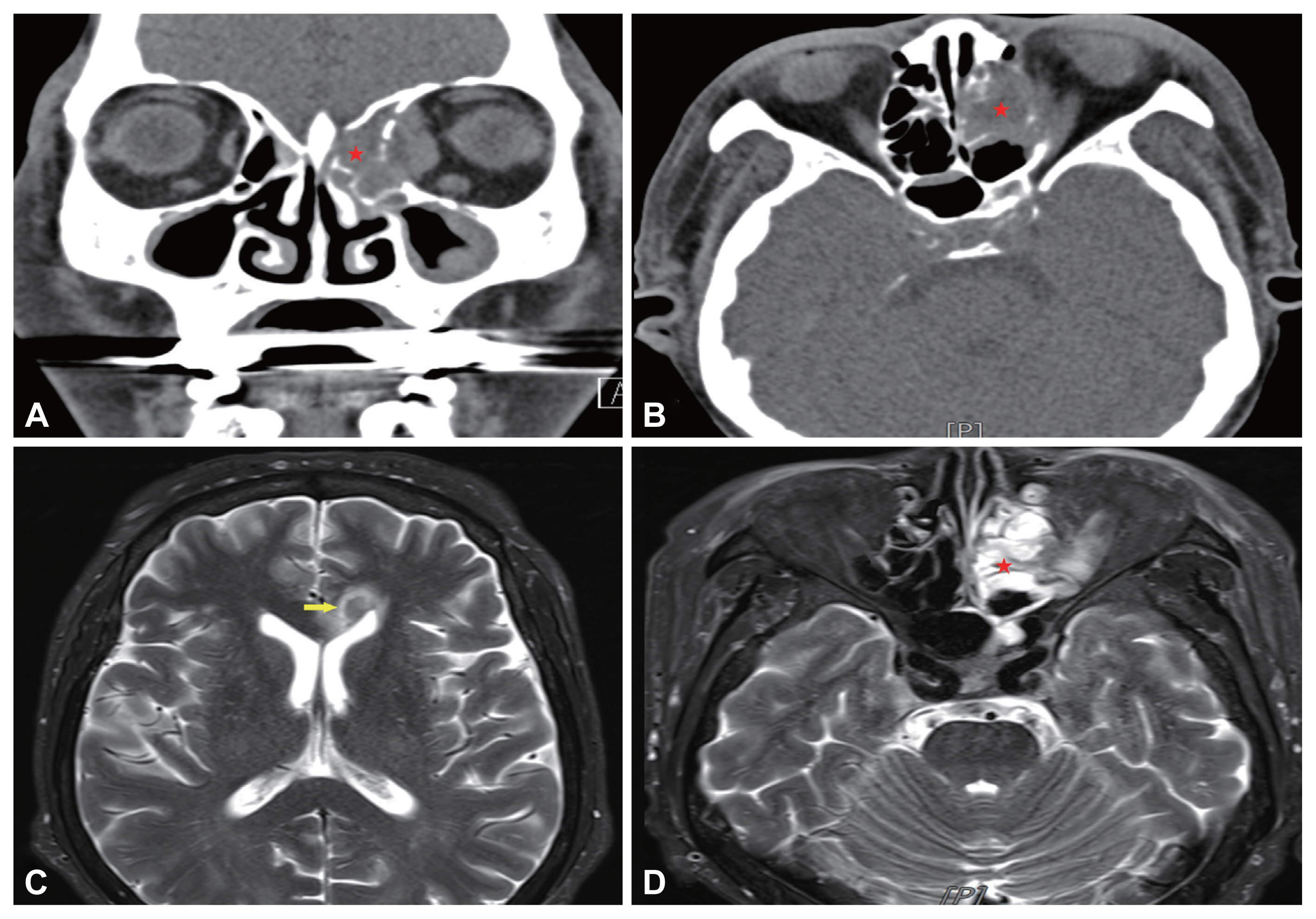
Fig. 2 Pre-operative patient’s paranasal sinus computed tomography view. A: Coronal view. B: Axial view. The 3.4-cm heterogenous mass was located inside of left ethmoid sinus. The lesion was accompanied by left orbital medial wall erosion and involved the medial rectus mscule (indicated by stars). C, D: Pre-operative patient’s paranasal sinus magnetic resonance imaging T2-weighted view. C: The 8-mm rim enhanced lesion was observed in left. aspect of genu of the corpus callosum (indicated by arrow). D: The heterogeneous enhancing lesion inside the left ethmoid sinus has a clear boundary with the inside of the nasal cavity, but confirmed as a pattern invading the left orbital wall (indicated by star).
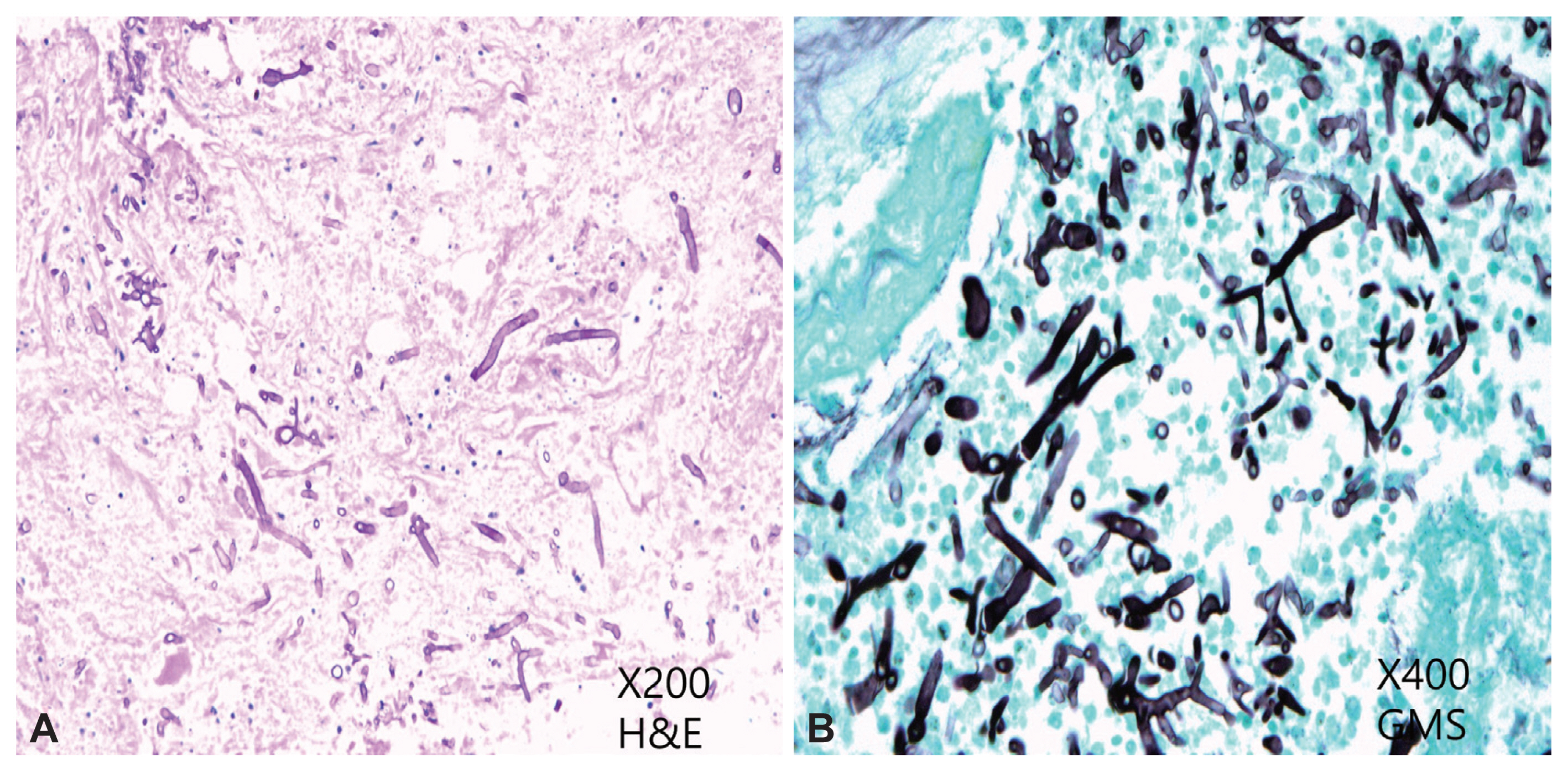
Fig. 3 Pathology of left. Nasal cavity lesion biopsy. A number of irregular shaped of ribbon-like hyphae are observed in H&E stain ×200 (A), GMS stain ×400 (B). H&E, Hematoxyling and Eosin; GMS, Grocott’s Methenamine Silver.
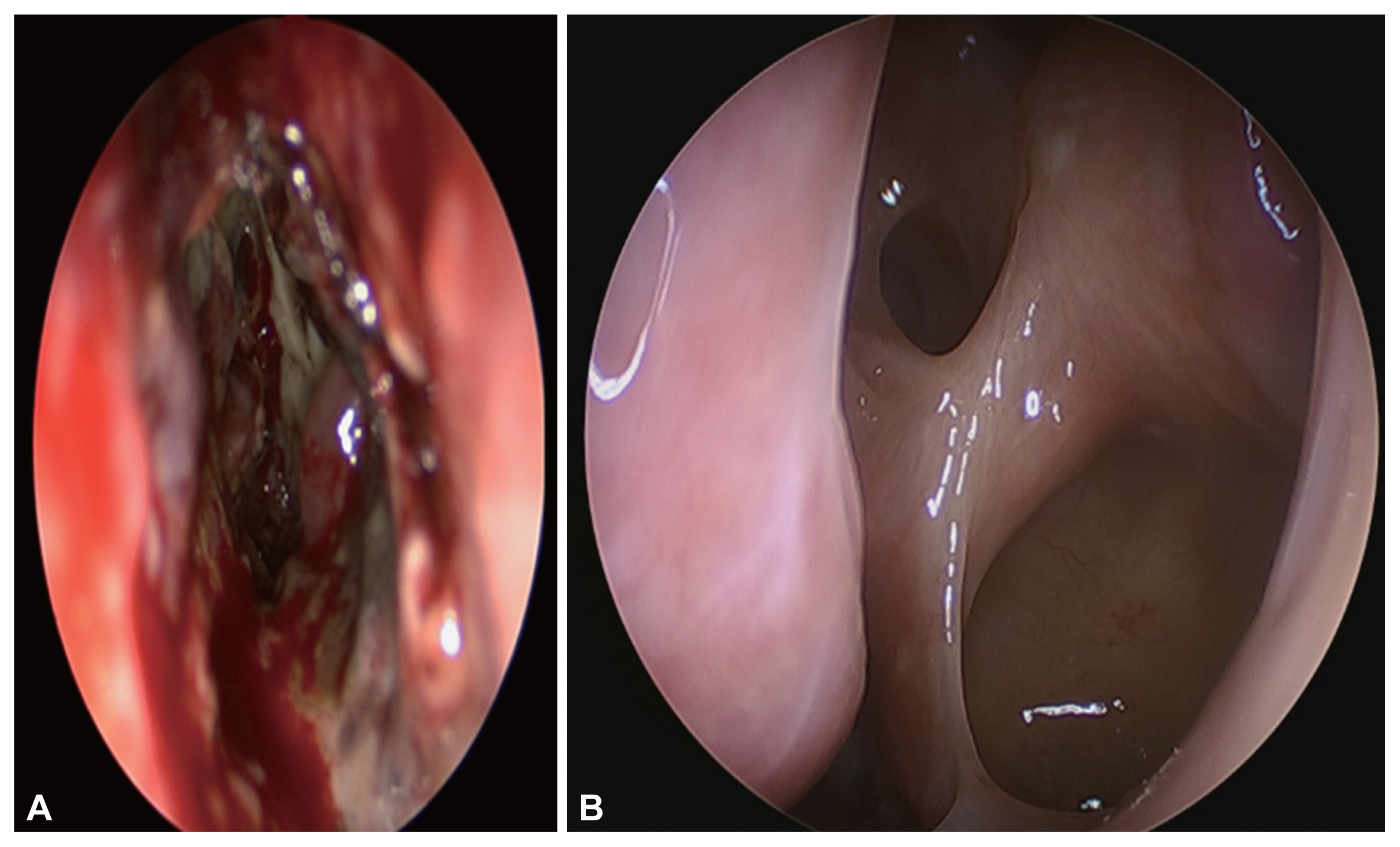
Fig. 4 Intra-operative. The dark brown mud-like discharge was shown from the inside of the ethmoid sinus. A: Necrotic granulation tissue with thick purulent discharge was attached to the cribriform plate and lamina papyracea. B: Three months after surgery, there were no abnormal findings on patient’s nasal cavity.
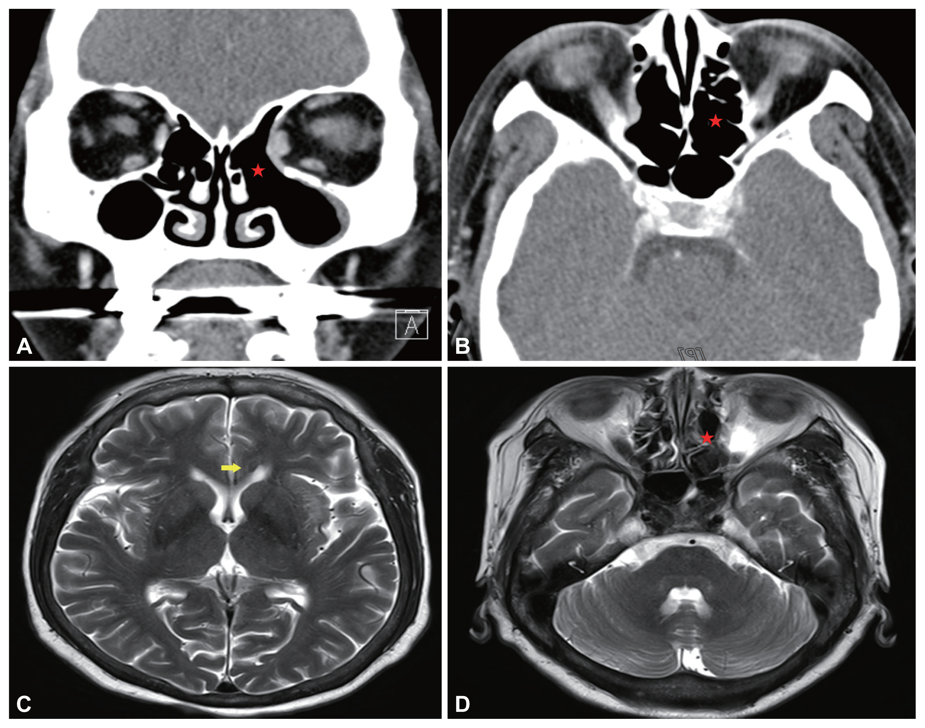
Fig. 5 Post-operative patient’s paranasal computed tomography view. A: Coronal view. B: Axial view. There is no lesion and no recurrence (indicated by stars). Nine months after treatment, the rim-enhanced lesion in left aspect of genu of the corpus callosum (indicated by arrow) (C) and ethmoid sinus (indicated by star) (D) was decreased compare with pre-operative magnetic resonance imaging.
Reference
-
1. Skiada A, Pagano L, Groll A, Zimmerli S, Dupont B, Lagrou K, et al. Zygomycosis in Europe: analysis of 230 cases accrued by the registry of the European Confederation of Medical Mycology (ECMM) working group on Zygomycosis between 2005 and 2007. Clin Microbiol Infect. 2011; 17(12):1859–67.2. Sipsas NV, Gamaletsou MN, Anastasopoulou A, Kontoyiannis DP. Therapy of mucormycosis. J Fungi (Basel). 2018; 4(3):90.3. Jeong W, Keighley C, Chen S. The epidemiology, management and outcomes of invasive mucormycosis in the 21st century: a systematic review. In : Proceedings of the 27th European Congress of Clinical Microbiology and Infectious Diseases; 2017 Apr 22–25; Vienna, Austria ECCMID. 2017. p. 1445.4. Talmi YP, Goldschmied-Reouven A, Bakon M, Barshack I, Wolf M, Horowitz Z, et al. Rhino-orbital and rhino-orbito-cerebral mucormycosis. Otolaryngol Head Neck Surg. 2002; 127(1):22–31.5. Thurtell MJ, Chiu AL, Goold LA, Akdal G, Crompton JL, Ahmed R, et al. Neuro-ophthalmology of invasive fungal sinusitis: 14 consecutive patients and a review of the literature. Clin Exp Ophthalmol. 2013; 41(6):567–76.6. Anaissie EJ, Mattiuzzi GN, Miller CB, Noskin GA, Gurwith MJ, Mamelok RD, et al. Treatment of invasive fungal infections in renally impaired patients with amphotericin B colloidal dispersion. Antimicrob Agents Chemother. 1998; 42(3):606–11.7. Spellberg B, Ibrahim A, Roilides E, Lewis RE, Lortholary O, Petrikkos G, et al. Combination therapy for mucormycosis: why, what, and how? Clin Infect Dis. 2012; 54(Suppl 1):S73–8.8. DiBartolo MA, Kelley PS. Rhino-orbital-cerebral mucormycosis (ROCM): a comprehensive case review. Aviat Space Environ Med. 2011; 82(9):913–6.9. Blitzer A, Lawson W, Meyers BR, Biller HF. Patient survival factors in paranasal sinus mucormycosis. Laryngoscope. 1980; 90(4):635–48.10. DelGaudio JM, Swain RE Jr, Kingdom TT, Muller S, Hudgins PA. Computed tomographic findings in patients with invasive fungal sinusitis. Arch Otolaryngol Head Neck Surg. 2003; 129(2):236–40.
- Full Text Links
-
- Actions
-
Cited
- CITED
-
- Close
- Share
-
- Similar articles
-
- Clinical Manifestation in Rhino-Orbito-Cerebral Mucormycosis
- Two Cases of Rhino-Orbito-Cerebral Mucormycosis
- A Case of Rhino-orbito-Cerebral Mucormycosis Presenting with Recurrent Transient Ischemic Attacks(TIAs)
- Two Cases of Rhino-orbito-cerebral Mucormycosis
- Rhino-Orbital-Cerebral Mucormycosis: 2 case reports
