A remnant choledochal cyst after choledochal cyst excision treated with a lumen-apposing metal stent: a case report
- Affiliations
-
- 1Department of Internal Medicine and Liver Research Institute, Seoul National University Hospital, Seoul National University College of Medicine, Seoul, Korea
- KMID: 2531951
- DOI: http://doi.org/10.5946/ce.2019.176
Abstract
- A lumen-apposing metal stent (LAMS) is a saddle-shaped stent with large flanges at both ends, thereby preventing stent migration and helping with approximation of the adjacent structures. We report the case of a 25-year-old female with remnant choledochal cyst which was successfully treated with LAMS after initial treatment failure with a plastic stent. Although complete excision of the cyst is the definite treatment of choledochal cysts, endoscopic ultrasonography-guided cystoduodenostomy can be considered in cases wherein surgery is not feasible and dysplasia is not present. LAMS may be preferred to plastic stents for effective resolution of remnant choledochal cyst and prevention of ascending infection.
Figure
-

Fig. 1. (A) Abdominal computed tomography and (B) magnetic resonance cholangiopancreatography taken at initial presentation. Huge cystic dilatation of the common bile duct with bilateral intrahepatic duct dilatation and abrupt luminal diameter change in the peripheral duct was observed, suggestive of choledochal cyst, type IVa.
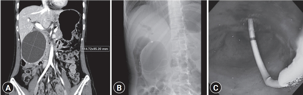
Fig. 2. (A) Abdominal computed tomography showed the remnant choledochal cyst after choledochal cyst excision. (B, C) Endoscopic ultrasonography-guided cystoduodenostomy at the second portion of the duodenum with a plastic stent was performed.
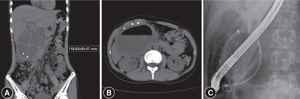
Fig. 3. Abdominal computed tomography showed (A) an increase in the size of the remnant choledochal cyst and (B) wall thickening with air-fluid level suggestive of cyst infection. (C) Endoscopic retrograde cholangiopancreatography showed the distorted pancreatic duct compressed by the remnant choledochal cyst. Endoscopic retrograde pancreatic drainage was done. Bile duct cannulation failed.

Fig. 4. (A, B) Cystoduodenostomy stent revision was done with a lumen-apposing metal stent. The previously inserted pancreatic stent was also noted.

Fig. 5. Drainage of the remnant choledochal cyst with lumen-apposing metal stent (LAMS). (A) Abdominal computed tomography taken 6 months after LAMS insertion showed an interval decrease in the size of the infected cyst. LAMS was subsequently removed. (B) Abdominal computed tomography taken 1 year after LAMS removal showed resolution of the remnant cyst.
Reference
-
1. Todani T, Watanabe Y, Toki A, et al. Classification of congenital biliary cystic disease: special reference to type Ic and IVA cysts with primary ductal stricture. J Hepatobiliary Pancreat Surg. 2003; 10:340–344.2. Søreide K, Søreide JA. Bile duct cyst as precursor to biliary tract cancer. Ann Surg Oncol. 2007; 14:1200–1211.3. Lee SE, Jang JY, Lee YJ, et al. Choledochal cyst and associated malignant tumors in adults: a multicenter survey in South Korea. Arch Surg. 2011; 146:1178–1184.4. Todani T, Tabuchi K, Watanabe Y, et al. Carcinoma arising in the wall of congenital bile duct cysts. Cancer. 1979; 44:1134–1141.5. Mussetto A, Fugazza A, Fuccio L, et al. Current uses and outcomes of lumen-apposing metal stents. Ann Gastroenterol. 2018; 31:535–540.6. Hammad T, Khan MA, Alastal Y, et al. Efficacy and safety of lumen-apposing metal stents in management of pancreatic fluid collections: are they better than plastic stents? A systematic review and meta-analysis. Dig Dis Sci. 2018; 63:289–301.7. Bank JS, Adler DG. Lumen apposing metal stents: a review of current uses and outcomes. Gastrointestinal Intervention. 2017; 6:9–14.8. Singham J, Schaeffer D, Yoshida E, et al. Choledochal cysts: analysis of disease pattern and optimal treatment in adult and paediatric patients. HPB (Oxford). 2007; 9:383–387.9. Singham J, Yoshida EM, Scudamore CH. Choledochal cysts. Part 3 of 3: management. Can J Surg. 2010; 53:51–56.10. Xia HT, Yang T, Liang B, et al. Treatment and outcomes of adults with remnant intrapancreatic choledochal cysts. Surgery. 2016; 159:418–425.11. Tan MY, Madhavan KK. An endoscopic approach for drainage of a remnant intra-pancreatic choledochal cyst–a rare complication following choledochal cyst excision causing gastric outlet obstruction. HPB (Oxford). 2018; 20(Suppl 2):S751.12. Yırgın H, Öter V, Aziret M, et al. Remnant choledochal cyst; report of a case. CausaPedia. 2017; 6:170–175.13. DeSimone ML, Asombang AW, Berzin TM. Lumen apposing metal stents for pancreatic fluid collections: recognition and management of complications. World J Gastrointest Endosc. 2017; 9:456–463.14. Bang JY, Navaneethan U, Hasan MK, et al. Non-superiority of lumen-apposing metal stents over plastic stents for drainage of walled-off necrosis in a randomised trial. Gut. 2019; 68:1200–1209.15. Wang K, Zhu J, Xing L, et al. Assessment of efficacy and safety of EUS-guided biliary drainage: a systematic review. Gastrointest Endosc. 2016; 83:1218–1227.16. Khashab MA, Messallam AA, Penas I, et al. International multicenter comparative trial of transluminal EUS-guided biliary drainage via hepatogastrostomy vs. choledochoduodenostomy approaches. Endosc Int Open. 2016; 4:E175–E181.17. Guo J, Feng L, Sun S, et al. Risk factors for infection after endoscopic ultrasonography-guided drainage of specific types of pancreatic and peripancreatic fluid collections (with video). Surg Endosc. 2016; 30:3114–3120.18. Brimhall B, Han S, Tatman PD, et al. Increased incidence of pseudoaneurysm bleeding with lumen-apposing metal stents compared to double-pigtail plastic stents in patients with peripancreatic fluid collections. Clin Gastroenterol Hepatol. 2018; 16:1521–1528.
- Full Text Links
-
- Actions
-
Cited
- CITED
-
- Close
- Share
-
- Similar articles
-
- A case of type IVa choledochal cyst
- Development of Cholangiocarcinoma Arising from Remnant Intrapancreatic Cyst 15 Years after Choledochal Cyst Excision
- Type IV-A Choledochal Cyst with Intrahepatic Bile Duct Stricture
- A Choledochal Cyst Combined with a Hilar Cholangiocarcinoma
- Type IVB Choledochal Cyst : A case report
