Pediatr Emerg Med J.
2022 Jun;9(1):41-43. 10.22470/pemj.2022.00430.
Enterobius vermicularis appendiceal infestation: a case of false-positive ultrasound-confirmed appendicitis
- Affiliations
-
- 1Department of Pediatrics, Brooke Army Medical Center, San Antonio, TX, United States
- 2Department of Pediatrics, Baylor College of Medicine, Children’s Hospital of San Antonio, San Antonio, TX, United States
- KMID: 2531528
- DOI: http://doi.org/10.22470/pemj.2022.00430
Abstract
- This case report discusses a school-aged boy who presented to the emergency department with diffuse abdominal pain and fever. After investigation, he was found to have Enterobius vermicularis infestation of the appendix without appendicitis. The novelty of this case is the video clip of a live worm-like finding on ultrasound in the boy. The video for this case serves as a reminder for emergency physicians or pediatricians to consider parasitic etiologies in children with abdominal pain, absent some of the classic manifestations like perianal pruritis.
Keyword
Figure
-
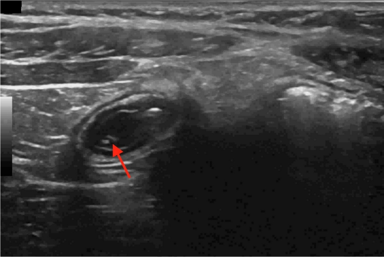
Fig. 1. A still image of the abdominal ultrasound. It was performed by an ultrasound technician, and read by a pediatric radiologist. The appendix measured up to 8 mm in diameter. There was mildly increased echogenicity of the periappendiceal fat and no evidence of perforation. These findings support a diagnosis of appendicitis. Within the lumen, there is a tiny live worm-like finding (arrow) presumed to be a parasite by the radiologist.
- Full Text Links
-
- Actions
-
Cited
- CITED
-
- Close
- Share
-
- Similar articles
-
- A Case of Acute Appendicitis Associated with Enterobius Vermicularis
- Laparoscopic Appendectomy for Acute Appendicitis Caused by Enterobius Vermicularis
- Perianal Granuloma Caused by a Female Pinworm (Enterobius vermicularis): A case report
- Appendiceal Diverticulitis
- Present status of Enterobius vermicularis infestation among children in kindergartens
