Discovery of a trans-sellar vascular supply for the pituitary gland
- Spinelli CP
 1
1 - Iwanaga J2,3
- Hur MS4
- Dumont AS2
- Tubbs RS2,3,4,5,6,
- Affiliations
-
- 1Tulane University & Ochsner Clinic Neurosurgery Program, Tulane University School of Medicine, New Orleans, LA, USA
- 2Department of Neurosurgery, Tulane University School of Medicine, New Orleans, LA, USA
- 3Department of Neurology, Tulane University School of Medicine, New Orleans, LA, USA
- 4Department of Anatomy, Catholic Kwandong University College of Medicine, Gangneung, Korea
- 5Department of Neurosurgery and Ochsner Neuroscience Institute, Ochsner Health System, New Orleans, LA, USA
- 6Department of Anatomical Sciences, St. George’s University, St. George’s, Grenada
- 7Department of Surgery, Tulane University School of Medicine, New Orleans, LA, USA
- 8Department of Structural & Cellular Biology, Tulane University School of Medicine, New Orleans, LA, USA
- 9University of Queensland, Brisbane, Australia
- KMID: 2531197
- DOI: http://doi.org/10.5115/acb.21.255
Abstract
- The vasculature of the pituitary gland is discussed briefly and the details of an anatomical discovery of the vessels supplying the pituitary gland provided. Twenty latex injected cadaveric heads were dissected. Any vessels that were found to penetrate the sella turcica and travel to the pituitary gland were documented and measured. Additionally, 25 adult skulls were evaluated for the presence, size, and sites of bony foramina in the floor of sella turcica. Trans-sellar vessels were identified in 65% of specimens. There was a mean of 1.5 vessels per specimen consisting usually of a mixture of veins and arteries. The mean diameter of these vessels was 0.3 mm and the mean length from the sella turcica to the pituitary gland was 2.3 mm. These vessels were concentrated in the most concave part of the sella turcica. In bony specimens, the mean number of transsellar foramina was four. The diameter of these foramina ranged from 0.3 to 0.6 mm in size. The trans-sellar foramina were concentrated near the center part of the sella turcica and had no regular pattern. The pituitary gland receives at least some blood supply and drainage via vessels traveling along the septum of the sphenoidal sinuses and through the sella turcica. Knowledge of such vessels might lead to a better understanding of the vascular supply and drainage of the pituitary gland and would be useful during skull base approaches such as trans-nasal approaches to the pituitary gland.
Keyword
Figure
-

Fig. 1 Schematic drawing of the various vessels supplying the pituitary gland.

Fig. 2 Lateral view of the pituitary gland noting the trans-sellar blood vessels (arrow) traveling along the septum of sphenoidal sinuses.

Fig. 3 Posterior view of the cadaveric specimen with the pituitary gland lifted up and pulled forward. The left black arrow notes a trans-sellar artery, which is off the midline. The right black arrow notes a trans-sellar vein with minimal latex within it but when traced inferiorly, this vessel arose from veins of the mucosa of the septum of sphenoidal sinuses. Inferiorly, the thin wall over the sinus is removed showing the intact mucosa and veins (yellow arrow) in its posterior aspect. These veins were confluent with the veins of the mucosa of the septum. For reference, note the transected internal carotid arteries (ICA), oculomotor nerve (CNIII), and cavernous sinus (CS). The white arrow marks the inferior hypophyseal artery.

Fig. 4 Posterior cadaveric view noting a midline trans-sellar vein (arrow) with the pituitary gland lifted up and pulled anteriorly.

Fig. 5 Medial view with the pituitary gland lift up and noting trans-sellar arteries (arrows) traveling along the septum of sphenoidal sinuses.
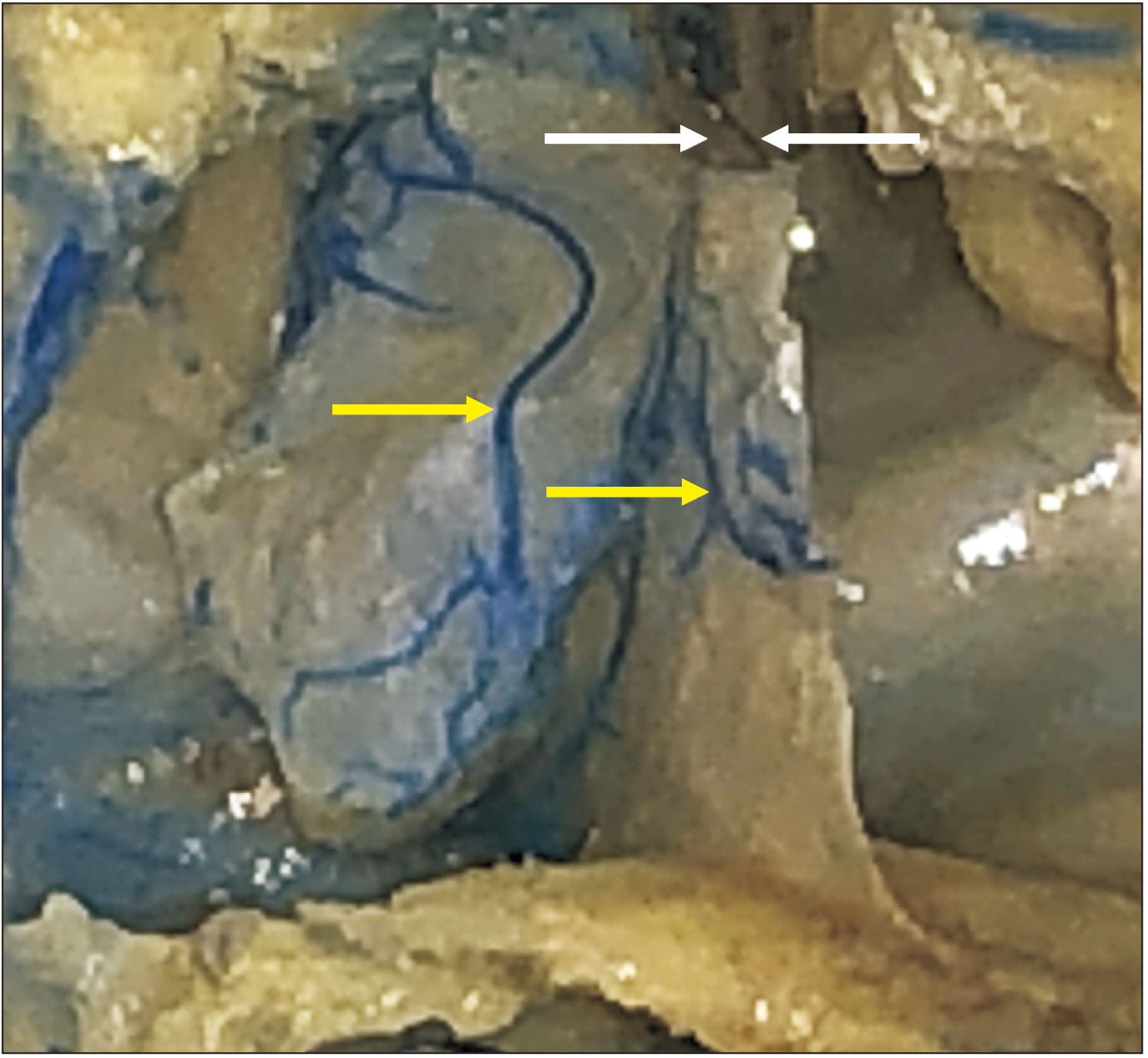
Fig. 6 Cadaveric dissection after removal of the posterior wall of the sphenoidal sinus. The left yellow arrow marks a large vein running in the mucosa of the sphenoidal sinus and the right yellow arrow marks a vein on the septum of sinuses. The left white arrow marks the sella turcica. The right white arrow shows a trans-sellar vein arising from the vessels in the septum of sphenoidal sinuses.

Fig. 7 Superior view of a dry bone specimen noting a single trans-sellar foramen in the sella turcica (arrow).

Fig. 8 Superior view of a dry bone specimen noting multiple trans-sellar foramina in the sella turcica (arrows).

Fig. 9 Anterior view with the sphenoidal sinus opened to show the relationship between its septum (arrows) and the sella turcica. All sphenoidal sinuses with trans-sellar vessels were of the sellar type- that is the sphenoidal sinus was well developed and located inferior to the sella turcica.
Reference
-
References
1. Cironi KA, Decater T, Iwanaga J, Dumont AS, Tubbs RS. 2020; Arterial supply to the pituitary gland: a comprehensive review. World Neurosurg. 142:206–11. DOI: 10.1016/j.wneu.2020.06.221. PMID: 32634634.
Article2. McCormack IG, Xu L, Nerva J, Berry JF, Melgar M, Wysiadecki G, Walocha J, Iwanaga J, Dumont AS, Tubbs RS. 2021; Anatomy of the dorsal meningeal artery including its variations: application to skull base surgery and diagnostic and interventional imaging. World Neurosurg. 155:e41–8. DOI: 10.1016/j.wneu.2021.07.132. PMID: 34365050.
Article3. Cappello ZJ, Minutello K, Dublin AB. 2022. Anatomy, head and neck, nose paranasal sinuses. StatPearls. StatPearls Publishing;Treasure Island: DOI: 10.1016/j.wneu.2021.07.132.4. Alsaied AS. Gendeh BS, editor. 2017. Paranasal sinus anatomy: what the surgeon needs to know. Paranasal Sinuses. IntechOpen;London: DOI: 10.5772/intechopen.69089.
Article5. Harju T, Alanko J, Numminen J. 2019; Pituitary apoplexy following endoscopic nasal surgery: a case report. SAGE Open Med Case Rep. 7:2050313X19855867. DOI: 10.1177/2050313X19855867. PMID: 31217974. PMCID: PMC6558528.
Article6. Fyrmpas G, Constantinidis J, Foroglou N, Selviaridis P. 2010; Pituitary apoplexy following endoscopic sinus surgery. J Laryngol Otol. 124:677–9. DOI: 10.1017/S0022215109991915. PMID: 19930782.
Article7. Briet C, Salenave S, Chanson P. 2015; Pituitary apoplexy. Endocrinol Metab Clin North Am. 44:199–209. DOI: 10.1016/j.ecl.2014.10.016. PMID: 25732655. PMCID: PMC1763528.
Article8. Agrawal B, Dziurzynski K, Salamat MS, Baskaya M. 2012; The temporal association of sphenoid sinus mucosal thickening on MR imaging with pituitary apoplexy. Turk Neurosurg. 22:785–90. DOI: 10.5137/1019-5149.JTN.4273-11.1. PMID: 23208917.
Article9. Liu JK, Couldwell WT. 2006; Pituitary apoplexy in the magnetic resonance imaging era: clinical significance of sphenoid sinus mucosal thickening. J Neurosurg. 104:892–8. DOI: 10.3171/jns.2006.104.6.892. PMID: 16776332.10. Arita K, Kurisu K, Tominaga A, Sugiyama K, Ikawa F, Yoshioka H, Sumida M, Kanou Y, Yajin K, Ogawa R. 2001; Thickening of sphenoid sinus mucosa during the acute stage of pituitary apoplexy. J Neurosurg. 95:897–901. DOI: 10.3171/jns.2001.95.5.0897. PMID: 11702884.
Article11. Carrau RL, Vescan AD, Snyderman CH, Kassam AB. Myers EN, Carrau RL, Eibling DE, Ferguson BJ, Ferris RL, Gillman GS, Golla S, Grandis JR, Hirsch BE, Johnson JT, Raz Y, Rosen CA, Schaitkin BM, Snyderman CH, Toh EH, editors. 2008. Endoscopic management of cerebrospinal fluid leaks. Operative Otolaryngology: Head and Neck Surgery. 2nd ed. Saunders;Philadelphia: DOI: 10.1016/B978-1-4160-2445-3.50108-4.
Article12. Tanaka A, Furubayashi T, Arai M, Inoue D, Kimura S, Kiriyama A, Kusamori K, Katsumi H, Yutani R, Sakane T, Yamamoto A. 2018; Delivery of oxytocin to the brain for the treatment of autism spectrum disorder by nasal application. Mol Pharm. 15:1105–111. DOI: 10.1021/acs.molpharmaceut.7b00991. PMID: 29338251.
Article13. Iwanaga J, Singh V, Ohtsuka A, Hwang Y, Kim HJ, Moryś J, Ravi KS, Ribatti D, Trainor PA, Sañudo JR, Apaydin N, Şengül G, Albertine KH, Walocha JA, Loukas M, Duparc F, Paulsen F, Del Sol M, Adds P, Hegazy A, Tubbs RS. 2021; Acknowledging the use of human cadaveric tissues in research papers: recommendations from anatomical journal editors. Clin Anat. 34:2–4. DOI: 10.1002/ca.23671. PMID: 32808702.
Article
- Full Text Links
-
- Actions
-
Cited
- CITED
-
- Close
- Share
-
- Similar articles
-
- Intrasellar Schwannomas
- Sellar Chordoma Mimicking Pituitary Adenoma
- Pituitary Metastasis of Bronchial Carcinoid Tumor Mimicking Pituitary Adenoma: a Case Report
- A Case of Pituitary Fossa Chondrosarcoma Mimicking Sellar and Suprasellar Mass
- Pituitary Ependymoma, 10-Year Follow-Up after Partial Resection and Radiation Therapy
