Temporomandibular joint ankylosis in Williams syndrome patient: an insight on the function of elastin in temporomandibular joint disorder
- Affiliations
-
- 1Department of Oral and Maxillofacial Surgery, Jeju National University College of Medicine, Jeju, Korea
- 2Department of Oral and Maxillofacial Surgery, Seoul National University Dental Hospital, Seoul, Korea
- 3Department of Oral and Maxillofacial Surgery, Seoul National University School of Dentistry, Seoul, Korea
- KMID: 2531055
- DOI: http://doi.org/10.5125/jkaoms.2022.48.3.178
Abstract
- Williams–Beuren syndrome (WS) is a rare genetic disorder that results from microdeletion at chromosome 7, which harbors the elastin gene. Clinical findings include arteriopathy, aortic stenosis, hypertension, and laxities and contractures in different joints throughout the body. While many components of the temporomandibular joint (TMJ) normally contain elastin, there are few reports on TMJ manifestations of WS. This study reports a TMJ ankylosis case in a WS patient and shares insight on a possible link between development of TMJ ankylosis and elastin deficiency in WS patients. A WS patient presented with bilateral TMJ ankylosis and was successfully treated with TMJ gap arthroplasty. Hypermobility of TMJ and lack of elastin in retrodiscal tissue can induce anterior disc displacement without reduction. Due to lack of elastin, which has a significant role in the compensatory and reparatory mechanism of TMJ, WS patients might be prone to TMJ ankylosis.
Figure
-

Fig. 1 A. FISH (fluorescence in situ hybridization) assay diagnostic of Williams–Beuren syndrome (WS). B. Facial features: micrognathia, short nose, long philtrum, thick lips, and long face and neck typical of WS. C. Initial maximal mouth opening (MMO) of 11 mm. Oligodontia is also notable. D. Intraoperative MMO of 40 mm after release of temporomandibular joint (TMJ) ankylosis. E. Section of patient TMJ stained with H&E showing typical mixed bony and fibrous tissue of TMJ ankylosis.
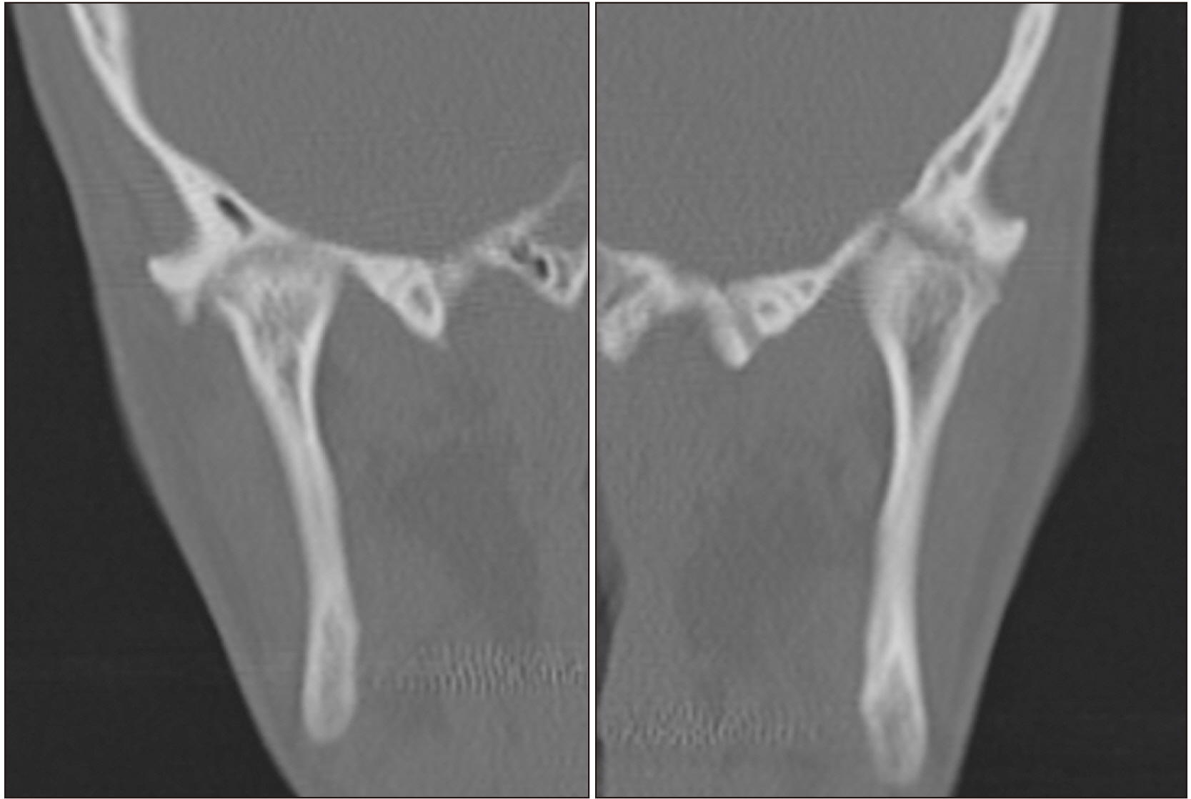
Fig. 2 Preoperative computed tomography of bilaterally ankylosed temporomandibular joints on coronal view.
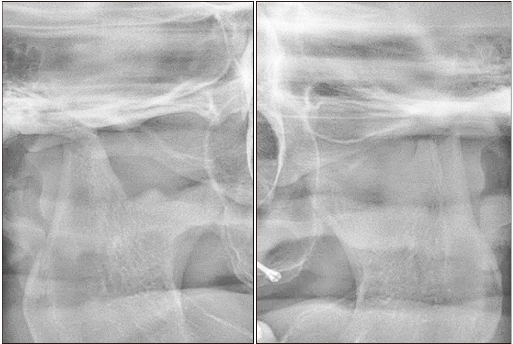
Fig. 3 Postoperative panoramic radiograph showing a gap between the glenoid fossa and the reduced condylar head on both sides.
Reference
-
References
1. Martens MA, Wilson SJ, Reutens DC. 2008; Research review: Williams syndrome: a critical review of the cognitive, behavioral, and neuroanatomical phenotype. J Child Psychol Psychiatry. 49:576–608. https://doi.org/10.1111/j.1469-7610.2008.01887.x. DOI: 10.1111/j.1469-7610.2008.01887.x. PMID: 18489677.
Article2. Copes LE, Pober BR, Terilli CA. 2016; Description of common musculoskeletal findings in Williams syndrome and implications for therapies. Clin Anat. 29:578–89. https://doi.org/10.1002/ca.22685. DOI: 10.1002/ca.22685. PMID: 26749433.
Article3. Coombs MC, Petersen JM, Wright GJ, Lu SH, Damon BJ, Yao H. 2017; Structure-function relationships of temporomandibular retrodiscal tissue. J Dent Res. 96:647–53. https://doi.org/10.1177/0022034517696458. DOI: 10.1177/0022034517696458. PMID: 28530471. PMCID: PMC5444618.
Article4. Leonardi R, Villari L, Bernasconi G, Caltabiano M. 2001; Histochemical study of the elastic fibers in pathologic human temporomandibular joint discs. J Oral Maxillofac Surg. 59:1186–92. https://doi.org/10.1053/joms.2001.26723. DOI: 10.1053/joms.2001.26723. PMID: 11573179.
Article5. Keith DA. 1979; Elastin in the bovine mandibular joint. Arch Oral Biol. 24:211–5. https://doi.org/10.1016/0003-9969(79)90142-0. DOI: 10.1016/0003-9969(79)90142-0. PMID: 289359.
Article6. Gross A, Bumann A, Hoffmeister B. 1999; Elastic fibers in the human temporo-mandibular joint disc. Int J Oral Maxillofac Surg. 28:464–8. DOI: 10.1016/S0901-5027(99)80064-2. PMID: 10609752.
Article7. de Bont LG, Liem RS, Boering G. 1985; Ultrastructure of the articular cartilage of the mandibular condyle: aging and degeneration. Oral Surg Oral Med Oral Pathol. 60:631–41. https://doi.org/10.1016/0030-4220(85)90367-6. DOI: 10.1016/0030-4220(85)90367-6. PMID: 3865135.
Article
- Full Text Links
-
- Actions
-
Cited
- CITED
-
- Close
- Share
-
- Similar articles
-
- Treatment of Temporomandibular Joint Disorder by Alloplastic Total Temporomandibular Joint Replacement
- A study on simultation of the mandibular movement of the patients with temporomandibular joint disorder
- Temporomandibular joint bony ankylosis following postoperative radiotherapy for maxillary cancer
- One-stage total reconstruction of temporomandibular joint ankylosis and facial asymmetry
- Difficult airway management in a case with lingual tonsil hypertrophy and temporo-mandibular joint partial ankylosis
