Case 1: A 44-Year-Old Woman Presented With Unexplained Painful Left Leg Swelling
- Affiliations
-
- 1Division of Cardiology, Department of Internal Medicine, Daejeon St. Mary’s Hospital, The Catholic University of Korea, Daejeon, Korea
- 2Division of Cardiology, Department of Internal Medicine, Yeouido St. Mary’s Hospital, The Catholic University of Korea, Seoul, Korea
- KMID: 2530506
- DOI: http://doi.org/10.3346/jkms.2022.37.e194
Figure
-
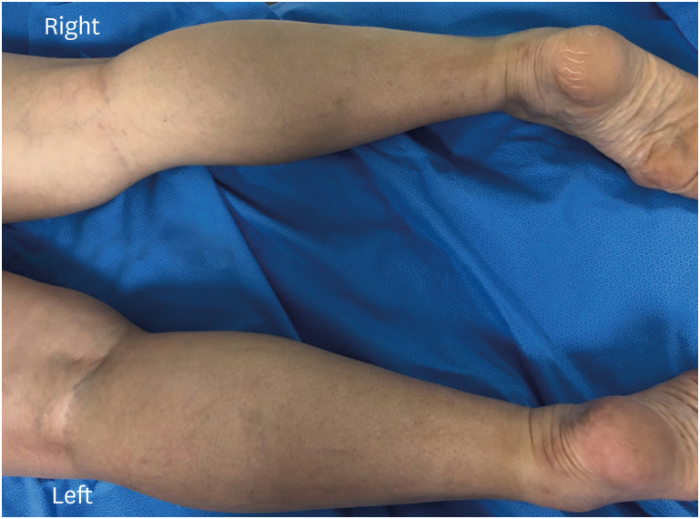
Fig. 1 Photograph of the patient’s lower extremity (prone position). Despite anticoagulation therapy, the patient’s left whole lower extremity shows diffuse swelling with mild erythema.
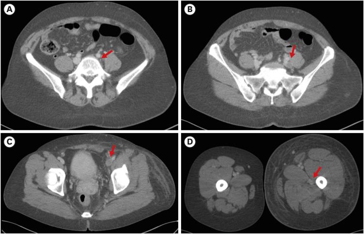
Fig. 2 Initial CT of lower extremity. CT scan shows that the left iliac vein is externally compressed (arrow) between the right common iliac artery and spinal body (A), resulting in complete occlusion of the left iliac vein (B) with extensive thrombus (arrows) involving the left iliac vein (B), left femoral vein (C), and left popliteal vein (D). The patient’s left lower extremity exhibits marked swelling.(B, C, D) Arrows indicate thrombus in the left iliac, left femoral, and left popliteal vein, respectively.CT = computed tomography.
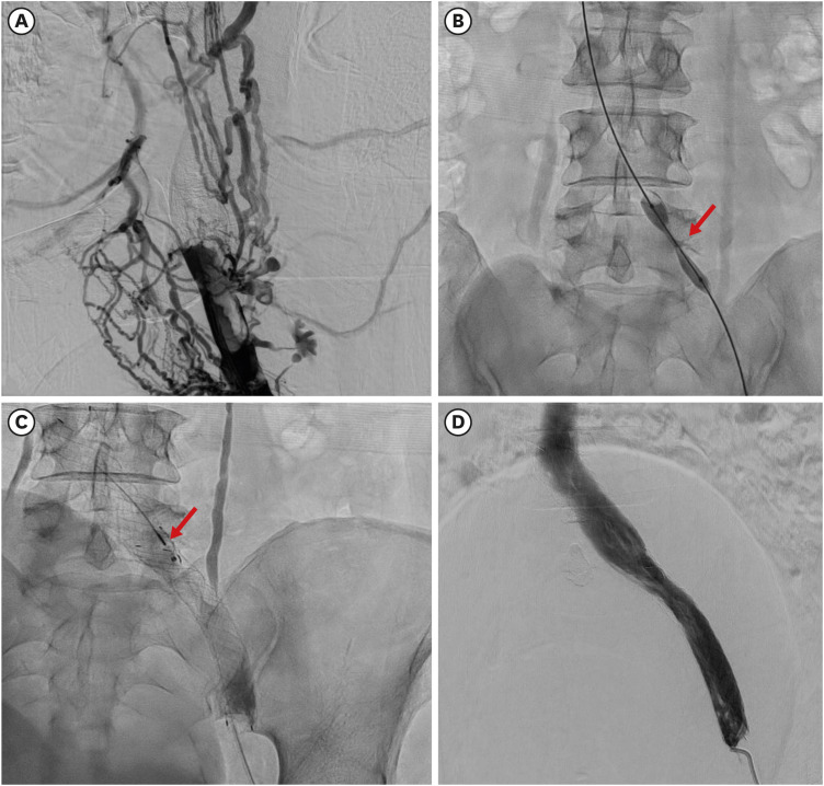
Fig. 3 Venography shows (A) the multiple large thrombi and complete occlusion of the left iliac and common femoral vein with extensive venous collaterals. (B) After advancing a guidewire into the inferior vena cava, balloon angioplasty showed a “waist” in the balloon (arrow) at the site of left common iliac vein compression by the right common iliac artery and spinal body. (C) Following balloon angioplasty, stents were implanted from the left common iliac vein to the femoral vein. Because post-stent venography revealed significant residual thrombi in the stent lumen, catheter-directed thrombolysis with alteplase (arrow) was performed. (D) The following day, venography shows well-positioned, patent stents and restored flow from the femoral vein to the inferior vena cava.
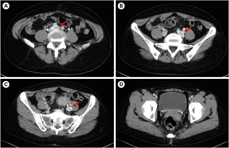
Fig. 4 The follow-up CT of the lower extremity at 6-months after endovascular intervention. CT shows complete resolution of thrombus in the entire left lower extremity and well-positioned vein stents (arrows in A, B, and C) without stenosis.CT = computed tomography.
Reference
-
1. Rodrigues LD, Bertanha M, El Dib R, Moura R. Association between deep vein thrombosis and stent patency in symptomatic iliac vein compression syndrome: Systematic review and meta-analysis. J Vasc Surg Venous Lymphat Disord. 2021; 9(1):275–284. PMID: 32827731.
Article2. Bashar K, Shalan A, Sharafat Ali S, Tang T, Tiwari A. Endovascular versus medical treatment of venous compression syndrome of the iliac vein - a systematic review. Vasa. 2021; 50(1):22–29. PMID: 33047662.
Article3. Mickley V, Schwagierek R, Rilinger N, Görich J, Sunder-Plassmann L. Left iliac venous thrombosis caused by venous spur: treatment with thrombectomy and stent implantation. J Vasc Surg. 1998; 28(3):492–497. PMID: 9737459.
Article4. Gloviczki P, Lawrence PF. Iliac vein stenting and contralateral deep vein thrombosis. J Vasc Surg Venous Lymphat Disord. 2017; 5(1):5–6. PMID: 27987610.
Article5. Kakkos SK, Gohel M, Baekgaard N, Bauersachs R, Bellmunt-Montoya S, Black SA, et al. Editor’s Choice - European Society for Vascular Surgery (ESVS) 2021 Clinical Practice Guidelines on the management of venous thrombosis. Eur J Vasc Endovasc Surg. 2021; 61(1):9–82. PMID: 33334670.
Article6. Jaff MR, McMurtry MS, Archer SL, Cushman M, Goldenberg N, Goldhaber SZ, et al. Management of massive and submassive pulmonary embolism, iliofemoral deep vein thrombosis, and chronic thromboembolic pulmonary hypertension: a scientific statement from the American Heart Association. Circulation. 2011; 123(16):1788–1830. PMID: 21422387.
Article7. May R, Thurner J. The cause of the predominantly sinistral occurrence of thrombosis of the pelvic veins. Angiology. 1957; 8(5):419–427. PMID: 13478912.
Article8. Taheri SA, Williams J, Powell S, Cullen J, Peer R, Nowakowski P, et al. Iliocaval compression syndrome. Am J Surg. 1987; 154(2):169–172. PMID: 3631389.
Article
- Full Text Links
-
- Actions
-
Cited
- CITED
-
- Close
- Share
-
- Similar articles
-
- Unilateral Leg Swelling Caused by a Ganglion Cyst on the Hip Joint
- May-Thurner Syndrome Appearing as Recurrent Swelling and Cellulitis in the Left Leg and Foot
- Multiple Solitary Plasmacytomas Presenting with Painful Erythematous Swelling of the Upper Eyelid
- Lung Cancer Presented as Painful Swelling of Lower Legs
- Psoriatic Onycho-pachydermo-periostitis of the Fingertips: A Report of Two Cases
