Pulsatile Compression of StentGraft by Re-pressurizing Aneurysmal Sac With Type III Endoleak After Endovascular Aneurysm Repair: A Case Report
- Affiliations
-
- 1Department of Cardiology, Gil Medical Center, Gachon University College of Medicine, Incheon, Korea
- KMID: 2530457
- DOI: http://doi.org/10.4070/kcj.2021.0333
Figure
-
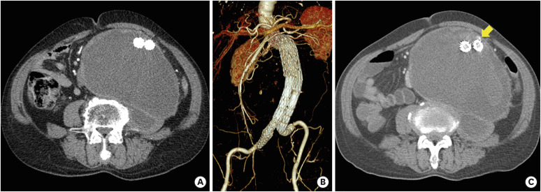
Figure 1 The CT images. (A and B) The CT angiography (performed 2 months ago) showed no endoleak and no interval change of the aneurysmal sac size (10×16 cm). (C) In this presentation, an increase in aneurysmal sac size (12×16 cm) and contrast leakage (arrow) in front of the left limb of the stent-graft were observed.CT = computed tomography.
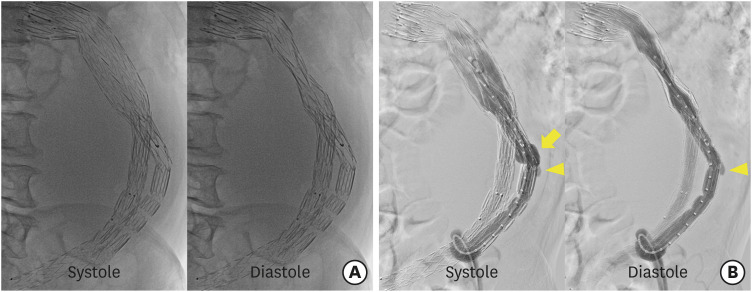
Figure 2 The fluoroscopic and aortographic images. (A) The fluoroscopy showed a diastolic compression of the main body of stent-graft. (B) The aortography showed that the left limb extension (implanted 2 years ago) for type IIIa endoleak was focally bulged at the systolic phase (arrow), which was considered an active-seal phenomenon of the AFX device (Endologix, Inc., Irvine, CA, USA). Another focal bulging at the connection part of the modular components was also observed (arrowhead), indicating a suspicious type IIIa endoleak.
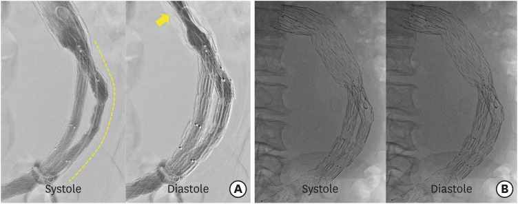
Figure 3 The completion aortographic and fluoroscopic images. (A) The completion aortography showed that the focal bulging of left limb disappeared after the additional placement of a stent-graft (dashed line), but the pulsatile compression of the main body persisted (arrow). (B) After 7 days, the fluoroscopy revealed that the pulsatile compression of the stent-graft completely disappeared.
- Full Text Links
-
- Actions
-
Cited
- CITED
-
- Close
- Share
-
- Similar articles
-
- Late Type 3b Endoleak Mimicking Type 2 Endoleak after Endovascular Aortic Aneurysm Repair
- Endovascular Treatment of Type II Endoleak Following Thoracic Endovascular Aortic Repair for Thoracic Aortic Aneurysm: Case Report of Squeeze Technique to Reach the Aneurysmal Sac
- Successful Endovascular Treatment of Delayed Type Ib Endoleak with Aortic Rupture after Endovascular Repair of Abdominal Aortic Aneurysm
- Late Type III Endoleak after Loss of Component Overlap after EVAR with AFX2 Device: A Case Report
- Surgical Experience of Persistent Type 2 Endoleaks with Aneurysmal Sac Enlargement after Endovascular Aneurysm Repair
