Korean J Gastroenterol.
2022 May;79(5):222-227. 10.4166/kjg.2022.031.
Primary Esophageal Malignant Melanoma in Korea: Clinical features, Management and Prognosis
- Affiliations
-
- 1Department of Internal Medicine, Chonnam National University Medical School, Gwangju, Korea
- KMID: 2530330
- DOI: http://doi.org/10.4166/kjg.2022.031
Abstract
- Primary esophageal melanoma is a rare disease with a poor prognosis. To date, 18 cases have been reported in Korea. Four patients visited the Chonnam National University Hwasun Hospital with dysphagia, followed by epigastric pain and discomfort, odynophagia, and weight loss. Esophagogastroduodenoscopy revealed a black pigmented polypoid mass, protruding mass, or black-pigmented flat lesions. Two patients had distant metastases and lymphadenopathies in imaging studies. Two patients underwent esophagectomy and intrathoracic esophagogastrostomy. One patient was treated with chemotherapy and interferon-alpha. The other patient declined further treatment. The routine histology using H&E revealed brown-colored atypical melanocytes. Immunohistochemical staining exhibited strong reactivity for Melan-A, S-100, and HMB-45 proteins. The biopsy specimens were interpreted to be malignant melanoma. One patient had multiple distant metastases 13 months after surgery. The other patient had no recurrence for 33 months after surgery. The patient treated with chemotherapy and interferon-alpha showed disease progression in the follow-up examination. Primary esophageal melanoma in Korea is a rare disease characterized by aggressive behavior, early metastasis, and poor prognosis.
Figure
-
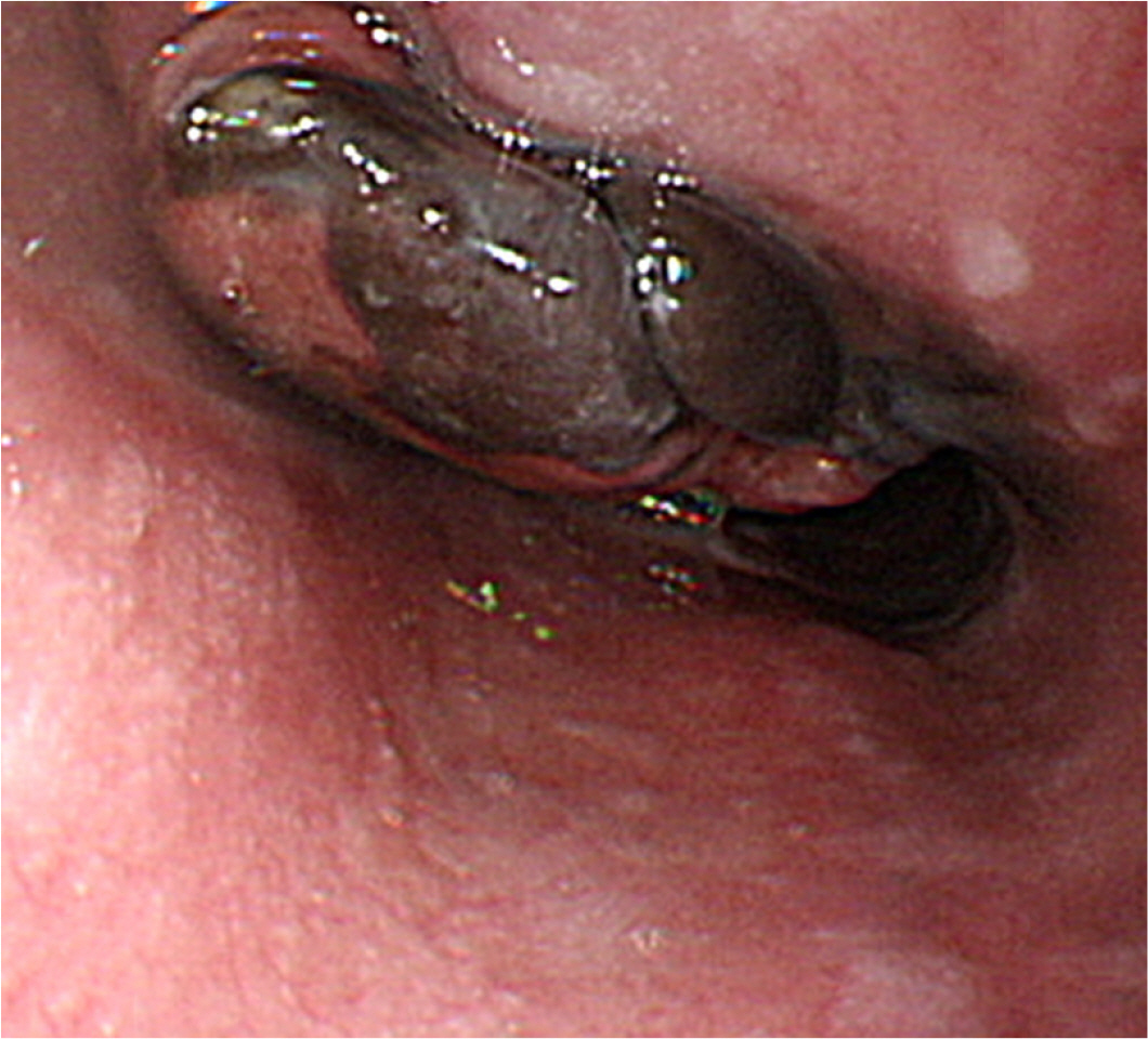
Fig. 1 Endoscopic findings show a black pigmented polypoid mass.

Fig. 2 Chest computed tomography shows the eccentric irregular wall thickening and enhancement in (A) the mid-esophagus (arrow) and multiple necrotizing lymphadenopathies at (B) the carina (arrow).

Fig. 3 Pathological findings. (A) In the endoscopic biopsy specimens, brown-colored atypical melanocytes are observed (arrows) (hematoxylin and eosin stain [H&E], ×200). Immunohistochemical staining is positive for (B) Melan A (×200), (C) S-100 (×200), and (D) HMB45 (×200) proteins.

Fig. 4 Endoscopy findings show (A) a protruding mass without black pigmentation, and black pigmented and (B) flat lesion in the esophagus.
Reference
-
1. Iwanuma Y, Tomita N, Amano T, et al. 2012; Current status of primary malignant melanoma of the esophagus: clinical features, pathology, management and prognosis. J Gastroenterol. 47:21–28. DOI: 10.1007/s00535-011-0490-y. PMID: 22048255.
Article2. Mihajlovic M, Vlajkovic S, Jovanovic P, Stefanovic V. 2012; Primary mucosal melanomas: a comprehensive review. Int J Clin Exp Pathol. 5:739–753. PMID: 23071856. PMCID: PMC3466987.3. Zheng J, Mo H, Ma S, Wang Z. 2014; Clinicopathological findings of primary esophageal malignant melanoma: report of six cases and review of literature. Int J Clin Exp Pathol. 7:7230–7235. PMID: 25400820. PMCID: PMC4230078.4. Kim YB, Woo JS, Kim CY, et al. 1991; Primary malignant melanoma of the esophagus. Korean J Gastroenterol. 23:971–976. DOI: 10.4166/kjg.2011.57.4.262. PMID: 21519182.
Article5. Park SJ, Hwang JH, Cho YS, et al. 1997; A operated case of primary melanoma of the esophagus. Korean J Gastrointest Endosc. 17:663–667.6. Jung BJ, Shin YM, Oh HT, et al. 1997; Esophagus, stomach & intestine; a case of primary malignant melanoma of the esophagus. Korean J Gastrointest Endosc. 17:163–166.7. Lee SA, Choi YH, Jo WM, et al. 1998; Primary malignant melanoma of the esophagus: 1 cases report. Korean J Thorac Cardiovasc Surg. 31:544–548.8. Park JK, Lee SH, Kim SH, et al. 1998; Primary malignant melanoma of the esophagus - a case report -. Korean J Thorac Cardiovasc Surg. 31:1106–1109. DOI: 10.12659/AJCR.894041,. PMID: 26212619. PMCID: PMC4520419.9. Lee SH, Park SH, Kim HG, Kim CB. 1998; Primary malignant melanoma of the esophagus. Yonsei Med J. 39:468–473. DOI: 10.3349/ymj.1998.39.5.468. PMID: 9821797.
Article10. Lee HJ, Choi ST, Eun JR, et al. 2001; A case of pimary malignant melanoma originated from esophageal melanosis. Korean J Med. 61:71–77.11. Lee JW, Park MS, Kim KW, et al. 2007; Primary malignant melanoma of the esophagus: a case report. J Korean Radiol Soc. 57:37–41. DOI: 10.3348/jkrs.2007.57.1.37. PMID: 26212619. PMCID: PMC4520419.
Article12. Lee SH, Park YH, Ryu BR, Kim HT, Jeong SH. 2004; Primary malignant melanoma of the esophagus. Korean J Med. 66:234–236. DOI: 10.1111/j.1442-2050.2000.00140.x. PMID: 17297270. PMCID: PMC2693554.
Article13. Park JS, Choi SJ, Cha CK, et al. 2006; A case of primary malignant melanoma of the esophagus treated by esophagectomy. Korean J Gastrointest Endosc. 33:220–225.14. Lee SA, Hwang JJ, Choi YH. 2007; Surgical treatment of primary malignant melanoma of the esophagus: a case report. J Korean Med Sci. 22:149–152. DOI: 10.3346/jkms.2007.22.1.149. PMID: 17297270. PMCID: PMC2693554.
Article15. Kim SH, Kim JK, Kim JI, et al. 2009; A case of primary malignant melanoma of the esophagus. Korean J Gastrointest Endosc. 38:133–136. DOI: 10.1093/jjco/hyn018. PMID: 30887427.16. Kang MJ, Yi SY. 2009; Nevus-like appearance of primary malignant melanoma of the esophagus. Gastroenterol Res Pract. 2009:285753. DOI: 10.1155/2009/285753. PMID: 19644559. PMCID: PMC2716486.
Article17. Kim SG. 2011; Primary malignant melanoma of the esophagus. Korean J Gastroenterol. 57:262–264. DOI: 10.4166/kjg.2011.57.4.262. PMID: 21519182.
Article18. Lee J, Kim JY, Bae JY, et al. 2015; Primary malignant melanoma of the esophagus treated by early diagnosis and surgical resection. Korean J Helicobacter Up Gastrointest Res. 15:127–131. DOI: 10.7704/kjhugr.2015.15.2.127.
Article19. Choi HH, Kim SS, Shin OR. 2018; Asymptomatic scattered pigmented flat lesions in the esophagus. Gastroenterology. 154:e8–e9. DOI: 10.1053/j.gastro.2017.06.057. PMID: 28712752.
Article20. Greene FL, Page DL, Fleming ID, et al. 2002. AJCC cancer staging manual. 6th ed. Springer Science & Business Media, 2002;New York: DOI: 10.1007/978-1-4757-3656-4.
- Full Text Links
-
- Actions
-
Cited
- CITED
-
- Close
- Share
-
- Similar articles
-
- Esophageal Metastasis of Malignant Melanoma in a 66-year-old Female Patient
- A case of pimary malignant melanoma originated from esophageal melanosis
- Primary Malignant Melanoma of the Esophagus: 1 cases report
- Primary malignant melanoma of the esophagus
- Primary Malignant Melanoma of the Central Nervous System: Case Report
