The Differentiation of Pluripotent Stem Cells towards Endothelial Progenitor Cells – Potential Application in Pulmonary Arterial Hypertension
- Affiliations
-
- 1Department of Cell Biology, Institute of Basic Medical Sciences, Chinese Academy of Medical Sciences & Peking Union Medical College, Beijing, China
- 2Department of Physiology, and Department of Cardiology of the Second Affiliated Hospital, Zhejiang University School of Medicine, Hangzhou, China
- KMID: 2529784
- DOI: http://doi.org/10.15283/ijsc21044
Abstract
- Background and Objectives
Endothelial progenitor cells (EPCs) and endothelial cells (ECs) have been applied in the clinic to treat pulmonary arterial hypertension (PAH), a disease characterized by disordered pulmonary vasculature. However, the lack of sufficient transplantable cells before the deterioration of disease condition is a current limitation to apply cell therapy in patients. It is necessary to differentiate pluripotent stem cells (PSCs) into EPCs and identify their characteristics.
Methods and Results
Comparing previously reported methods of human PSCs-derived ECs, we optimized a highly efficient differentiation protocol to obtain cells that match the phenotype of isolated EPCs from healthy donors. The protocol is compatible with chemically defined medium (CDM), it could produce a large number of clinically applicable cells with low cost. Moreover, we also found PSCs-derived EPCs express CD133, have some characteristics of mesenchymal stem cells and are capable of homing to repair blood vessels in zebrafish xenograft assays. In addition, we further revealed that IPAH PSCs-derived EPCs have higher expression of proliferation-related genes and lower expression of immune-related genes than normal EPCs and PSCs-derived EPCs through microarray analysis.
Conclusions
In conclusion, we optimized a highly efficient differentiation protocol to obtain PSCs-derived EPCs with the phenotypic and molecular characteristics of EPCs from healthy donors which distinguished them from EPCs from PAH.
Keyword
Figure
-

Fig. 1 Identifying induction factors for PSCs-derived EPCs. (A) A schematic diagram of inducing human PSCs-derived ECs. (B) The impact of cell seeding number on differentia-tion efficiency of PSCs-derived EC was analysed by flow cytometry. (C) In the second step of PSCs-derived EC differentiation, flow cytometry analysis showed adding BMP4 promoted PSCs-derived EC differentia-tion efficiency. (D, E) In the second step of EC differentiation, increasing SB431542 dose (5∼20 μM) promoted PSCs-derived EC differentia-tion efficiency. (F, G) The inhibitor Y27632 enhanced 202-iPSCs-deri-ved EC differentiation efficiency in the first step of differentiation in which Y27632 was used for three days. Statistics of CD31+CD34+ cells, CD31+ cells, CD34+ cells and CD43+ cells. Data are represented as the mean±SD. n=3∼4, the experiments were repeated more than three independent times. Student’s t test was performed (*p<0.05, **p< 0.01, ***p<0.001).

Fig. 2 DMSO improves EC and HE cell differentiation efficiency. (A, B) PSCs-derived EC (H1EC) differentia-tion efficiency increased when the cells were treated with DMSO in the first step for one day or three days. Statistics of CD31+CD34+ cells, CD31+ cells, CD34+ cells and CD43+ cells were analysed by flow cytometry. (C, D) Improved protocol for the HE differentiation of PSCs. A schematic digram of the stepwise induction process was shown. DMSO treatment for three days in the first step promoted the differentiation efficiency as analysed by flow cyto-metry. Statistics of APLNR+ cells, CD31+CD34+ cells, CD31+ cells, CD34+ cells and CD43+cells. Data were represented as the mean±SD, n=3∼4, and the experiments were repeated more than three times. Student’s t test was performed (ns: not significant, **p<0.01,****p< 0.0001).

Fig. 3 Improved protocol for the highly efficient differentiation rate of PSCs-derived ECs. (A) Schematic diagram of inducing human PSCs-derived ECs via a mesoderm interme-diate. (B) Representative yields of APLNR+ cells as analysed by flow cytometry. (C) Representative yields of CD34+CD31+ cells and CD43+ cells were analysed by flow cytome-try after 7 days of differentiation. (D) Statistics of CD31+CD34+ and CD43+ cells. Data were represented as the mean±SD, n=3, and the experiments were repeated three independent times.

Fig. 4 The functional characteristics of PSCs-derived ECs. (A) Uptake assay of Dil-acetylated LDL by the cells (scale bars: 50 μm). (B) Tube formation assay of H1-derived ECs after 4 hours and 12 hours (scale bars: 500 μm). (C) Brief outline of Zebrafish experiments. (D) Vascular competence of PSCs-derived ECs in a zebrafish xenograft model. Representative image of ECs-derived vessel-type structures (in red) within embryonic zebrafish (Flk: GFP; in green) 2 days after implantation, with magnification of the vessel. Scale bars are 300 μm. (E) Using a zebrafish model for gene therapy. Data were represented as the mean±SD, n=3, the experiments were repeated three times with 20∼30 fish per condition. Student’s t test was performed (*p<0.05). (F) CD34+EPCs (labelled with FITC-CD34, green) were injected into the ventral end of the duct of Cuvier of Zebra fish, the cells stained with green fluorescence incorporated into the vasculature of kdr1:mcherry (red) line at 24 hour and 48 hour.

Fig. 5 PSCs-derived ECs have characteristics of EPCs. (A) Comparison of the cell morphology of PSCs-derived ECs (H7EC, 202EC) with peripheral blood-derived normal EPCs (normal EPC1, normal EPC2) and IPAH-EPCs (IPAH EPC1, IPAH EPC2) (scale bars: 50 μm). (B) qRT-PCR of NRP1, which was reported to promote ECFC proliferation. GAPDH was used as an internal control. (C) ECs from DAY 5 or DAY 7 were analysed by the expression of the genes CD133, EFNB2, and EPHB4. GAPDH was used as an internal control. Data were represented as the mean±SD, n=3, and the experiments were repeated three times and analysed with student’s t test (*p<0.05, **p< 0.01, ***p<0.001).

Fig. 6 Bioinformatics analysis further reveals the characteristics of PSCs-derived EPCs. (A, B) Heatmap of EC-related genes from the 2D_MG_H1EC and 2D_MG_HUVEC datasets from GSE93511 and our microarray data in IPAH-EPCs (IPAH1, IPAH2, IPAH3), normal EPCs (Con1, Con2, Con3) and PSCs-derived EPCs (H7EC, H9EC, 202EC). PROM1, SPN and PTPRC were not expressed, and EFNB2 had a higher expression level in HUVECs (A), but PROM1, SPN and PTPRC were highly expressed in PSCs-derived EPCs, normal EPCs, IPAH-EPCs and 2D-MGH1EC (B). Moreover, EFNB2 also had a higher expression level in PSCs-derived EPCs than in normal EPCs and IPAH-EPCs (B). (C, D) Heatmap of homing-related genes from the 2D_MG_H1EC and 2D_MG_HUVEC datasets from GSE93511 and our microarray data in IPAH-EPCs (IPAH1, IPAH2, IPAH3), normal EPCs (Con1, Con2, Con3) and PSCs-derived EPCs (H7EC, H9EC, 202EC). IGF2, CXCL12 and CD90 (THY1) were not expressed in HUVECs (C), but had higher expression levels in PSCs-derived EPCs, normal EPCs, IPAH-EPCs (D) and 2D-MG-H1EC (C). IGFBP3 had a lower expression level in IPAH-EPCs than in PSCs-derived EPCs and normal EPCs (D).
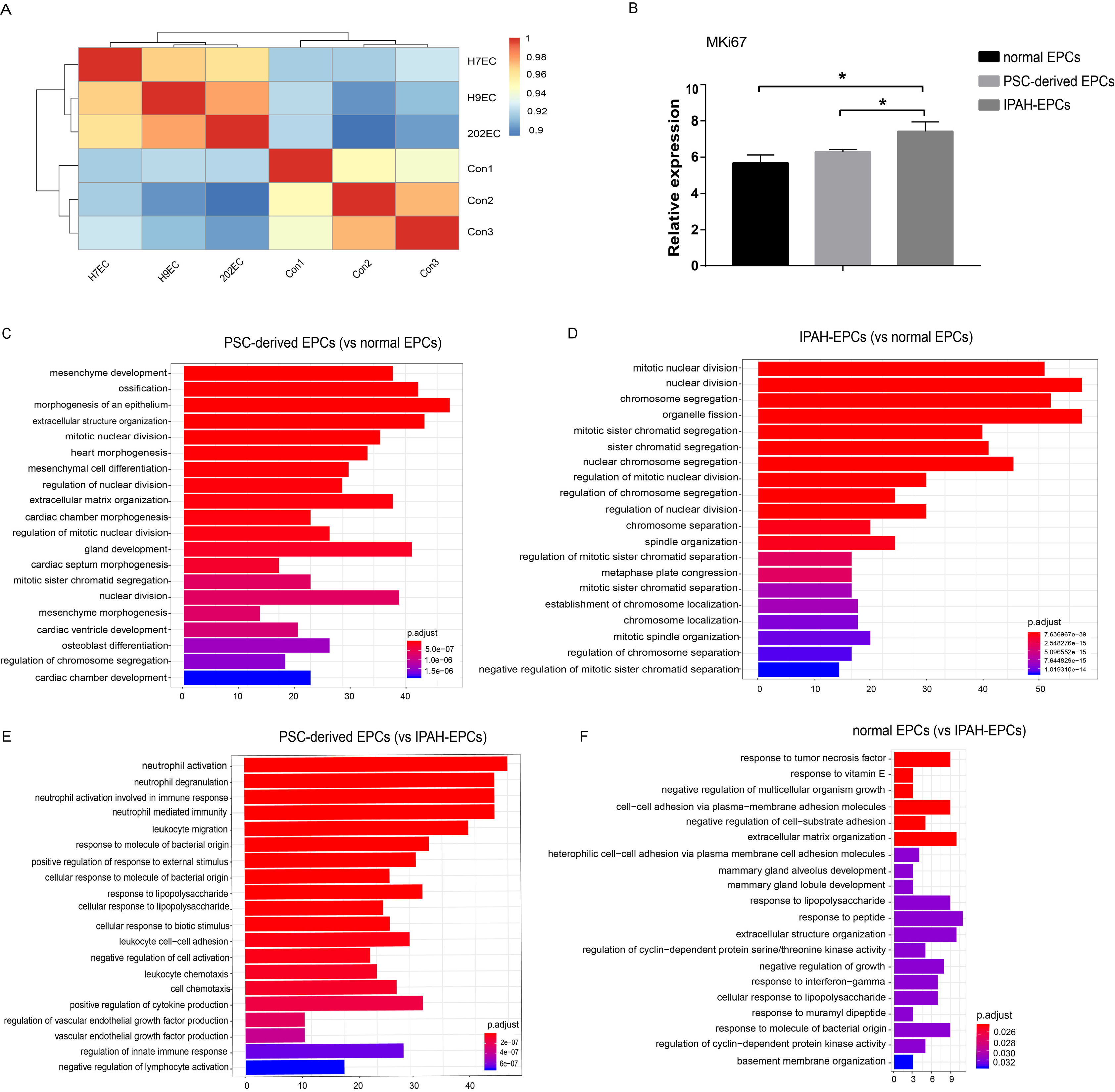
Fig. 7 PSCs-derived EPCs have special molecular characteristics compared with normal EPCs and IPAH-EPCs (IPAH1, IPAH2 and IPAH3) based on our microarray data. (A) Correlation analysis between normal EPCs (Con1, Con2, Con3) and PSCs-derived EPCs (H7EC, H9EC, 202EC). (B) MKi67 relative expression from microarray data. Data were represented as the mean±SD. n=3, Student’s t test was performed (*p<0.05). (C, D) GOBP analysis (TOP20) of PSCs-derived EPCs and IPAH-EPCs relative to normal EPCs. (E, F) GOBP analysis (TOP20) of PSCs-derived EPCs and normal EPCs relative to IPAH-EPCs.
Reference
-
References
1. Niskanen H, Tuszynska I, Zaborowski R, Heinäniemi M, Ylä-Herttuala S, Wilczynski B, Kaikkonen MU. 2018; Endothelial cell differentiation is encompassed by changes in long range interactions between inactive chromatin regions. Nucleic Acids Res. 46:1724–1740. DOI: 10.1093/nar/gkx1214. PMID: 29216379. PMCID: PMC5829566.
Article2. Zhao W, Zhao L, Liao J, Luo Y, He L. 2019; Early risk assessment of circulating endothelial progenitor cells and plasma stromal cell-derived factor-1 for nondisabling ischemic cerebrovascular events. BMC Neurol. 19:22. DOI: 10.1186/s12883-019-1250-5. PMID: 30755169. PMCID: PMC6371535. PMID: d1eac0c7980f43839bbb40d45011f554.
Article3. Ward MR, Stewart DJ, Kutryk MJ. 2007; Endothelial progenitor cell therapy for the treatment of coronary disease, acute MI, and pulmonary arterial hypertension: current perspectives. Catheter Cardiovasc Interv. 70:983–998. DOI: 10.1002/ccd.21302. PMID: 18044749.
Article4. Sekiguchi H, Ii M, Losordo DW. 2009; The relative potency and safety of endothelial progenitor cells and unselected mononuclear cells for recovery from myocardial infarction and ischemia. J Cell Physiol. 219:235–242. DOI: 10.1002/jcp.21672. PMID: 19115244.
Article5. Zhang J, Chu LF, Hou Z, Schwartz MP, Hacker T, Vickerman V, Swanson S, Leng N, Nguyen BK, Elwell A, Bolin J, Brown ME, Stewart R, Burlingham WJ, Murphy WL, Thomson JA. 2017; Functional characterization of human pluripotent stem cell-derived arterial endothelial cells. Proc Natl Acad Sci U S A. 114:E6072–E6078. DOI: 10.1073/pnas.1702295114. PMID: 28696312. PMCID: PMC5544294.
Article6. Medina RJ, O'Neill CL, O'Doherty TM, Wilson SE, Stitt AW. 2012; Endothelial progenitors as tools to study vascular disease. Stem Cells Int. 2012:346735. DOI: 10.1155/2012/346735. PMID: 22550504. PMCID: PMC3329655.
Article7. Medina RJ, O'Neill CL, Sweeney M, Guduric-Fuchs J, Gardiner TA, Simpson DA, Stitt AW. 2010; Molecular analysis of endothelial progenitor cell (EPC) subtypes reveals two distinct cell populations with different identities. BMC Med Genomics. 3:18. DOI: 10.1186/1755-8794-3-18. PMID: 20465783. PMCID: PMC2881111.
Article8. Belair DG, Whisler JA, Valdez J, Velazquez J, Molenda JA, Vickerman V, Lewis R, Daigh C, Hansen TD, Mann DA, Thomson JA, Griffith LG, Kamm RD, Schwartz MP, Murphy WL. 2015; Human vascular tissue models formed from human induced pluripotent stem cell derived endothelial cells. Stem Cell Rev Rep. 11:511–525. DOI: 10.1007/s12015-014-9549-5. PMID: 25190668. PMCID: PMC4411213.
Article9. James D, Nam HS, Seandel M, Nolan D, Janovitz T, Tomishima M, Studer L, Lee G, Lyden D, Benezra R, Zaninovic N, Rosenwaks Z, Rabbany SY, Rafii S. 2010; Expansion and maintenance of human embryonic stem cell-derived endothelial cells by TGFbeta inhibition is Id1 dependent. Nat Biotechnol. 28:161–166. DOI: 10.1038/nbt.1605. PMID: 20081865. PMCID: PMC2931334.
Article10. Lian X, Bao X, Al-Ahmad A, Liu J, Wu Y, Dong W, Dunn KK, Shusta EV, Palecek SP. 2014; Efficient differentiation of human pluripotent stem cells to endothelial progenitors via small-molecule activation of WNT signaling. Stem Cell Reports. 3:804–816. DOI: 10.1016/j.stemcr.2014.09.005. PMID: 25418725. PMCID: PMC4235141.
Article11. Nguyen MTX, Okina E, Chai X, Tan KH, Hovatta O, Ghosh S, Tryggvason K. 2016; Differentiation of human embryonic stem cells to endothelial progenitor cells on laminins in defined and xeno-free systems. Stem Cell Reports. 7:802–816. DOI: 10.1016/j.stemcr.2016.08.017. PMID: 27693424. PMCID: PMC5063508.
Article12. Patsch C, Challet-Meylan L, Thoma EC, Urich E, Heckel T, O'Sullivan JF, Grainger SJ, Kapp FG, Sun L, Christensen K, Xia Y, Florido MH, He W, Pan W, Prummer M, Warren CR, Jakob-Roetne R, Certa U, Jagasia R, Freskgård PO, Adatto I, Kling D, Huang P, Zon LI, Chaikof EL, Gerszten RE, Graf M, Iacone R, Cowan CA. 2015; Generation of vascular endothelial and smooth muscle cells from human pluripotent stem cells. Nat Cell Biol. 17:994–1003. DOI: 10.1038/ncb3205. PMID: 26214132. PMCID: PMC4566857.
Article13. Prasain N, Lee MR, Vemula S, Meador JL, Yoshimoto M, Ferkowicz MJ, Fett A, Gupta M, Rapp BM, Saadatzadeh MR, Ginsberg M, Elemento O, Lee Y, Voytik-Harbin SL, Chung HM, Hong KS, Reid E, O'Neill CL, Medina RJ, Stitt AW, Murphy MP, Rafii S, Broxmeyer HE, Yoder MC. 2014; Differentiation of human pluripotent stem cells to cells similar to cord-blood endothelial colony-forming cells. Nat Biotechnol. 32:1151–1157. DOI: 10.1038/nbt.3048. PMID: 25306246. PMCID: PMC4318247.
Article14. Park TS, Bhutto I, Zimmerlin L, Huo JS, Nagaria P, Miller D, Rufaihah AJ, Talbot C, Aguilar J, Grebe R, Merges C, Reijo-Pera R, Feldman RA, Rassool F, Cooke J, Lutty G, Zambidis ET. 2014; Vascular progenitors from cord blood-derived induced pluripotent stem cells possess augmented capacity for regenerating ischemic retinal vasculature. Circulation. 129:359–372. DOI: 10.1161/CIRCULATIONAHA.113.003000. PMID: 24163065. PMCID: PMC4090244.
Article15. Park SJ, Lee JH, Lee SG, Lee JE, Seo J, Choi JJ, Jung TH, Chung EB, Kim HN, Ju J, Song YH, Chung HM, Lee DR, Moon SH. 2019; Functional equivalency in human somatic cell nuclear transfer-derived endothelial cells. Stem Cells. 37:623–630. DOI: 10.1002/stem.2986. PMID: 30721559.
Article16. Han X, Chen H, Huang D, Chen H, Fei L, Cheng C, Huang H, Yuan GC, Guo G. 2018; Mapping human pluripotent stem cell differentiation pathways using high throughput single-cell RNA-sequencing. Genome Biol. 19:47. DOI: 10.1186/s13059-018-1426-0. PMID: 29622030. PMCID: PMC5887227.
Article17. Toshner M, Voswinckel R, Southwood M, Al-Lamki R, Howard LS, Marchesan D, Yang J, Suntharalingam J, Soon E, Exley A, Stewart S, Hecker M, Zhu Z, Gehling U, Seeger W, Pepke-Zaba J, Morrell NW. 2009; Evidence of dysfunction of endothelial progenitors in pulmonary arterial hypertension. Am J Respir Crit Care Med. 180:780–787. DOI: 10.1164/rccm.200810-1662OC. PMID: 19628780. PMCID: PMC2778151.
Article18. Foris V, Kovacs G, Marsh LM, Bálint Z, Tötsch M, Avian A, Douschan P, Ghanim B, Klepetko W, Olschewski A, Olschewski H. 2016; CD133+ cells in pulmonary arterial hypertension. Eur Respir J. 48:459–469. DOI: 10.1183/13993003.01523-2015. PMID: 27103380.19. Gomberg-Maitland M, Bull TM, Saggar R, Barst RJ, Elgazayerly A, Fleming TR, Grimminger F, Rainisio M, Stewart DJ, Stockbridge N, Ventura C, Ghofrani AH, Rubin LJ. 2013; New trial designs and potential therapies for pulmonary artery hypertension. J Am Coll Cardiol. 62(25 Suppl):D82–D91. DOI: 10.1016/j.jacc.2013.10.026. PMID: 24355645. PMCID: PMC4117578.
Article20. Ormiston ML, Toshner MR, Kiskin FN, Huang CJ, Groves E, Morrell NW, Rana AA. 2015; Generation and culture of blood outgrowth endothelial cells from human peripheral blood. J Vis Exp. (106):e53384. DOI: 10.3791/53384. PMID: 26780290. PMCID: PMC4758763.
Article21. Yu G, Wang LG, Han Y, He QY. 2012; clusterProfiler: an R package for comparing biological themes among gene clusters. OMICS. 16:284–287. DOI: 10.1089/omi.2011.0118. PMID: 22455463. PMCID: PMC3339379.
Article22. Zhang J, Schwartz MP, Hou Z, Bai Y, Ardalani H, Swanson S, Steill J, Ruotti V, Elwell A, Nguyen BK, Bolin J, Stewart R, Thomson JA, Murphy WL. 2017; A genome-wide analysis of human pluripotent stem cell-derived endothelial cells in 2D or 3D culture. Stem Cell Reports. 8:907–918. DOI: 10.1016/j.stemcr.2017.02.014. PMID: 28343999. PMCID: PMC5390115.
Article23. Duan F, Huang R, Zhang F, Zhu Y, Wang L, Chen X, Bai L, Guo W, Chang SC, Hu X, Na J. 2018; Biphasic modulation of insulin signaling enables highly efficient hematopoietic differentiation from human pluripotent stem cells. Stem Cell Res Ther. 9:205. DOI: 10.1186/s13287-018-0934-x. PMID: 30053898. PMCID: PMC6062919.
Article24. Wang H, Liu C, Liu X, Wang M, Wu D, Gao J, Su P, Nakahata T, Zhou W, Xu Y, Shi L, Ma F, Zhou J. 2018; MEIS1 regulates hemogenic endothelial generation, megakaryopoiesis, and thrombopoiesis in human pluripotent stem cells by targeting TAL1 and FLI1. Stem Cell Reports. 10:447–460. DOI: 10.1016/j.stemcr.2017.12.017. PMID: 29358086. PMCID: PMC5830947.
Article25. Bai H, Gao Y, Arzigian M, Wojchowski DM, Wu WS, Wang ZZ. 2010; BMP4 regulates vascular progenitor development in human embryonic stem cells through a Smad-dependent pathway. J Cell Biochem. 109:363–374. DOI: 10.1002/jcb.22410. PMID: 19950207. PMCID: PMC3065830.
Article26. Sadahiro T, Isomi M, Muraoka N, Kojima H, Haginiwa S, Kurotsu S, Tamura F, Tani H, Tohyama S, Fujita J, Miyoshi H, Kawamura Y, Goshima N, Iwasaki YW, Murano K, Saito K, Oda M, Andersen P, Kwon C, Uosaki H, Nishizono H, Fukuda K, Ieda M. 2018; Tbx6 induces nascent mesoderm from pluripotent stem cells and temporally controls cardiac versus somite lineage diversification. Cell Stem Cell. 23:382–395.e5. DOI: 10.1016/j.stem.2018.07.001. PMID: 30100166. PMCID: PMC6190602.
Article27. Orlova VV, van den Hil FE, Petrus-Reurer S, Drabsch Y, Ten Dijke P, Mummery CL. 2014; Generation, expansion and functional analysis of endothelial cells and pericytes derived from human pluripotent stem cells. Nat Protoc. 9:1514–1531. DOI: 10.1038/nprot.2014.102. PMID: 24874816.
Article28. Yoder MC, Mead LE, Prater D, Krier TR, Mroueh KN, Li F, Krasich R, Temm CJ, Prchal JT, Ingram DA. 2007; Redefining endothelial progenitor cells via clonal analysis and hematopoietic stem/progenitor cell principals. Blood. 109:1801–1809. DOI: 10.1182/blood-2006-08-043471. PMID: 17053059. PMCID: PMC1801067.
Article29. Kanayasu-Toyoda T, Tanaka T, Kikuchi Y, Uchida E, Matsuyama A, Yamaguchi T. 2016; Cell-surface MMP-9 protein is a novel functional marker to identify and separate proangiogenic cells from early endothelial progenitor cells derived from CD133(+) cells. Stem Cells. 34:1251–1262. DOI: 10.1002/stem.2300. PMID: 26824798.
Article30. Yuan Z, Kang L, Wang Z, Chen A, Zhao Q, Li H. 2018; 17β-estradiol promotes recovery after myocardial infarction by enhancing homing and angiogenic capacity of bone marrow-derived endothelial progenitor cells through ERα-SDF-1/CXCR4 crosstalking. Acta Biochim Biophys Sin (Shanghai). 50:1247–1256. DOI: 10.1093/abbs/gmy127. PMID: 30371725.
Article31. Zhuang Y, Chen X, Xu M, Zhang LY, Xiang F. 2009; Chemokine stromal cell-derived factor 1/CXCL12 increases homing of mesenchymal stem cells to injured myocardium and neovascularization following myocardial infarction. Chin Med J (Engl). 122:183–187. DOI: 10.3901/jme.2009.07.183. PMID: 19187644.32. Xiaowei C, Jia M, Xiaowei W, Yina Z. 2013; Overexpression of CXCL12 chemokine up-regulates connexin and integrin expression in mesenchymal stem cells through PI3K/Akt pathway. Cell Commun Adhes. 20:67–72. DOI: 10.3109/15419061.2013.791682. PMID: 23659290.
Article33. Ferrari D, Gulinelli S, Salvestrini V, Lucchetti G, Zini R, Manfredini R, Caione L, Piacibello W, Ciciarello M, Rossi L, Idzko M, Ferrari S, Di Virgilio F, Lemoli RM. 2011; Purinergic stimulation of human mesenchymal stem cells potentiates their chemotactic response to CXCL12 and increases the homing capacity and production of proinflammatory cytokines. Exp Hematol. 39:360–374. e1–e5. DOI: 10.1016/j.exphem.2010.12.001. PMID: 21145936.
Article34. Caplan AI. 2015; Adult mesenchymal stem cells and women's health. Menopause. 22:131–135. DOI: 10.1097/GME.0000000000000408. PMID: 25608271. PMCID: PMC4308553.
Article35. Jiang B, Yan L, Wang X, Li E, Murphy K, Vaccaro K, Li Y, Xu RH. 2019; Concise review: mesenchymal stem cells derived from human pluripotent cells, an unlimited and quality-controllable source for therapeutic applications. Stem Cells. 37:572–581. DOI: 10.1002/stem.2964. PMID: 30561809.
Article36. Lofqvist C, Chen J, Connor KM, Smith AC, Aderman CM, Liu N, Pintar JE, Ludwig T, Hellstrom A, Smith LE. 2007; IGFBP3 suppresses retinopathy through suppression of oxygen-induced vessel loss and promotion of vascular regrowth. Proc Natl Acad Sci U S A. 104:10589–10594. DOI: 10.1073/pnas.0702031104. PMID: 17567756. PMCID: PMC1965557.
Article37. Chang KH, Chan-Ling T, McFarland EL, Afzal A, Pan H, Baxter LC, Shaw LC, Caballero S, Sengupta N, Li Calzi S, Sullivan SM, Grant MB. 2007; IGF binding protein-3 regulates hematopoietic stem cell and endothelial precursor cell function during vascular development. Proc Natl Acad Sci U S A. 104:10595–10600. DOI: 10.1073/pnas.0702072104. PMID: 17567755. PMCID: PMC1965558.
Article38. Li J, Narayanan C, Bian J, Sambo D, Brickler T, Zhang W, Chetty S. 2018; A transient DMSO treatment increases the differentiation potential of human pluripotent stem cells through the Rb family. PLoS One. 13:e0208110. DOI: 10.1371/journal.pone.0208110. PMID: 30540809. PMCID: PMC6291069. PMID: b747acd0c6e0441ba715bbadeeeecf34.
Article39. Czysz K, Minger S, Thomas N. 2015; DMSO efficiently down regulates pluripotency genes in human embryonic stem cells during definitive endoderm derivation and increases the proficiency of hepatic differentiation. PLoS One. 10:e0117689. DOI: 10.1371/journal.pone.0117689. PMID: 25659159. PMCID: PMC4320104. PMID: 91b67dffeae5493fac82e9d59091272c.
Article40. Pal R, Mamidi MK, Das AK, Bhonde R. 2012; Diverse effects of dimethyl sulfoxide (DMSO) on the differentiation potential of human embryonic stem cells. Arch Toxicol. 86:651–661. DOI: 10.1007/s00204-011-0782-2. PMID: 22105179.
Article41. Vernardis SI, Terzoudis K, Panoskaltsis N, Mantalaris A. 2017; Human embryonic and induced pluripotent stem cells maintain phenotype but alter their metabolism after exposure to ROCK inhibitor. Sci Rep. 7:42138. DOI: 10.1038/srep42138. PMID: 28165055. PMCID: PMC5292706.
Article42. Motomura K, Okada N, Morita H, Hara M, Tamari M, Orimo K, Matsuda G, Imadome KI, Matsuda A, Nagamatsu T, Fujieda M, Sago H, Saito H, Matsumoto K. 2017; A Rho-associated coiled-coil containing kinases (ROCK) inhibitor, Y-27632, enhances adhesion, viability and differentiation of human term placenta-derived trophoblasts in vitro. PLoS One. 12:e0177994. DOI: 10.1371/journal.pone.0177994. PMID: 28542501. PMCID: PMC5438149.
Article43. Kurosawa H. 2012; Application of Rho-associated protein kinase (ROCK) inhibitor to human pluripotent stem cells. J Biosci Bioeng. 114:577–581. DOI: 10.1016/j.jbiosc.2012.07.013. PMID: 22898436.
Article44. Maldonado M, Luu RJ, Ramos ME, Nam J. 2016; ROCK inhibitor primes human induced pluripotent stem cells to selectively differentiate towards mesendodermal lineage via epithelial-mesenchymal transition-like modulation. Stem Cell Res. 17:222–227. DOI: 10.1016/j.scr.2016.07.009. PMID: 27591478.
Article45. Ingram DA, Mead LE, Tanaka H, Meade V, Fenoglio A, Mortell K, Pollok K, Ferkowicz MJ, Gilley D, Yoder MC. 2004; Identification of a novel hierarchy of endothelial progenitor cells using human peripheral and umbilical cord blood. Blood. 104:2752–2760. DOI: 10.1182/blood-2004-04-1396. PMID: 15226175.
Article46. Gu M, Shao NY, Sa S, Li D, Termglinchan V, Ameen M, Karakikes I, Sosa G, Grubert F, Lee J, Cao A, Taylor S, Ma Y, Zhao Z, Chappell J, Hamid R, Austin ED, Gold JD, Wu JC, Snyder MP, Rabinovitch M. 2017; Patient-specific iPSC-derived endothelial cells uncover pathways that protect against pulmonary hypertension in BMPR2 mutation carriers. Cell Stem Cell. 20:490–504.e5. DOI: 10.1016/j.stem.2016.08.019. PMID: 28017794. PMCID: PMC5500296.
Article
- Full Text Links
-
- Actions
-
Cited
- CITED
-
- Close
- Share
-
- Similar articles
-
- Comparative Evaluation for Potential Differentiation of Endothelial Progenitor Cells and Mesenchymal Stem Cells into Endothelial-Like Cells
- Endothelial progenitor cells and mesenchymal stem cells from human cord blood
- Generation of hematopoietic stem cells from human embryonic stem cells using a defined, stepwise, serum-free, and serum replacement-free monolayer culture method
- A Simple Method for Generating Cerebral Organoids from Human Pluripotent Stem Cells
- Liver Stem Cells
