Clinical Manifestations and Surgical Treatment Outcomes of Paranasal Sinus Osteoma
- Affiliations
-
- 1Department of Otorhinolaryngology Haed & Neck Suregery, Yonsei University College of Medicine, Seoul, Republic of Korea
- KMID: 2528315
- DOI: http://doi.org/10.18787/jr.2021.00388
Abstract
- Background and Objectives
Osteomas are the most common benign tumors of the nasal cavity and paranasal sinuses (PNSs). In this study, clinical features and imaging findings were analyzed in patients with osteoma confirmed by ostiomeatal unit (OMU) computed tomography (CT) and PNS CT, and the surgical treatment performed at our hospital was introduced.
Methods
The Severance Clinical Research Analysis Portal (SCRAP) service of Severance Hospital was used to collect research data. A total of 128 cases of osteomas of the nasal cavity or PNSs confirmed by OMU CT or PNS CT was retrospectively reviewed, including the location and size of the osteoma, clinical features, accompanying findings on imaging tests, and cases of surgical treatment.
Results
In this study, osteomas were found in about 0.55% of patients who underwent computed tomography. Osteomas were most frequently found in the ethmoid sinus, followed by the frontal sinus, fronto-ethmoid sinus, maxillary sinus, intranasal sphenoid sinus, and maxillary sinus-ethmoid sinus. Patients with osteomas complained of symptoms such as rhinorrhea, postnasal drip, nasal congestion, hyposmia, headache, visual disturbance, and lacrimal duct obstruction.
Conclusion
Surgical treatment was considered for patients presenting with severe headache, visual field symptoms, or accompanying rhinosinusitis. Surgery was performed by endoscopic or external approaches depending on location and size of the osteoma.
Figure
-
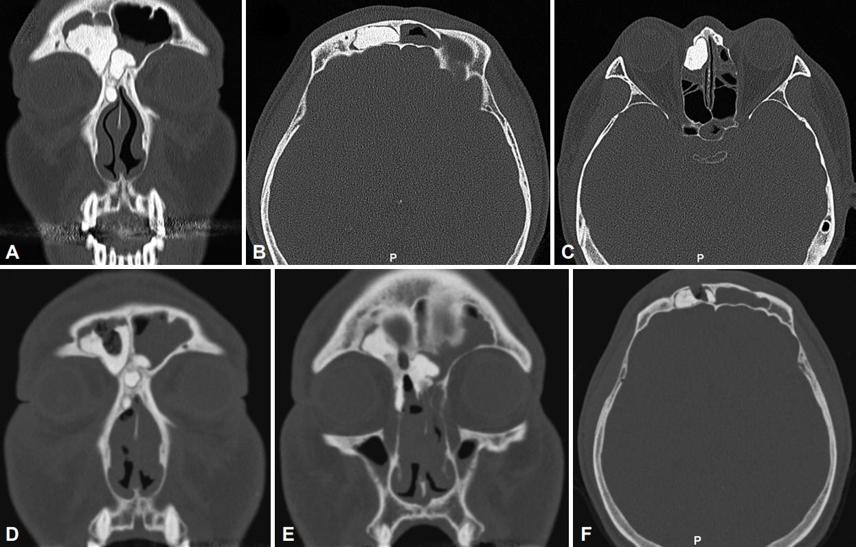
Fig. 1. CT scan of the 42-year-old male patient. A-C: Pre-operative CT scan. An osteoma filling the right frontal sinus and posterior ethmoid sinus was discovered. D-F: Post-operative CT scan at 15 months follow-up. A, D, E: Coronal view. B, C, F: Axial view. CT, computed tomography
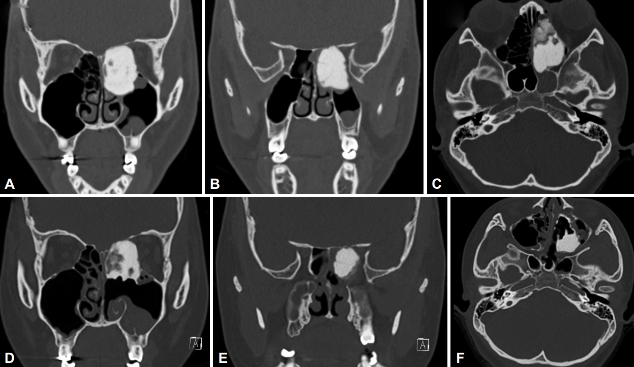
Fig. 2. CT scan of 25-year-old female patient. A-C: Pre-operative CT scan. An osteoma of 2.48×4.18×3.22 cm in size was compressing the optic nerve and forming a sphenoid sinus mucocele. D-F: Post-operative CT scan at 13 months follow-up. A, B, D, E: Coronal view. C, F: Axial view. CT, computed tomography.
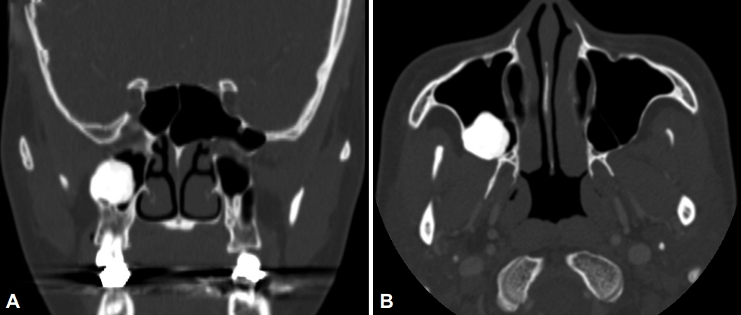
Fig. 3. CT scan of a 47-year-old female patient who visited outpatient clinic due to incidental finding of osteoma. An osteoma about 2 cm in size of posterior maxillary sinus was noted. A: Coronal view. B: Axial view. CT, computed tomography
Reference
-
References
1. Earwaker J. Paranasal sinus osteomas: a review of 46 cases. Skeletal Radiol. 1993; 22(6):417–23.
Article2. Erdogan N, Demir U, Songu M, Ozenler NK, Uluç E, Dirim B. A prospective study of paranasal sinus osteomas in 1,889 cases: changing patterns of localization. Laryngoscope. 2009; 119(12):2355–9.
Article3. Eggesbø HB. Imaging of sinonasal tumours. Cancer Imaging. 2012; 12:136–52.
Article4. Çelenk F, Baysal E, Karata ZA, Durucu C, Mumbuç S, Kanlıkama M. Paranasal sinus osteomas. J Craniofac Surg. 2012; 23(5):e433–7.
Article5. Karunaratne YG, Gunaratne DA, Floros P, Wong EH, Singh NP. Frontal sinus osteoma: from direct excision to endoscopic removal. J Craniofac Surg. 2019; 30(6):e494.6. Lund VJ, Stammberger H, Nicolai P, Castelnuovo P, Beal T, Beham A, et al. European position paper on endoscopic management of tumours of the nose, paranasal sinuses and skull base. Rhinol Suppl. 2010; 22:1–143.7. Buyuklu F, Akdogan MV, Ozer C, Cakmak O. Growth characteristics and clinical manifestations of the paranasal sinus osteomas. Otolaryngol Head Neck Surg. 2011; 145(2):319–23.
Article8. Koivunen P, Löppönen H, Fors AP, Jokinen K. The growth rate of osteomas of the paranasal sinuses. Clin Otolaryngol Allied Sci. 1997; 22(2):111–4.
Article9. Pons Y, Blancal JP, Vérillaud B, Sauvaget E, Ukkola-Pons E, Kania R, et al. Ethmoid sinus osteoma: diagnosis and management. Head Neck. 2013; 35(2):201–4.
Article10. Lee DH, Jung SH, Yoon TM, Lee JK, Joo YE, Lim SC. Characteristics of paranasal sinus osteoma and treatment outcomes. Acta Otolaryngol. 2015; 135(6):602–7.
Article11. Mansour AM, Salti H, Uwaydat S, Dakroub R, Bashshour Z. Ethmoid sinus osteoma presenting as epiphora and orbital cellulitis: case report and literature review. Surv Ophthalmol. 1999; 43(5):413–26.12. McHugh JB, Mukherji SK, Lucas DR. Sino-orbital osteoma: a clinicopathologic study of 45 surgically treated cases with emphasis on tumors with osteoblastoma-like features. Arch Pathol Lab Med. 2009; 133(10):1587–93.
Article13. Janovic A, Antic S, Rakocevic Z, Djuric M. Paranasal sinus osteoma: is there any association with anatomical variations? Rhinology. 2013; 51(1):54–60.
Article14. Jack LS, Smith TL, Ng JD. Frontal sinus osteoma presenting with orbital emphysema. Ophthalmic Plast Reconstr Surg. 2009; 25(2):155–7.
Article15. Nguyen S, Nadeau S. Giant frontal sinus osteomas: demographic, clinical presentation, and management of 10 cases. Am J Rhinol Allergy. 2019; 33(1):36–43.
Article16. Chiu AG, Schipor I, Cohen NA, Kennedy DW, Palmer JN. Surgical decisions in the management of frontal sinus osteomas. Am J Rhinol. 2005; 19(2):191–7.
Article17. Sofokleous V, Maragoudakis P, Kyrodimos E, Giotakis E. Management of paranasal sinus osteomas: a comprehensive narrative review of the literature and an up-to-date grading system. Am J Otolaryngol. 2020; 42(5):102644.
Article18. Georgalas C, Goudakos J, Fokkens WJ. Osteoma of the skull base and sinuses. Otolaryngol Clin North Am. 2011; 44(4):875–90.
Article19. Karligkiotis A, Pistochini A, Turri-Zanoni M, Terranova P, Volpi L, Battaglia P, et al. Endoscopic endonasal orbital transposition to expand the frontal sinus approaches. Am J Rhinol Allergy. 2015; 29(6):449–56.
Article
- Full Text Links
-
- Actions
-
Cited
- CITED
-
- Close
- Share
-
- Similar articles
-
- Pneumocephalus and Porencephaly Associated with a Frontoethmoid Sinus Osteoma: A Case Report
- A Huge Osteoma Originated From the Frontal Sinus: Case Report
- A Case of Osteoma in the Nasal Cavity
- Intranasal Endoscopic Removal of Osteoma in Ethmoid Sinus
- Osteoma of the Frontal Sinus with Secondary Subdural Empyema Formation
