The First Application of Transcatheter Caval Valve Implantation for Severe Tricuspid Regurgitation in a Patient With High Surgical Risk
- Affiliations
-
- 1Division of Cardiology, Department of Internal Medicine, Incheon St. Mary’s Hospital, College of Medicine, The Catholic University of Korea, Incheon, Korea
- 2Division of Cardiology, Department of Internal Medicine, Seoul St. Mary’s Hospital, College of Medicine, The Catholic University of Korea, Seoul, Korea
- KMID: 2526961
- DOI: http://doi.org/10.4070/kcj.2021.0392
Abstract
- No abstract available.
Figure
-
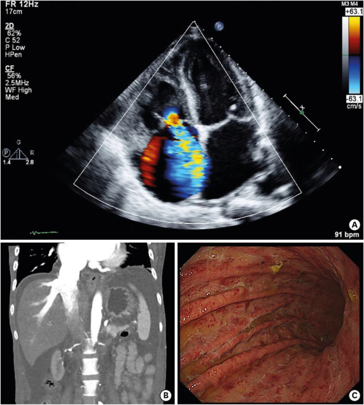
Figure 1 Echocardiogram, abdominal enhanced CT, and gastroscopy.(A) Apical 4 chamber view shows incomplete coaptation of tricuspid valve with severe tricuspid regurgitation by color doppler echocardiogram. (B) Abdominal enhanced CT scan illustrates significant contrast reflux into the hepatic veins and inferior vena cava. (C) Gastroscopy reveals submucosal congestion and erythema in the gastric body, suggesting congestive gastropathy.CT = computed tomography.
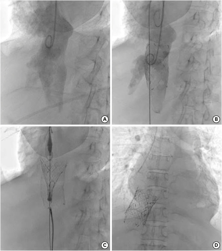
Figure 2 CAVI using self-expandable valve.(A) Cavogram by using a pigtail catheter through the right jugular vein reveals the locations of the RA, IVC, and hepatic veins. (B) After the placement of a 20 Fr sheath through the left femoral vein, a small pre-shaped Safari wire was located at the right atrium. After then, a large-diameter bare-metal stent (Niti-S® 30×60 mm; TaeWoong Medical) was placed from the junction of the IVC and RA to distal IVC beyond hepatic veins for the supporting frame to prevent valve migration. (C) Pulsta® (30×38 mm; TaeWoong Medical) valve was gradually deployed in the IVC-RA junction with about 5 mm RA protrusion. During the implantation process, a snare was used to hold the Niti- S® stent not to be dragged up. (D) Final fluoroscopy shows successful CAVI without any complication.CAVI = caval valve implantation; IVC = inferior vena cava; RA = right atrium.
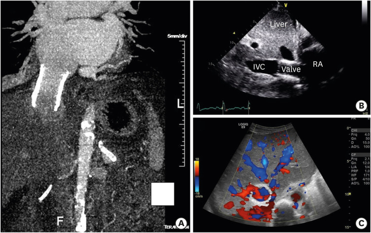
Figure 3 CT, echocardiogram, and liver sonogram after CAVI.(A) CT (post CAVI, reconstructed image) scan reveals blocking contrast reflux into hepatic vein compared to CT before CAVI. (B) Echocardiographic subcostal view shows implanted caval valve. (C) Liver sonogram demostrates mild hepatic venous stenosis without obstruction.CAVI = caval valve implantation; CT = computed tomography; IVC = inferior vena cava; RA = right atrium.

Figure 4 Pre-CAVI and post-CAVI chest X-rays.(A) Chest X-ray shows cardiomegaly and pleural effusion. (B) Chest X-ray after CAVI shows improving cardiomegaly and pleural effusion.CAVI=caval valve implantation.
Reference
-
1. Lauten A, Figulla HR, Unbehaun A, et al. Interventional treatment of severe tricuspid regurgitation: early clinical experience in a multicenter, observational, first-in-man study. Circ Cardiovasc Interv. 2018; 11:e006061. PMID: 29445001.2. O’Neill B, Wang DD, Pantelic M, et al. Transcatheter caval valve implantation using multimodality imaging: roles of TEE, CT, and 3D printing. JACC Cardiovasc Imaging. 2015; 8:221–225. PMID: 25677894.
- Full Text Links
-
- Actions
-
Cited
- CITED
-
- Close
- Share
-
- Similar articles
-
- TricValve in Severe Tricuspid Regurgitation: A Case Series Illustrating The Role of CT Angiography and Treatment Outcome
- Permanent Pacemaker Lead Induced Severe Tricuspid Regurgitation in Patient Undergoing Multiple Valve Surgery
- Tricuspid Valve Imaging and Right Ventricular Function Analysis Using Cardiac CT and MRI
- Repair of Posttraumatic Tricuspid Regurgitation Using Artificial Chordae and an Annuloplasty Ring
- A Case of Severe Aortic Stenosis Patient With High Operative Risk Treated by Transcatheter Aortic-Valve Implantation
