Hyperbaric oxygen therapy and prostaglandin usage for shred injury of the fingers: a case report
- Affiliations
-
- 1Department of Orthopedic Surgery, Yonsei University Wonju College of Medicine, Wonju, Korea
- KMID: 2526649
- DOI: http://doi.org/10.12790/ahm.21.0132
Abstract
- A 53-year-old woman came to the emergency department because her right hand had been stuck in a potato-shredding machine for 30 minutes. The 2nd, 3rd, and 4th fingers were shredded into multiple slices deep into the phalangeal bone, which showed good circulation, and the wounds were cleaned with massive saline irrigation. The slices of each finger were put together to re-form the finger, which was sutured with nylon, and the circulation of the fingers remained good. Three weeks of gentamicin, cefazolin, and hyperbaric oxygen therapy were used for acute traumatic ischemia since color change of the fingers was observed. Six weeks of prostaglandin was used to promote recovery of circulation. The patient was able to grasp with minimal pain and to perform flexion and extension, and the wound completely healed. Radiography showed the bone union process, and digital infrared thermal imaging showed relatively good circulation.
Figure
-

Fig. 1. A 53-year-old female patient injured her 1st to 4th fingers in a shredding machine. (A) The 1st to 4th fingers were inserted into the machine. (B) Initial X-ray with the machine. (C) Initial three-dimensional computed tomography with the machine. (D, E) After machine removal and massive irrigation; the 2nd, 3rd, and 4th fingers in the (D) dorsal aspect and (E) volar aspect.

Fig. 2. After debridement and suturing. (A) Plantar aspect. (B) Dorsal aspect. (C) Fingertips. (D) X-ray images at postoperative 2 weeks; anteroposterior, oblique, and lateral. (E) Postoperative computed tomography, which shows multiple simple fractures at the right 1st, 2nd, 3rd, and 4th distal phalanges and the 3rd middle phalanx without displacement.
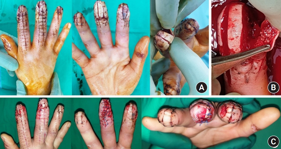
Fig. 3. (A) Wound dehiscence on the distal phalanx of the 3rd finger. (B) Second debridement and suturing of the distal phalanx of the 3rd finger. (C) After the second debridement and suturing.
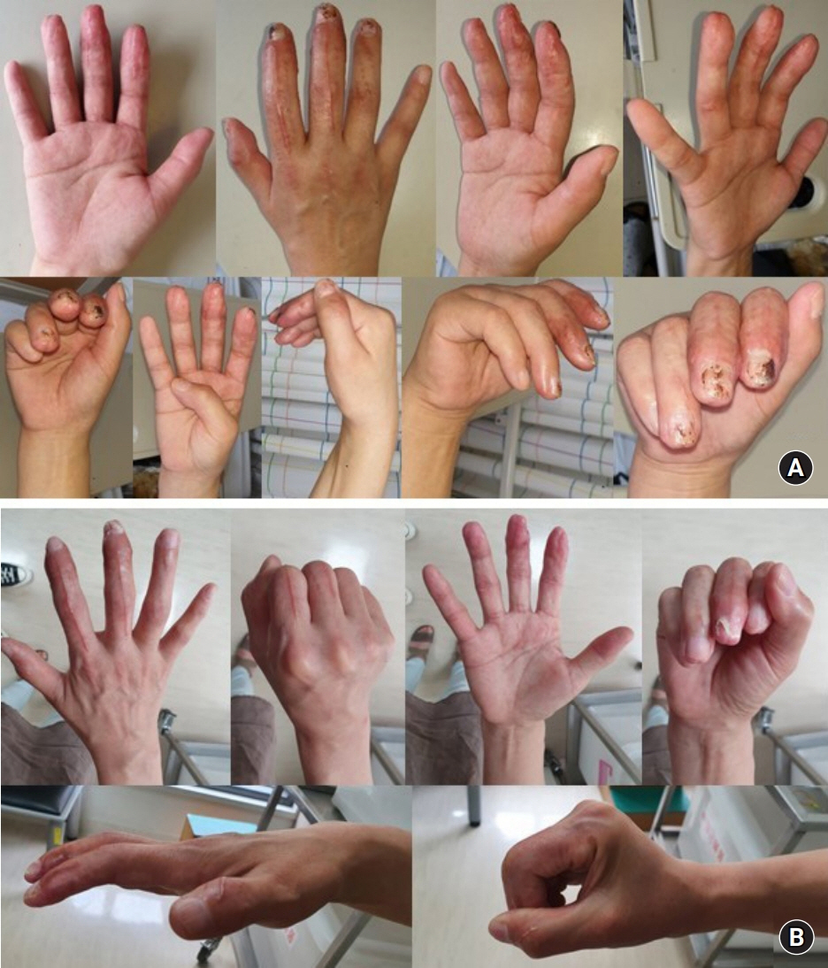
Fig. 4. (A) The wound was fully healed; new fingernails grew at postoperative 8 weeks. Active range of motion (ROM) at postoperative 8 weeks. (B) Active ROM on postoperative 6 months.
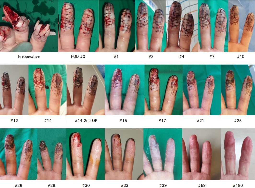
Fig. 5. Wound healing process of the volar aspect of the right 2nd and 3rd fingers from the day of trauma to postoperative 6 months. Ischemia and necrosis all normalized over time. POD, postoperative day.
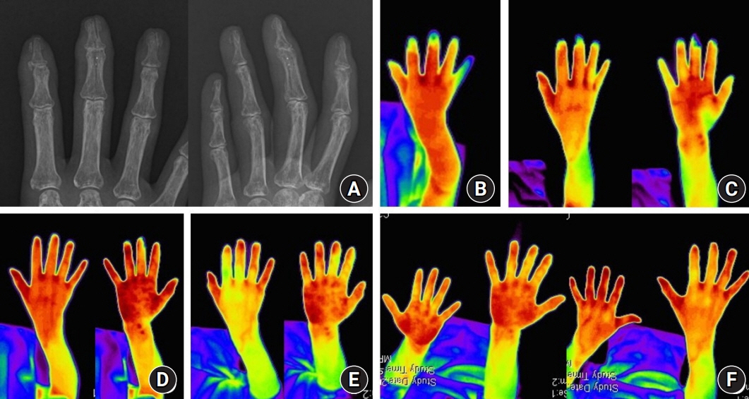
Fig. 6. (A) X-ray images at postoperative 3 months, which show that the complete fractures at the 2nd to 4th distal phalanges and the 3rd middle phalanges are in the healing process. (B) The result of digital infrared thermal imaging (DITI) at postoperative 2 weeks. Infrared shows temperature difference between distal and proximal parts of the dorsal aspect of the right 3rd finger (temperature difference, 2.58°C). Infrared shows the temperature difference between the distal and proximal parts of the volar aspect of the right 3rd finger (temperature difference, 2.54°C). These findings are suggestive of a circulation defect in the distal part of the dorsal and volar aspects of the right 3rd finger compared to the proximal part. (C) DITI at postoperative 3 weeks. Infrared shows the temperature difference between the distal and proximal parts of the dorsal aspect of the right 3rd finger (temperature difference, 2.00°C). Infrared shows the temperature difference between the distal and proximal parts of the volar aspect of the right 3rd finger (temperature difference, 3.67°C). These findings are suggestive of improvement of the circulation defect in the distal part of dorsum and aggravation of the circulation defect in the volar aspect of the right 3rd finger compared to the proximal part. (D) DITI at postoperative 5 weeks. Infrared shows the temperature difference between the distal and proximal parts of the volar aspect of the right 3rd finger (temperature difference, 1.33°C). These findings are suggestive of a circulation defect in the distal part of the dorsal and volar aspects of the right 3rd finger compared to the proximal part (up to normal level). (E) DITI at postoperative 2 months. Infrared shows an higher temperature in the distal part than in the proximal part of the dorsal aspect of the right 2nd, 3rd fingers and the volar aspect of the right 3rd finger. These findings are suggestive of neovascularization of end arteries on the dorsal side of the 2nd, 3rd fingers, and the volar side of the 3rd finger. (F) DITI at postoperative 6 months. The temperature difference of the 2nd, 3rd, and 4th injured fingers was less than 1°C, indicating complete revascularization. There is no evidence of a circulation defect or complex regional pain syndrome.
Reference
-
References
1. Dellinger EP, Miller SD, Wertz MJ, Grypma M, Droppert B, Anderson PA. Risk of infection after open fracture of the arm or leg. Arch Surg. 1988; 123:1320–7.
Article2. Boerama I, Meyne NG, Brummelkamp WH, Bouma S, Mensch MH, Kamermans F, Stern Hanf M, Van Aalderen. [Life without blood]. Ned Tijdschr Geneeskd. 1960; 104:949–54. In Dutch.3. Levy JM, Joseph RB, Bodell LS, Nykamp PW, Hessel SJ. Prostaglandin E1 in hand angiography. AJR Am J Roentgenol. 1983; 141:1043–6.
Article4. Aburakawa Y, Kawabe J, Okada M, et al. Prostacyclin stimulated integrin-dependent angiogenic effects of endothelial progenitor cells and mediated potent circulation recovery in ischemic hind limb model. Circ J. 2013; 77:1053–62.
Article5. Cross WW 3rd, Swiontkowski MF. Treatment principles in the management of open fractures. Indian J Orthop. 2008; 42:377–86.
Article6. Kline DG, Hackett ER, Davis GD, Myers MB. Effect of mobilization on the blood supply and regeneration of injured nerves. J Surg Res. 1972; 12:254–66.
Article7. Ulubayram K, Nur Cakar A, Korkusuz P, Ertan C, Hasirci N. EGF containing gelatin-based wound dressings. Biomaterials. 2001; 22:1345–56.
Article8. Kota S, Jahangir MA, Ahmed M, et al. Development and evaluation of ofloxacin topical gel containing wound healing modifiers from natural sources. Der Pharm Lett. 2015; 7:226–33.9. Igarashi R, Takenaga M, Matsuda T. Distribution of lipid microsphere preparations. Adv Drug Deliv Rev. 1996; 20:147–54.
Article10. Behnke AR, Forbes HS, Motley EP. Circulatory and visual effects of oxygen at 3 atmospheres pressure. Am J Physiol. 1936; 114:436–42.
Article
- Full Text Links
-
- Actions
-
Cited
- CITED
-
- Close
- Share
-
- Similar articles
-
- Treatment of radiation-induced cystitis with hyperbaric oxygen
- A Basic Survey for Regional Capability of Hyperbaric Oxygen Therapy to Multiple Fire Victims
- Early experience of hyperbaric oxygen therapy in radiation-induced cystitis
- Hyperbaric Oxygen Therapy on Pyoderma Gangrenosum - A case report
- Hyperbaric Oxygen Therapy in Pyoderma Gangrenosum
