Korean J Orthod.
2022 Jan;52(1):53-65. 10.4041/kjod.2022.52.1.53.
Proposed parameters of optimal central incisor positioning in orthodontic treatment planning: A systematic review
- Affiliations
-
- 1Dental School, Department of Medical and Surgical Specialties, Radiological Sciences and Public Health, University of Brescia, Brescia, Italy
- 2Division of Orofacial Pain, College of Dentistry, University of Kentucky, Lexington, KY, USA
- 3Orthodontics, Division of Paediatric Dentistry and Orthodontics, Faculty of Dentistry, The University of Hong Kong, Hong Kong SAR
- KMID: 2524902
- DOI: http://doi.org/10.4041/kjod.2022.52.1.53
Abstract
Objective
Planning of incisal position is crucial for optimal orthodontic treatment outcomes due to its consequences on facial esthetics and occlusion. A systematic summary of the proposed parameters is presented.
Methods
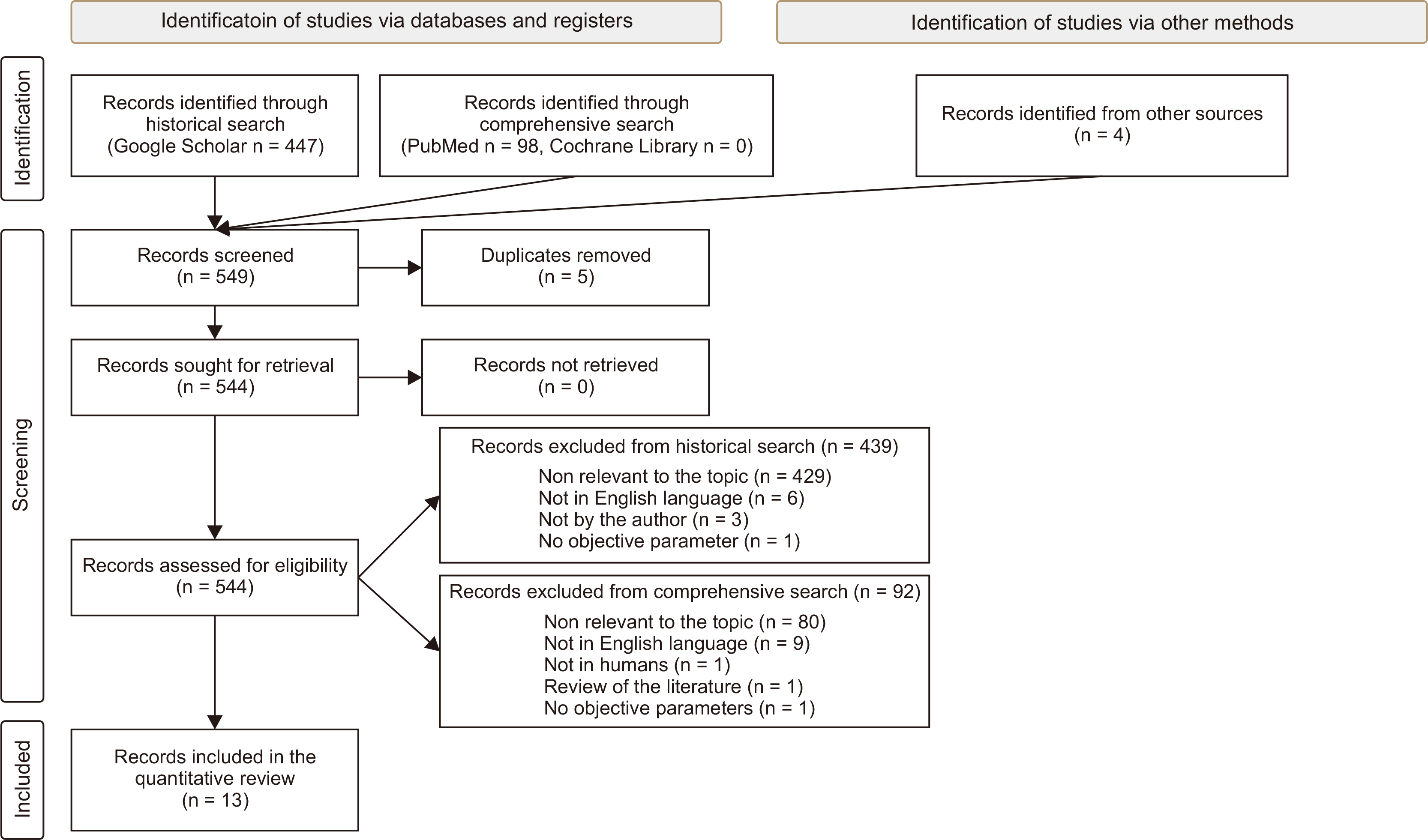
Studies on Google Scholar© , PubMed© , and Cochrane Library, providing quantitative information on optimal central incisor position were included.
Results
Upper incisors supero-inferior position (4–5 mm to upper lip, 67–73 mm to axial plane through pupils), antero-posterior position (3–4 mm to Nasion-A, 3–6 mm to A-Pogonion, 9–12 mm to true vertical line, 5 mm to A-projection, 9–10 mm to coronal plane through pupils), bucco-lingual angulation (4–7° to occlusal plane perpendicular on models, 20–22° to Nasion-A, 57–58° to upper occlusal plane, 16–20° to coronal plane through pupils, 108–110° to anterior-posterior nasal spine), mesio-distal angulation (5° to occlusal plane perpendicular on models). Lower incisors supero-inferior position (41–48 mm to soft-tissue mandibular plane), antero-posterior position (3–4 mm to Nasion-B, 1–3 mm to A-Pogonion, 12–15 mm to true vertical line, 6–8 mm to coronal plane through pupils), bucco-lingual angulation (1-4° to occlusal plane perpendicular on models, 87–94° to mandibular plane, 68° to Frankfurt plane, 22–25° to Nasion-B, 105° to occlusal plane, 64° to lower occlusal plane, 21° to A-Pogonion), mesiodistal angulation (2° to occlusal plane perpendicular on models).
Conclusions
Although these findings can provide clinical guideline, they derive from heterogeneous studies in terms of subject characteristics and reference methods. Therefore, the optimal incisal position remains debatable.
Keyword
Figure
Reference
-
1. Arnett WG, McLaughlin RP. 2004. Facial and dental planning for orthodontists and oral surgeons. Mosby;Edinburgh: DOI: 10.1016/j.ajodo.2004.06.006. PMID: 15356488.2. Huang YP, Li WR. 2015; Correlation between objective and subjective evaluation of profile in bimaxillary protrusion patients after orthodontic treatment. Angle Orthod. 85:690–8. DOI: 10.2319/070714-476.1. PMID: 25347046. PMCID: PMC8611736.
Article3. Sriphadungporn C, Chamnannidiadha N. 2017; Perception of smile esthetics by laypeople of different ages. Prog Orthod. 18:8. DOI: 10.1186/s40510-017-0162-4. PMID: 28317085. PMCID: PMC5357618.
Article4. Johal A, Alyaqoobi I, Patel R, Cox S. 2015; The impact of orthodontic treatment on quality of life and self-esteem in adult patients. Eur J Orthod. 37:233–7. DOI: 10.1093/ejo/cju047. PMID: 25214505.
Article5. Zhou Y, Wang Y, Wang X, Volière G, Hu R. 2014; The impact of orthodontic treatment on the quality of life a systematic review. BMC Oral Health. 14:66. DOI: 10.1186/1472-6831-14-66. PMID: 24913619. PMCID: PMC4060859.
Article6. Keele KD. 2014. Leonardo da Vinci's elements of the science of man. Academic Press;London:7. Ricketts RM. 1982; The biologic significance of the divine proportion and Fibonacci series. Am J Orthod. 81:351–70. DOI: 10.1016/0002-9416(82)90073-2. PMID: 6960724.
Article8. Little AC, Jones BC, DeBruine LM. 2011; Facial attractiveness: evolutionary based research. Philos Trans R Soc Lond B Biol Sci. 366:1638–59. DOI: 10.1098/rstb.2010.0404. PMID: 21536551. PMCID: PMC3130383.
Article9. Mchorris WH. 1979; Occlusion with particular emphasis on the functional and parafunctional role of anterior teeth. Part 2. J Clin Orthod. 13:684–701. PMID: 298284.10. Patil HD, Nehete AB, Gulve ND, Shah KR, Aher SD. 2018; Evaluation of upper incisor position and its comparison with lip posture in orthodontically treated patients. J Dent Med Sci. 17:53–60.11. Krishnan V, Daniel ST, Lazar D, Asok A. 2008; Characterization of posed smile by using visual analog scale, smile arc, buccal corridor measures, and modified smile index. Am J Orthod Dentofacial Orthop. 133:515–23. DOI: 10.1016/j.ajodo.2006.04.046. PMID: 18405815.
Article12. Machado AW, Moon W, Gandini LG Jr. 2013; Influence of maxillary incisor edge asymmetries on the perception of smile esthetics among orthodontists and laypersons. Am J Orthod Dentofacial Orthop. 143:658–64. DOI: 10.1016/j.ajodo.2013.02.013. PMID: 23631967.
Article13. Zarif Najafi H, Oshagh M, Khalili MH, Torkan S. 2015; Esthetic evaluation of incisor inclination in smiling profiles with respect to mandibular position. Am J Orthod Dentofacial Orthop. 148:387–95. DOI: 10.1016/j.ajodo.2015.05.016. PMID: 26321336.14. Cao L, Zhang K, Bai D, Jing Y, Tian Y, Guo Y. 2011; Effect of maxillary incisor labiolingual inclination and anteroposterior position on smiling profile esthetics. Angle Orthod. 81:121–9. DOI: 10.2319/033110-181.1. PMID: 20936964.
Article15. Tosun H, Kaya B. 2020; Effect of maxillary incisors, lower lip, and gingival display relationship on smile attractiveness. Am J Orthod Dentofacial Orthop. 157:340–7. DOI: 10.1016/j.ajodo.2019.04.030. PMID: 32115112.
Article16. Tweed CH. 1954; The Frankfort-Mandibular Incisor Angle (FMIA) in orthodontic diagnosis, treatment planning and prognosis. Angle Orthod. 24:121–69.17. Pitts TR. 2017; Bracket positioning for smile arc protection. J Clin Orthod. 51:142–56. PMID: 28646818.18. Moore RN, Moyer BA, DuBois LM. 1990; Skeletal maturation and craniofacial growth. Am J Orthod Dentofacial Orthop. 98:33–40. DOI: 10.1016/0889-5406(90)70029-C. PMID: 2363404.
Article19. Masoud MI, Bansal N, C Castillo J, Manosudprasit A, Allareddy V, Haghi A, et al. 2017; 3D dentofacial photogrammetry reference values: a novel approach to orthodontic diagnosis. Eur J Orthod. 39:215–25. DOI: 10.1093/ejo/cjw055. PMID: 28339510.
Article20. Andrews LF. 1976; The diagnostic system: occlusal analysis. Dent Clin North Am. 20:671–90. PMID: 1067200.21. Dickens ST, Sarver DM, Proffit WR. 2002; Changes in frontal soft tissue dimensions of the lower face by age and gender. World J Orthod. 3:313–20.22. Mamandras AH. 1988; Linear changes of the maxillary and mandibular lips. Am J Orthod Dentofacial Orthop. 94:405–10. DOI: 10.1016/0889-5406(88)90129-1. PMID: 3189242.
Article23. Sharma P, Arora A, Valiathan A. 2014; Age changes of jaws and soft tissue profile. ScientificWorldJournal. 2014:301501. DOI: 10.1155/2014/301501. PMID: 25506064. PMCID: PMC4258316.
Article24. Behrents RG. 1985. Craniofacial growth series. Growth in the aging craniofacial skeleton. University of Michigan;Ann Arbor:25. Sabri R. 2005; The eight components of a balanced smile. J Clin Orthod. 39:155–67. quiz 154PMID: 15888949.26. Dong JK, Jin TH, Cho HW, Oh SC. 1999; The esthetics of the smile: a review of some recent studies. Int J Prosthodont. 12:9–19. PMID: 10196823.27. Chetan P, Tandon P, Singh GK, Nagar A, Prasad V, Chugh VK. 2013; Dynamics of a smile in different age groups. Angle Orthod. 83:90–6. DOI: 10.2319/040112-268.1. PMID: 22889201.
Article28. Câmara CA. 2010; Aesthetics in orthodontics: six horizontal smile lines. Dental Press J Orthod. 15:118–31. DOI: 10.1590/S2176-94512010000100014.29. Devreese H, De Pauw G, Van Maele G, Kuijpers-Jagtman AM, Dermaut L. 2007; Stability of upper incisor inclination changes in Class II division 2 patients. Eur J Orthod. 29:314–20. DOI: 10.1093/ejo/cjm011. PMID: 17483493.
Article30. Wysong A, Kim D, Joseph T, MacFarlane DF, Tang JY, Gladstone HB. 2014; Quantifying soft tissue loss in the aging male face using magnetic resonance imaging. Dermatol Surg. 40:786–93. DOI: 10.1111/dsu.0000000000000035. PMID: 25111352.31. Huber KL, Suri L, Taneja P. 2008; Eruption disturbances of the maxillary incisors: a literature review. J Clin Pediatr Dent. 32:221–30. DOI: 10.17796/jcpd.32.3.m175g328l100x745. PMID: 18524273.32. National Heart, Lung, and Blood Institute. Study quality assessment tools. 2013. Study quality assessment tools. Quality assessment tool for observational cohort and cross-sectional studies. National Heart, Lung, and Blood Institute;Bethesda:33. Ricketts RM. 1960; A foundation for cephalometric communication. Am J Orthod. 46:330–57. DOI: 10.1016/0002-9416(60)90047-6.
Article34. Arnett GW, Jelic JS, Kim J, Cummings DR, Beress A, Worley CM Jr, et al. 1999; Soft tissue cephalometric analysis: diagnosis and treatment planning of dentofacial deformity. Am J Orthod Dentofacial Orthop. 116:239–53. DOI: 10.1016/S0889-5406(99)70234-9. PMID: 10474095.35. Ross VA, Isaacson RJ, Germane N, Rubenstein LK. 1990; Influence of vertical growth pattern on faciolingual inclinations and treatment mechanics. Am J Orthod Dentofacial Orthop. 98:422–9. DOI: 10.1016/S0889-5406(05)81651-8. PMID: 2239841.
Article36. Holdaway RA. 1956; Changes in relationship of points A and B during orthodontic treatment. Am J Orthod. 42:176–93. DOI: 10.1016/0002-9416(56)90112-9.
Article37. Steiner CC. 1953; Cephalometrics for you and me. Am J Orthod. 39:729–55. DOI: 10.1016/0002-9416(53)90082-7.
Article38. Knösel M, Engelke W, Attin R, Kubein-Meesenburg D, Sadat-Khonsari R, Gripp-Rudolph L. 2008; A method for defining targets in contemporary incisor inclination correction. Eur J Orthod. 30:374–80. DOI: 10.1093/ejo/cjn015. PMID: 18678757.
Article39. Downs WB. 1956; Analysis of the dentofacial profile. Angle Orthod. 26:191–212.40. Andrews LF. 1972; The six keys to normal occlusion. Am J Orthod. 62:296–309. DOI: 10.1016/S0002-9416(72)90268-0. PMID: 4505873.
Article41. Webb MA, Cordray FE, Rossouw PE. 2016; Upper-incisor position as a determinant of the ideal soft-tissue profile. J Clin Orthod. 50:651–62. PMID: 28045679.42. McNamara JA Jr. 1984; A method of cephalometric evaluation. Am J Orthod. 86:449–69. DOI: 10.1016/S0002-9416(84)90352-X. PMID: 6594933.
Article43. Knösel M, Jung K. 2011; On the relevance of "ideal" occlusion concepts for incisor inclination target definition. Am J Orthod Dentofacial Orthop. 140:652–9. DOI: 10.1016/j.ajodo.2010.12.021. PMID: 22051485.
Article44. Savoldi F, Massetti F, Tsoi JKH, Matinlinna JP, Yeung AWK, Tanaka R, et al. 2021; Anteroposterior length of the maxillary complex and its relationship with the anterior cranial base: a study on human dry skulls using cone beam computed tomography. Angle Orthod. 91:88–97. DOI: 10.2319/020520-82.1. PMID: 33289836. PMCID: PMC8032287.
Article45. Zataráin B, Avila J, Moyaho A, Carrasco R, Velasco C. 2016; Lower incisor inclination regarding different reference planes. Acta Odontol Latinoam. 29:115–22. PMID: 27731480.46. Hixon EH. 1956; The norm concept and cephalometrics. Am J Orthod. 42:898–906. DOI: 10.1016/0002-9416(56)90190-7.
Article47. Moorrees CFA, Kean MR. 1958; Natural head position, a basic consideration in the interpretation of cephalometric radiographs. Am J Phys Anthropol. 16:213–34. DOI: 10.1002/ajpa.1330160206.
Article48. Machado AW. 2014; 10 commandments of smile esthetics. Dental Press J Orthod. 19:136–57. DOI: 10.1590/2176-9451.19.4.136-157.sar. PMID: 25279532. PMCID: PMC4296640.
Article49. Maddalone M, Losi F, Rota E, Baldoni MG. 2019; Relationship between the position of the incisors and the thickness of the soft tissues in the upper jaw: cephalometric evaluation. Int J Clin Pediatr Dent. 12:391–7. DOI: 10.5005/jp-journals-10005-1667. PMID: 32440043. PMCID: PMC7229373.
Article50. Jeelani W, Fida M, Shaikh A. 2018; The maxillary incisor display at rest: analysis of the underlying components. Dental Press J Orthod. 23:48–55. DOI: 10.1590/2177-6709.23.6.048-055.oar. PMID: 30672985. PMCID: PMC6340193.
Article51. Peck S, Peck L, Kataja M. 1992; The gingival smile line. Angle Orthod. 62:91–100. discussion 101–2. DOI: 10.1043/0003-3219(1992)062<0091:TGSL>2.0.CO;2. PMID: 1626754.52. Morley J. 1999; The role of cosmetic dentistry in restoring a youthful appearance. J Am Dent Assoc. 130:1166–72. DOI: 10.14219/jada.archive.1999.0370. PMID: 10491926.
Article53. Machado AW, McComb RW, Moon W, Gandini LG Jr. 2013; Influence of the vertical position of maxillary central incisors on the perception of smile esthetics among orthodontists and laypersons. J Esthet Restor Dent. 25:392–401. DOI: 10.1111/jerd.12054. PMID: 24180675.
Article54. Sarver DM, Ackerman MB. 2003; Dynamic smile visualization and quantification: part 1. Evolution of the concept and dynamic records for smile capture. Am J Orthod Dentofacial Orthop. 124:4–12. DOI: 10.1016/S0889-5406(03)00306-8. PMID: 12867893.
Article55. Spear F. 2016; Diagnosing and treatment planning inadequate tooth display. Br Dent J. 221:463–72. DOI: 10.1038/sj.bdj.2016.773. PMID: 27767133.
Article56. Sarver DM. 2001; The importance of incisor positioning in the esthetic smile: the smile arc. Am J Orthod Dentofacial Orthop. 120:98–111. DOI: 10.1067/mod.2001.114301. PMID: 11500650.
Article57. Saver DM. 2001. Craniofacial growth series. The importance of incisor positioning in the esthetic smile: the smile arc. University of Michigan;Ann Arbor: p. 19–54.58. Bell WH, Jacobs JD, Quejada JG. 1986; Simultaneous repositioning of the maxilla, mandible, and chin. Treatment planning and analysis of soft tissues. Am J Orthod. 89:28–50. DOI: 10.1016/0002-9416(86)90110-7. PMID: 3455794.
Article59. Ackerman JL, Proffit WR. 1997; Soft tissue limitations in orthodontics: treatment planning guidelines. Angle Orthod. 67:327–36. DOI: 10.1043/0003-3219(1997)067<0327:STLIOT>2.3.CO;2. PMID: 9347106.60. Agostino P, Butti AC, Poggio CE, Salvato A. 2007; Perception of the maxillary incisor position with respect to the protrusion of nose and chin. Prog Orthod. 8:230–9. PMID: 18030369.61. Ghaleb N, Bouserhal J, Bassil-Nassif N. 2011; Aesthetic evaluation of profile incisor inclination. Eur J Orthod. 33:228–35. DOI: 10.1093/ejo/cjq059. PMID: 20716642.
Article62. Stromboni Y. 1979; Facial aesthetics in orthodontic treatment with and without extractions. Eur J Orthod. 1:201–6. DOI: 10.1093/ejo/1.3.201. PMID: 296944.
Article63. Savoldi F, Papoutsi A, Dianiskova S, Dalessandri D, Bonetti S, Tsoi JKH, et al. 2018; Resistance to sliding in orthodontics: misconception or method error? A systematic review and a proposal of a test protocol. Korean J Orthod. 48:268–80. DOI: 10.4041/kjod.2018.48.4.268. PMID: 30003061. PMCID: PMC6041452.
Article64. Savoldi F, Paganelli C. 2019; In vitro evaluation of loop design influencing the sliding of orthodontic wires: a preliminary study. J Appl Biomater Funct Mater. 17:2280800018787072. DOI: 10.1177/2280800018787072. PMID: 30009658.
Article65. Diels RM, Kalra V, DeLoach N Jr, Powers M, Nelson SS. 1995; Changes in soft tissue profile of African-Americans following extraction treatment. Angle Orthod. 65:285–92. DOI: 10.1043/0003-3219(1995)065<0285:CISTPO>2.0.CO;2. PMID: 7486243.66. Kusnoto J, Kusnoto H. 2001; The effect of anterior tooth retraction on lip position of orthodontically treated adult Indonesians. Am J Orthod Dentofacial Orthop. 120:304–7. DOI: 10.1067/mod.2001.116089. PMID: 11552130.
Article67. Caplan MJ, Shivapuja PK. 1997; The effect of premolar extractions on the soft-tissue profile in adult African American females. Angle Orthod. 67:129–36. DOI: 10.1043/0003-3219(1997)067<0129:TEOPEO>2.3.CO;2. PMID: 9107377.68. Mirabella D, Bacconi S, Gracco A, Lombardo L, Siciliani G. 2008; Upper lip changes correlated with maxillary incisor movement in 65 orthodontically treated adult patients. World J Orthod. 9:337–48. PMID: 19146015.69. Jacobs JD. 1978; Vertical lip changes from maxillary incisor retraction. Am J Orthod. 74:396–404. DOI: 10.1016/0002-9416(78)90061-1. PMID: 281142.
Article70. Kim SJ, Kim KH, Yu HS, Baik HS. 2014; Dentoalveolar compensation according to skeletal discrepancy and overjet in skeletal Class III patients. Am J Orthod Dentofacial Orthop. 145:317–24. DOI: 10.1016/j.ajodo.2013.11.014. PMID: 24582023.
Article71. Ul Huqh MZ, Hassan R, Zainal Abidin S, IA Karobari M, Yaqoob MA. 2020; Rickett's and Holdaway analysis following extraction of four premolars and orthodontic treatment in bimaxillary protrusion female Malays. J Int Oral Health. 12:58–65. DOI: 10.4103/jioh.jioh_155_19.
Article72. Oliver BM. 1982; The influence of lip thickness and strain on upper lip response to incisor retraction. Am J Orthod. 82:141–9. DOI: 10.1016/0002-9416(82)90492-4. PMID: 6961784.
Article73. Mackley RJ. 1993; An evaluation of smiles before and after orthodontic treatment. Angle Orthod. 63:183–9. discussion 190DOI: 10.1043/0003-3219(1993)063<0183:AEOSBA>2.0.CO;2. PMID: 8214786.74. Pikdoken L, Erkan M, Usumez S. 2009; Editor's summary, Q & A, reviewer's critique: gingival response to mandibular incisor extrusion. Am J Orthod Dent Orthop. 135:432.e1–432.e6. DOI: 10.1016/j.ajodo.2008.07.014.75. Murakami T, Yokota S, Takahama Y. 1989; Periodontal changes after experimentally induced intrusion of the upper incisors in Macaca fuscata monkeys. Am J Orthod Dentofacial Orthop. 95:115–26. DOI: 10.1016/0889-5406(89)90390-9. PMID: 2916468.
Article76. Erkan M, Pikdoken L, Usumez S. 2007; Gingival response to mandibular incisor intrusion. Am J Orthod Dentofacial Orthop. 132:143.e9–13. DOI: 10.1016/j.ajodo.2006.10.015. PMID: 17693359.
Article77. Dorfman HS. 1978; Mucogingival changes resulting from mandibular incisor tooth movement. Am J Orthod. 74:286–97. DOI: 10.1016/0002-9416(78)90204-X. PMID: 281132.
Article78. Kalina E, Zadurska M, Sobieska E, Górski B. 2019; Relationship between periodontal status of mandibular incisors and selected cephalometric parameters: preliminary results. J Orofac Orthop. 80:107–15. DOI: 10.1007/s00056-019-00170-0. PMID: 31041493. PMCID: PMC6491396.
Article79. Nanda R. 2015. Esthetics and biomechnics in orthodontics. 2nd ed. Elsevier;St. Louis:80. Gütermann C, Peltomäki T, Markic G, Hänggi M, Schätzle M, Signorelli L, et al. 2014; The inclination of mandibular incisors revisited. Angle Orthod. 84:109–19. DOI: 10.2319/040413-262.1. PMID: 23985035. PMCID: PMC8683058.
Article81. Hernández-Sayago E, Espinar-Escalona E, Barrera-Mora JM, Ruiz-Navarro MB, Llamas-Carreras JM, Solano-Reina E. 2013; Lower incisor position in different malocclusions and facial patterns. Med Oral Patol Oral Cir Bucal. 18:e343–50. DOI: 10.4317/medoral.18434. PMID: 23229262. PMCID: PMC3613890.82. Simons ME, Joondeph DR. 1973; Change in overbite: a ten-year postretention study. Am J Orthod. 64:349–67. DOI: 10.1016/0002-9416(73)90243-1. PMID: 4521238.
Article83. Kau CH, Bakos K, Lamani E. 2020; Quantifying changes in incisor inclination before and after orthodontic treatment in class I, II, and III malocclusions. J World Fed Orthod. 9:170–4. DOI: 10.1016/j.ejwf.2020.08.002. PMID: 32948483.
Article84. Ishikawa H, Nakamura S, Iwasaki H, Kitazawa S, Tsukada H, Sato Y. 1999; Dentoalveolar compensation related to variations in sagittal jaw relationships. Angle Orthod. 69:534–8. DOI: 10.1043/0003-3219(1999)069<0534:DCRTVI>2.3.CO;2. PMID: 10593444.85. Chirivella P, Singaraju GS, Mandava P, Reddy VK, Neravati JK, George SA. 2017; Comparison of the effect of labiolingual inclination and anteroposterior position of maxillary incisors on esthetic profile in three different facial patterns. J Orthod Sci. 6:1–10. DOI: 10.4103/2278-0203.197387. PMID: 28197396. PMCID: PMC5278578.
Article86. Sackstein M. 2008; Display of mandibular and maxillary anterior teeth during smiling and speech: age and sex correlations. Int J Prosthodont. 21:149–51. PMID: 18546770.87. Misch CE. 2008. Contemporary implant dentistry. Mosby Elsevier;St. Louis:88. Savoldi F, Bonetti S, Dalessandri D, Mandelli G, Paganelli C. 2015; Incisal apical root resorption evaluation after low-friction orthodontic treatment using two-dimensional radiographic imaging and trigonometric correction. J Clin Diagn Res. 9:ZC70–4. DOI: 10.7860/JCDR/2015/14140.6841. PMID: 26676099. PMCID: PMC4668528.
Article89. Lee RL. Lundeen HC, Gibbs CH, editors. 1982. Anterior guidance. Advances in occlusion. John Wright PSG Inc.;Boston: p. 51–79.90. Misch CE. 2015. Dental implant prosthetics. Mosby Elsevier;St. Louis: DOI: 10.1016/B978-0-323-07845-0.00016-6.91. Kaley J, Phillips C. 1991; Factors related to root resorption in edgewise practice. Angle Orthod. 61:125–32. DOI: 10.1043/0003-3219(1991)061<0125:FRTRRI>2.0.CO;2. PMID: 2064070.92. Park JH, Hong JY, Ahn HW, Kim SJ. 2018; Correlation between periodontal soft tissue and hard tissue surrounding incisors in skeletal Class III patients. Angle Orthod. 88:91–9. DOI: 10.2319/060117-367.1. PMID: 29072859. PMCID: PMC8315704.
Article93. van der Beek MC, Hoeksma JB, Prahl-Andersen B. 1991; Vertical facial growth: a longitudinal study from 7 to 14 years of age. Eur J Orthod. 13:202–8. DOI: 10.1093/ejo/13.3.202. PMID: 1936138.
Article94. Millett D. 2021; The rationale for orthodontic retention: piecing together the jigsaw. Br Dent J. 230:739–49. DOI: 10.1038/s41415-021-3012-1. PMID: 34117429.
Article
- Full Text Links
- Actions
-
Cited
- CITED
-
- Close
- Share
- Similar articles
-
- Esthetic improvement in the patient with one missing maxillary central incisor restored with porcelain laminate veneers
- A study on the affecting factors on root resorption
- Orthodontic treatment of an impacted maxillary central incisor with dilacerations
- The positioning errors in bonding lingual brackets
- Strategic surgical-combined orthodontic treatment planning of patient with missing incisors on maxilla: a case report


