The Importance of Lamina Size Measurement and Proper Implants Selection before Laminoplasty : Two Case Reports
- Affiliations
-
- 1Department of Neurosurgery & Medical Research Institue, Pusan National University Hospital, Pusan National University School of Medicine, Busan, Republic of Korea
- 2Department of Neurosurgery, Dong-Eui Hospital, Busan, Republic of Korea
- 3Department of Neurosurgery Pusan National University Yangsan Hospital, Yangsan, Republic of Korea
- KMID: 2524661
- DOI: http://doi.org/10.7180/kmj.2021.36.2.169
Abstract
- Open door laminoplasty using plates is a safe and effective procedure for multi-level cord compression. To achieve stable laminar arch, various types of plate have been developed and used. Now, we introduce two rare complications related to the laminar shelf of plate. In the first case, we used the wider laminar shelf plate because the elevated lamina did not fit well into the usual laminar shelf. During follow-up, cord compression due to laminar shelf was observed. And in the second case, the laminar shelf of plate did not fit into the elevated lamina, so we inserted it with a little bit of force. But the patient’s symptom was not improved. On CT image, the inner cortical bone of the lamina was fractured. To prevent these complications, surgeons need to consider the thickness of the lamina and the size of the laminar shelf before surgery.
Figure
-
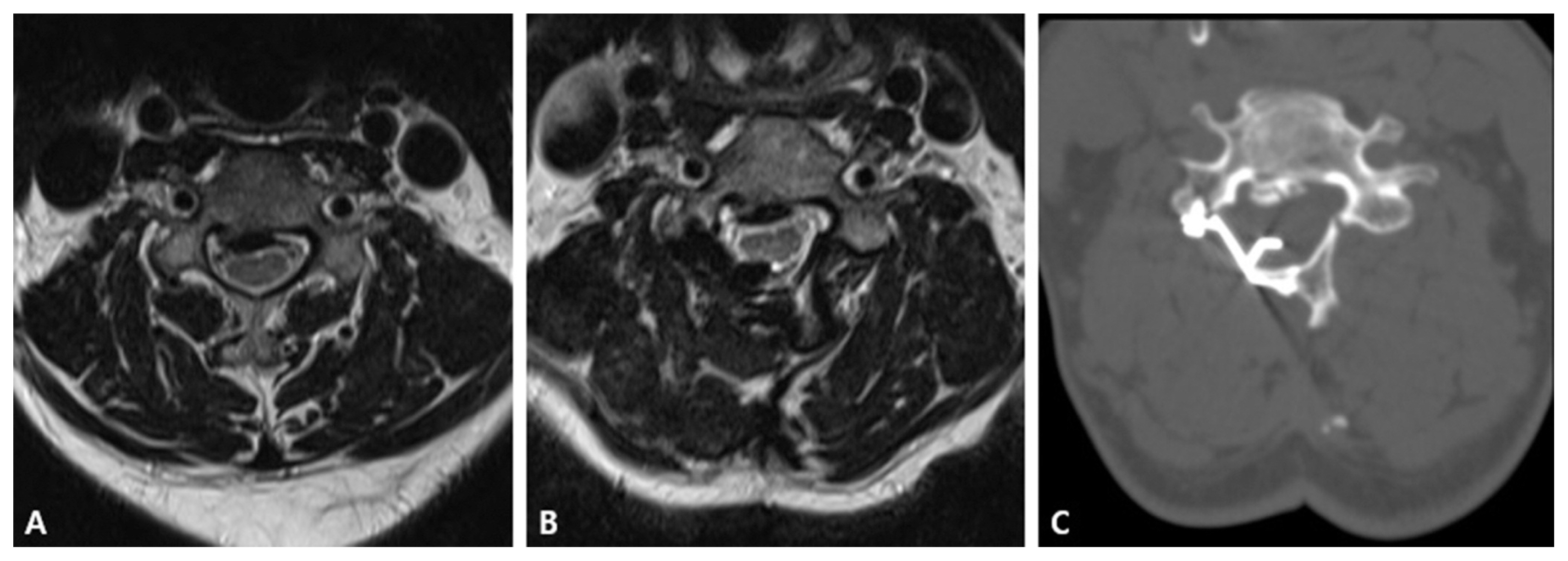
Fig. 1 (A) Spinal cord compression due to OPLL on the pre-operative magnetic resonance image. (B) Spinal cord compression and subtle signal intensity change of spinal cord were identified on the magnetic resonance image. (C) The wide lamina shelf of plate protruded into the epidural space on computed tomography image.
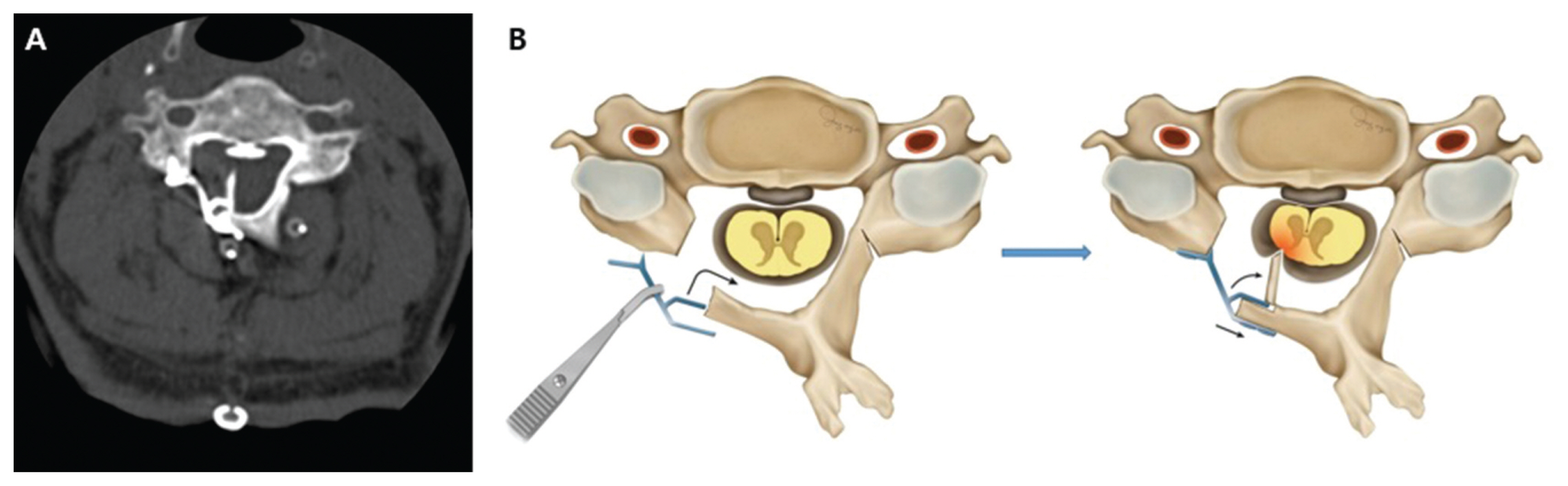
Fig. 2 (A) Inner cortical bone fracture with epidural space invasion was shown on the computed tomography. (B) An illustration cord compression caused by inner cortical bone fracture. Inner cortical bone fracture was caused by using a smaller laminar shelf plate than a laminar size.
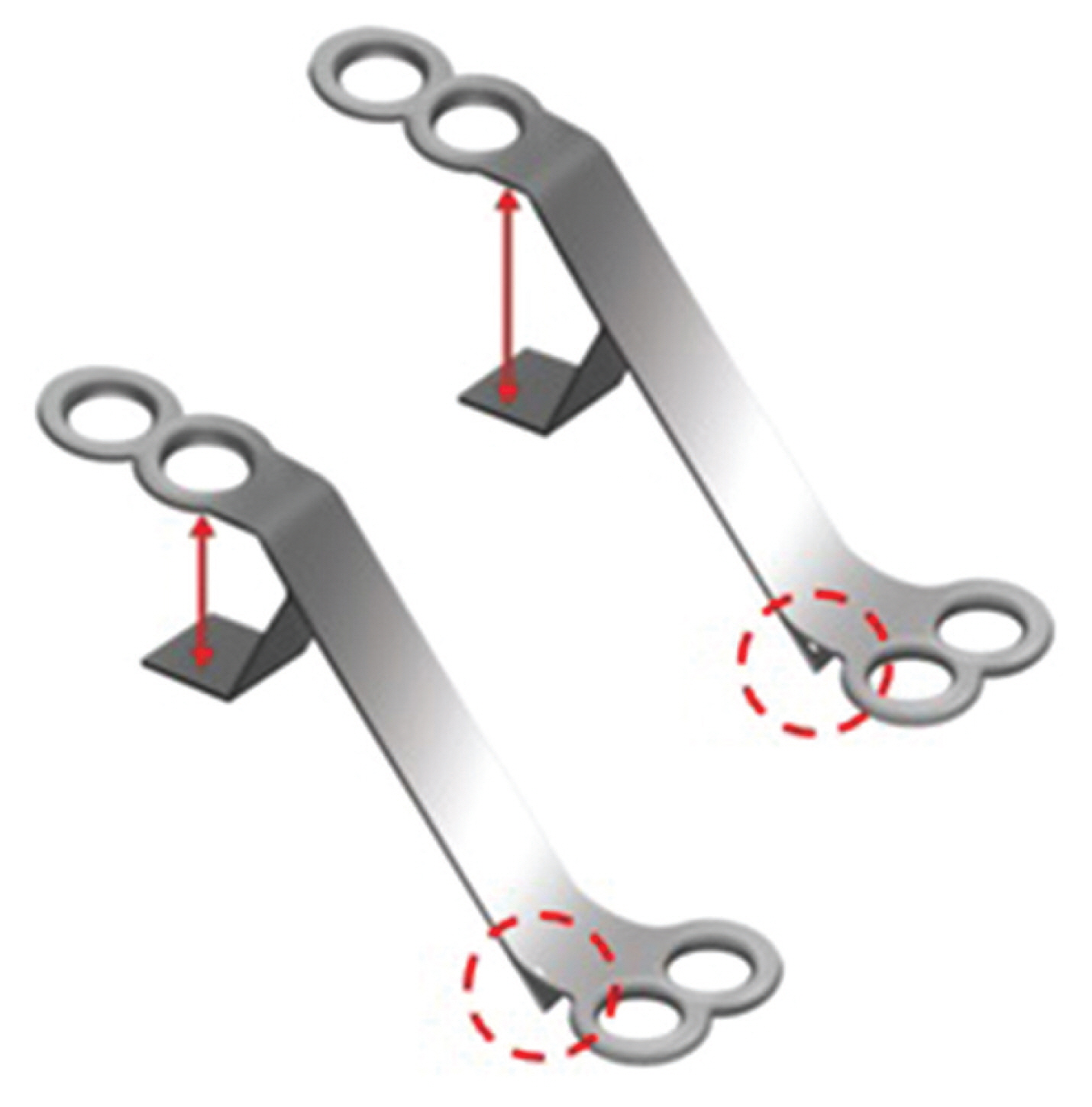
Fig. 3 An illustration of a plate with laminar shelf. It consists of two distinctive designs; lamina shelf (red arrow) and kickstand design (red dot circle) which allow stable positioning between the cut edge of the lamina and lateral mass.
Reference
-
1. Hur JW, Park YK, Kim BJ, Moon HJ, Kim JH. Risk Factors for Delayed Hinge Fracture after Plate-Augmented Cervical Open-Door Laminoplasty. J Korean Neurosurg Soc. 2016; 59:368–73.
Article2. Hirabayashi K, Watanabe K, Wakano K, Suzuki N, Satomi K, Ishii Y. Expansive open-door laminoplasty for cervical spinal stenotic myelopathy. Spine (Phila Pa 1976). 1983; 8:693–9.
Article3. Chen HC, Chang MC, Yu WK, Wang ST, Liu CL, Chen TH. Lateral mass anchoring screws for cervical laminoplasty: preliminary report of a novel technique. J Spinal Disord Tech. 2008; 21:387–92.4. Goto T, Ohata K, Takami T, Nishikawa M, Tsuyuguchi N, Morino M, et al. Hydroxyapatite laminar spacers and titanium mini-plates in cervical laminoplasty. J Neurosurg. 2002; 97:323–9.
Article5. Lin X, Chen K, Tang H, Huang X, Wei C, Xiao Z. Comparison of anchor screw fixation versus mini-plate fixation in unilateral expansive open-door laminoplasty for the treatment of multi-level cervical spondylotic myelopathy. Medicine (Baltimore). 2018; 97:e13534.
Article6. Nakano K, Harata S, Suetsuna F, Araki T, Itoh J. Spinous Process-Splitting Laminoplasty Using Hydroxyapatite Spinous Process Spacer. Spine. 1992; 17:S41–S3.
Article7. Lee DG, Lee SH, Park SJ, Kim ES, Chung SS, Lee CS, et al. Comparison of surgical outcomes after cervical laminoplasty: open-door technique versus French-door technique. J Spinal Disord Tech. 2013; 26:E198–203.8. Park AE, Heller JG. Cervical laminoplasty: use of a novel titanium plate to maintain canal expansion--surgical technique. J Spinal Disord Tech. 2004; 17:265–71.9. Wang LN, Wang L, Song YM, Yang X, Liu LM, Li T. Clinical and radiographic outcome of unilateral open-door laminoplasty with alternative levels centerpiece mini-plate fixation for cervical compressive myelopathy: a five-year follow-up study. Int Orthop. 2016; 40:1267–74.
Article10. Wang HQ, Mak KC, Samartzis D, El-Fiky T, Wong YW, Luo ZJ, et al. “Spring-back” closure associated with open-door cervical laminoplasty. Spine J. 2011; 11:832–8.
Article11. Kanemura A, Doita M, Iguchi T, Kasahara K, Kurosaka M, Sumi M. Delayed dural laceration by hydroxyapatite spacer causing tetraparesis following double-door laminoplasty. J Neurosurg Spine. 2008; 8:121–8.
Article12. Derenda M, Kowalina I. Cervical laminoplasty--review of surgical techniques, indications, methods of efficacy evaluation, and complications. Neurol Neurochir Pol. 2006; 40:422–32. discussion 33.13. Liu G, Buchowski JM, Riew KD. Screw Back-Out Following “Open-Door” Cervical Laminoplasty: A Review of 165 Plates. Asian Spine J. 2015; 9:849–54.
Article14. Chen H, Li H, Wang B, Li T, Gong Q, Song Y, et al. Facet joint disturbance induced by miniscrews in plated cervical laminoplasty: Dose it influence the clinical and radiologic outcomes? Medicine (Baltimore). 2016; 95:e 4666.
- Full Text Links
-
- Actions
-
Cited
- CITED
-
- Close
- Share
-
- Similar articles
-
- New Cervical Laminoplasty Polyethererketone Cage: Two Case Reports
- Cervical Expansive Laminoplasty with 90degrees Box-Shape Double Door Method
- Cervical Open-Door Laminoplasty by Hydroxyapatite Implant Insertion Without Suturing
- Clinical Analysis of the Size of the Orbital Implant and Prosthesis in Eviscerated Patients
- Novel Hybrid Hydroxyapatite Spacers Ensure Sufficient Bone Bonding in Cervical Laminoplasty
