Primary Subcapsular Reflux as an Etiology of Subcapsular Renal Abscess
- Affiliations
-
- 1Department of Pediatrics, Ajou University School of Medicine, Suwon, Republic of Korea
- 2Department of Nuclear Medicine, Ajou University School of Medicine, Suwon, Republic of Korea
- 3Department of Radiology, Ajou University School of Medicine, Suwon, Republic of Korea
- KMID: 2524516
- DOI: http://doi.org/10.3339/jkspn.2021.25.2.133
Abstract
- Herein, we report two rare cases of renal infection. The first case was renal subcapsular urine reflux in a 8-month-old girl with recurrent urinary tract infection and the second was subcapsular abscess in a 14-year-old girl with diabetes, who was successfully treated with percutaneous drainage. It has been suggested that renal subcapsular abscesses could be caused by the direct reflux of urine into the subcapsular space, rather than spread of infection from an existing parenchymal lesion, and that complete recovery can be achieved if percutaneous drainage is performed in a timely manner. We propose primary subcapsular reflux, in which urine directly refluxes upwards into the subcapsular space of the kidney, as one of the mechanisms for development of renal subcapsular abscesses.
Figure
-
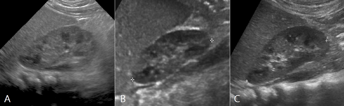
Fig. 1. Serial ultrasound examinations of the right kidney on May 7, 2018 (A), 6 weeks later on June 15 due to recurrent infection (B), and 3 years later on May 24, 2021 (C).

Fig. 2. Voiding cystourethrogram taken on October 5, 2018 showed grade II reflux, with leak of contrast medium to the entire subscapsular space of right kidney (A), and this was proved as subcapsular collection on the lateral view (B). There was no evidence of extrarenal leakage until urination was completed during examination (C).

Fig. 3. Renal computed tomography on January 25, 2021 showed abscess formation in the upper and lower portion of right kidney (A) and the follow-up exam on February 1, 2021 revealed that the size of abscesses had expanded (B).
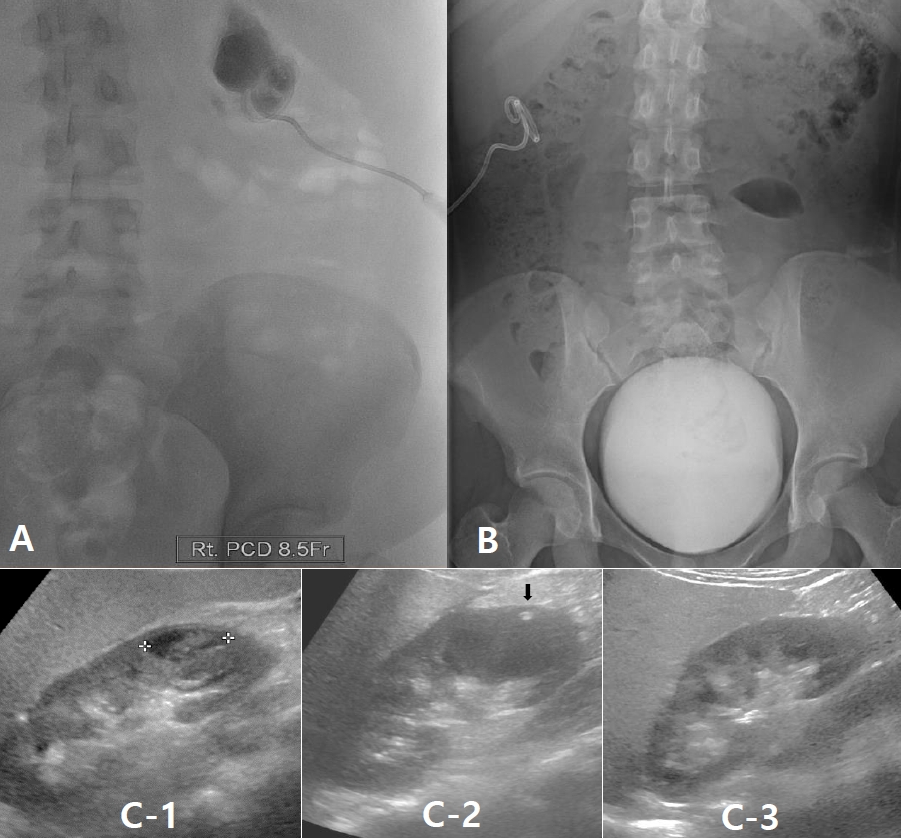
Fig. 4. A percutaneous drainage catheter was inserted into the subcapsular pocket to drain pus on February 2, 2021 (A) and VCUG was performed on 3 days after admission with no evidence of reflux stream (B). Renal ultrasonography on January 25, 2021 showed an abscess at the lower pole of the right kidney (C-1). This lesion disappeared 4 days after PCD, and the PCD catheter tip was noted (arrow) (C-2). Follow-up examination on February 24, 2021 showed complete recovery of the right kidney (C-3).
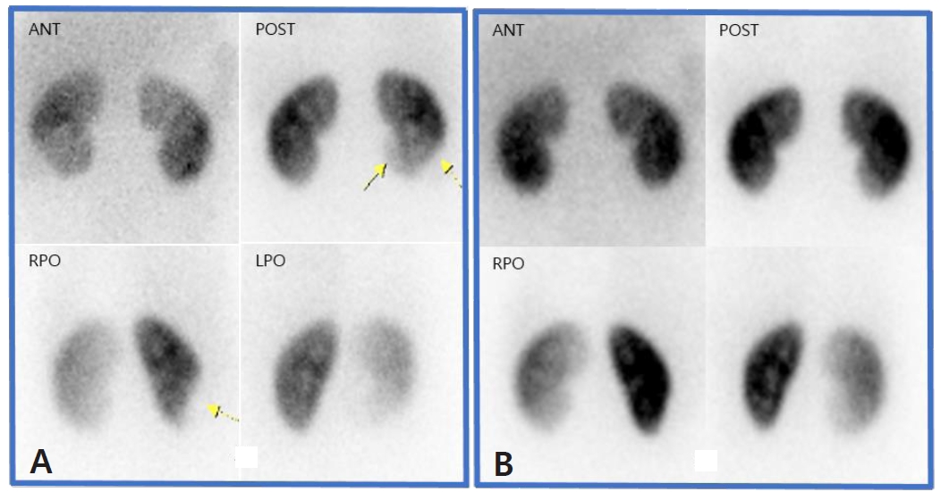
Fig. 5. Tc-99m DMSA renal scan on February 3, 2021 showed large photon defect in lower portion of right kidney (A), and the lesion completely disappeared on the follow-up exam after 6 months (B).
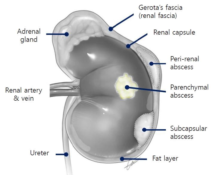
Fig. 6. Classification of renal abscesses: picture produced by the authors to facilitate understanding of the differential diagnosis of renal abscesses based on their locations.
Reference
-
References
1. Bhat YR. Renal subcapsular abscess. Indian Pediatr. 2007; 44:546–7.2. Lee JH, Earm EJ, Han JS, Kim SW, Lee JS. Clinical features of renal and perirenal abscess. The Korean Journal of Nephrology. 1990; 9:357–63.3. Choi SR, Kim CH, Kim SK, Oh JS. A case of multiple abscesses due to renal stone. Clin Exp Pediatr. 1985; 28:1245–9.4. Sim JH, Yim HE, Yoo KH. Two cases of renal and perinephric abscesses in children. Korean Soc Pediatr Nephrol. 2014; 18:116–22.
Article5. Jeong JY, Park YS, Ham SY. A case of renal abscess in healthy child. J Korean Pediatr Soc. 2000; 43:1012–5.6. Chi BH, Kim TH, Park SJ, Kim JY. A clinical survey of renal and perirenal abscess. Korean J Urogenit Tract Infect Inflamm. 2009; 4:190–5.7. Choi SY, Hwang DH, Yoon HS. A case of renal abscess in neonate. J Korean Soc Neonatol. 2005; 12:200–5.8. Finn DJ, Palestrant AM, DeWolf WC. Successful percutaneous management of renal abscess. J Urol. 1982; 127:425–6.
Article9. Seguias L, Srinivasan K, Mehta A. Pediatric renal abscess: a 10-year single-center retrospective analysis. Hosp Pediatr. 2012; 2:161–6.
Article10. Maitra NU, Conn IG. A unique case of spontaneous bilateral renal subcapsular urinoma secondary to high pressure chronic retention of urine. Urol Case Rep. 2017; 14:21–3.
Article11. Tsai JD, Huang FY, Lin CC, Tsai TC, Lee HC, Sheu JC, et al. Intermittent hydronephrosis secondary to ureteropelvic junction obstruction: clinical and imaging features. Pediatrics. 2006; 117:139–46.
Article12. Beetz R, Stein R, Rohatsch P, Brzezinska R, Thuroff JW. Acute perirenal extravasation of urine in an infant with non-refluxing megaureter. Pediatr Nephrol. 2004; 19:357–60.13. Titton RL, Gervais DA, Hahn PF, Harisinghani MG, Arellano RS, Mueller PR. Urine leaks and urinomas: diagnosis and imagingguided intervention. Radiographics. 2003; 23:1133–47.
Article
- Full Text Links
-
- Actions
-
Cited
- CITED
-
- Close
- Share
-
- Similar articles
-
- Two cases of subcapsular renal hematoma as a complication of acute pyelonephritis
- A Case of Subcapsular Liver Abscess Secondary to Perforating Ulcer of Gastric Cancer
- A Case of Renal Subcapsular Abscess Complicated with Psoas Abscess and Femoral Vein Thrombosis in Diabetic Patient
- A Case of Spontaneous Renal Subcapsular Hematoma and Acute Renal Failure Developed after Cesarean Section with Severe Preeclampsia
- Effect of Subcapsular Orchiectomy on Serum Testosterone in Rats
