Anat Cell Biol.
2021 Dec;54(4):518-521. 10.5115/acb.21.083.
Giant dural arteriovenous fistula in an infant
- Affiliations
-
- 1Department of Neurosurgery, Tulane Center for Clinical Neurosciences, Tulane University School of Medicine, New Orleans, LA, USA.
- 2Department of Neurology, Tulane Center for Clinical Neurosciences, Tulane University School of Medicine, New Orleans, LA, USA.
- 3Department of Structural & Cellular Biology, Tulane University School of Medicine, New Orleans, LA, USA.
- 4Department of Neurosurgery and Ochsner Neuroscience Institute, Ochsner Health System, New Orleans, LA, USA.
- 5Department of Anatomical Sciences, St. George's University, St. George's, Grenada.
- 6Department of Surgery, Tulane University School of Medicine, New Orleans, LA, USA.
- KMID: 2523581
- DOI: http://doi.org/10.5115/acb.21.083
Abstract
- Dural arteriovenous fistulas (dAVFs) are commonly encountered by the neurosurgeon. Herein, we present a case illustration of an infant presenting with an extremely large fistula that took up a significant part of the intracranial volume. A one-month-old female presented with irritability and failure to thrive. She was the product of a 35-week pregnancy and was delivered vaginally without complications or a difficult labor. Based on the findings of magnetic resonance imaging, the diagnosis of a giant dAVF involving the transerve-sigmoid sinuses was made. The patient was scheduled for an arteriogram but died before the procedure could be performed. Such a case illustrates how large some dAVF can become and at a very early age. As in the present case, the patient was minimally symptomatic. Therefore, the time to intervention after diagnosis is thus, sometimes, critical.
Figure
-
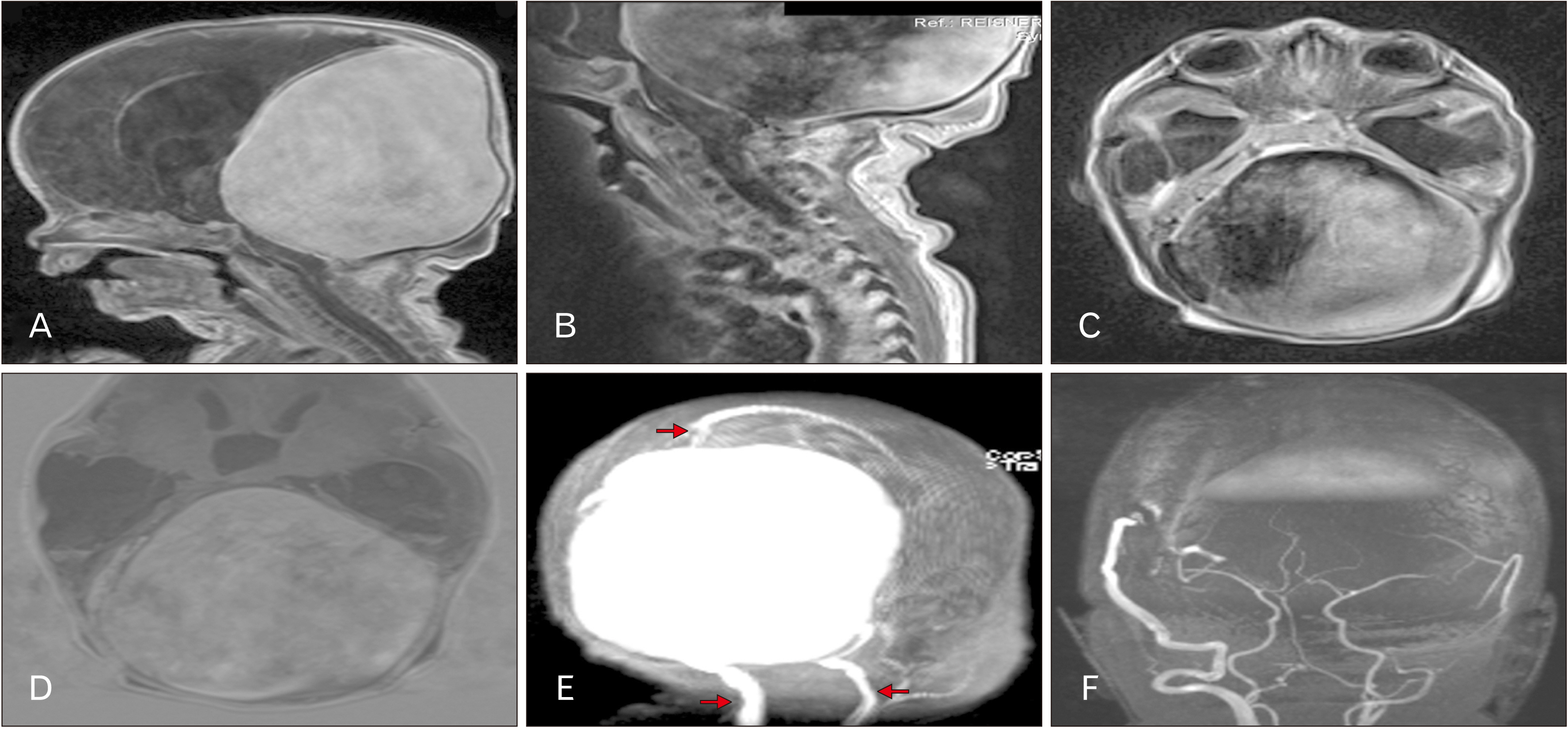
Fig. 1 (A) T1-weighted magnetic resonance imaging (MRI) noting the large intracranial mass. Hydrocephalus is evident in the lateral ventricles. (B) T1-weighted MRI noting the compression of the contents of the posterior cranial fossa. (C) T1-weighted MRI noting the large intracranial mass filling most of the posterior cranial fossa. Hydrocephalus is evident in the temporal horns. (D) Axial image noting the dilated ventricular system. Hydrocephalus is evident in the third ventricle. (E) Scout image illustrating the giant mass and dilated internal jugular veins (lower arrows). Note the superior sagittal sinus at the upper arrow. (F) Note the normal appearance of the components of the circle of Willis and vertebrobasilar system.
Reference
-
References
1. Li Q, Zhang Q, Huang QH, Fang YB, Zhang ZL, Xu Y, Liu JM. 2014; A pivotal role of the vascular endothelial growth factor signaling pathway in the formation of venous hypertension-induced dural arteriovenous fistulas. Mol Med Rep. 9:1551–8. DOI: 10.3892/mmr.2014.2037. PMID: 24626343. PMCID: PMC4020488.
Article2. Xu K, Yang X, Li C, Yu J. 2018; Current status of endovascular treatment for dural arteriovenous fistula of the transverse-sigmoid sinus: a literature review. Int J Med Sci. 15:1600–10. DOI: 10.7150/ijms.27683. PMID: 30588182. PMCID: PMC6299407.
Article3. Cognard C, Gobin YP, Pierot L, Bailly AL, Houdart E, Casasco A, Chiras J, Merland JJ. 1995; Cerebral dural arteriovenous fistulas: clinical and angiographic correlation with a revised classification of venous drainage. Radiology. 194:671–80. DOI: 10.1148/radiology.194.3.7862961. PMID: 7862961.
Article4. Borden JA, Wu JK, Shucart WA. 1995; A proposed classification for spinal and cranial dural arteriovenous fistulous malformations and implications for treatment. J Neurosurg. 82:166–79. DOI: 10.3171/jns.1995.82.2.0166. PMID: 7815143.
Article5. Adamczyk P, Amar AP, Mack WJ, Larsen DW. 2012; Recurrence of "cured" dural arteriovenous fistulas after Onyx embolization. Neurosurg Focus. 32:E12. DOI: 10.3171/2012.2.FOCUS1224. PMID: 22537121. PMCID: PMC4085989.
Article6. Dawson RC 3rd, Joseph GJ, Owens DS, Barrow DL. 1998; Transvenous embolization as the primary therapy for arteriovenous fistulas of the lateral and sigmoid sinuses. AJNR Am J Neuroradiol. 19:571–6. PMID: 9541321. PMCID: PMC8338260.7. Piechowiak E, Zibold F, Dobrocky T, Mosimann PJ, Bervini D, Raabe A, Gralla J, Mordasini P. 2017; Endovascular treatment of dural arteriovenous fistulas of the transverse and sigmoid sinuses using transarterial balloon-assisted embolization combined with transvenous balloon protection of the venous sinus. AJNR Am J Neuroradiol. 38:1984–9. DOI: 10.3174/ajnr.A5333. PMID: 28818827. PMCID: PMC7963627.
Article8. Giller CA, Barnett DW, Thacker IC, Hise JH, Berger BD. 2008; Multidisciplinary treatment of a large cerebral dural arteriovenous fistula using embolization, surgery, and radiosurgery. Proc (Bayl Univ Med Cent). 21:255–7. DOI: 10.1080/08998280.2008.11928405. PMID: 18628973. PMCID: PMC2446414.
Article9. Reynolds MR, Lanzino G, Zipfel GJ. 2017; Intracranial dural arteriovenous fistulae. Stroke. 48:1424–31. DOI: 10.1161/STROKEAHA.116.012784. PMID: 28432263. PMCID: PMC5435465.
Article10. Friedman JA, Pollock BE, Nichols DA, Gorman DA, Foote RL, Stafford SL. 2001; Results of combined stereotactic radiosurgery and transarterial embolization for dural arteriovenous fistulas of the transverse and sigmoid sinuses. J Neurosurg. 94:886–91. DOI: 10.3171/jns.2001.94.6.0886. PMID: 11409515.
Article11. Mullan S, Mojtahedi S, Johnson DL, Macdonald RL. 1996; Embryological basis of some aspects of cerebral vascular fistulas and malformations. J Neurosurg. 85:1–8. DOI: 10.3171/jns.1996.85.1.0001. PMID: 8683257.
Article12. Kaushik KS, Acharya UV, Ananthasivan R, Girishekar B, Reddy P. 2020; Fetal dural sinus malformation. Neurology. 95:452–3. DOI: 10.1212/WNL.0000000000010446. PMID: 32753437.
Article
- Full Text Links
-
- Actions
-
Cited
- CITED
-
- Close
- Share
-
- Similar articles
-
- Endovascular Treatment of Dural Sinus Malformation in Infant: A Case Report
- Endovascular Treatment of Spinal Dural and Epidural Arteriovenous Fistula as Complication of Lumbar Surgery
- Stereotactic radiosurgery for dural arteriovenous fistula
- A Case of Dural Arteriovenous Fistula of the Anterior Condylar Vein
- Occurrence of Metachronous Intracranial Dural Arteriovenous Fistula after Embolization of Intracranial Dural Arteriovenous Fistula: A Case Report
